Thermo Fisher Scientific › Electron Microscopy › Electron Microscopes › 3D Visualization, Analysis and EM Software › Use Case Gallery
Lamellar bodies (LBs) are surfactant rich organelles in alveolar type 2 cells. LBs disassemble into a lipid-protein network that reduces surface tension and facilitates gas exchange at the air-water interface in the alveolar cavity. Current knowledge of LB architecture is predominantly based on electron microscopy studies using disruptive sample preparation methods. We established a post-correlation on-lamella cryo-correlative light and electron microscopy approach for cryo-FIB milled lung cells to structurally characterize and validate LB identity in their unperturbed state using the well-established ABCA3-eGFP marker. In situ cryo-electron tomography revealed that LBs are composed of lipidic structures unique in organelle biology. We report open-ended membrane sheets frequently attached to the limiting membrane of LBs and so far undescribed dome-shaped protein complexes. We propose that LB biogenesis is driven by parallel membrane sheet import and the curvature of the limiting membrane to maximize lipid storage capacity.
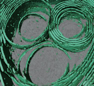
Renderings were created in Amira 2019.3. First, a subvolume was extracted and manually segmented based on image enhancement and thresholding. Based on this, a convoluted neural network was trained using the interface implemented in Amira 2019.3. The output was used to visualize membranes in the full tomogram.
For Research Use Only. Not for use in diagnostic procedures.