Thermo Fisher Scientific › Electron Microscopy › Electron Microscopes › 3D Visualization, Analysis and EM Software › Use Case Gallery
Accumulating evidence indicates the critical importance of cerebrovascular dysfunction in the pathogenesis of Alzheimer’s disease (AD). However, systematic comparative studies on the precise brain vasculature of wild-type and AD model mice are still rare. Using an image optimization method for analyzing Micro-Optical Sectioning Tomography (MOST) data, we generated cross-scale whole-brain 3D atlases that cover the entire vascular system from large vessels down to smallest capillaries at submicron resolution, for both wild-type mice and a transgenic (APP/PS1) mouse model of AD. In addition to distinct vascular patterns in different brain regions, we found that main vessels of the molecular layer of hippocampal dentate gyrus (DG-ml) undergo abrupt changes in both diameter and branch angle, spreading a unique comb-like pattern of capillaries. By using a quantitative analysis workflow, we identified in the hippocampus of AD mice an overall reduction of the mean vascular diameter, volume fraction and branch angle, with most significant impairment in the DG-ml. In addition, virtual endoscopy revealed irregular morphological features in the vessel lumen of the AD mice, potentially contributing to the impairment of blood flow. Our results demonstrate the capability of high-resolution cross-scale evaluation of brain vasculature, and underscore the importance of studying hippocampal microcirculation for understanding AD pathogenesis.
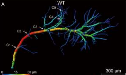
“Segment manually slice by slice with brush to get contours using the segmentation editor in Amira software in reference to the Allen Reference Atlas and the Franklin and Paxinos Mouse Atlas (…) Surface reconstruction of hippocampal formation in Amira software (…) Visualization of the cerebrovascular network: Take advantages of volume rendering module in Amira and the high-end GPU card, hardware-accelerated real-time rendering was performed for the 3D visualization. (…) The blood vessels of the whole mouse hippocampus were also visualized using Amira software after getting the segmented images. (…) Then the single vascular branch was manually extracted and reconstructed from the complicated hippocampal vascular network using the AMIRA segmentation program. The reconstruction of vascular network in Tg-AD and WT mice was also carried out following the technical route above. (…) Quantification of hippocampal vasculature: The calculation and analysis were conducted by using Amira (…).”
For Research Use Only. Not for use in diagnostic procedures.