Thermo Fisher Scientific › Electron Microscopy › Electron Microscopes › 3D Visualization, Analysis and EM Software › Use Case Gallery
Masseter muscle function influences mandibular bone homeostasis. As previously reported, bone resorption markers increased in the mouse mandibular condyle two days after masseter paralysis induced with botulinum toxin type A (BoNTA), followed by local bone loss. This study aimed to evaluate the bone quality of both the mandibular condyle and alveolar process in the mandible of adult mice during the early stage of a BoNTA‐induced masseter muscle atrophy, using a combined 3D histomorphometrics and shape analysis approach.
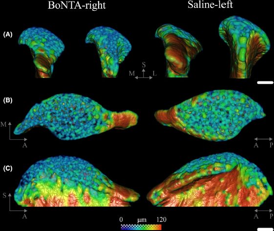
After 2 weeks, masseter mass was significantly reduced (P‐value <0.001). When compared to Saline‐left and untreated condyles, BoNTA‐right condyles showed significant bone loss (P‐value <0.001) and shape changes. No significant bone loss was observed in the alveolar processes of any of the groups (P‐value >0.05). Condyle bone quality deteriorates at an early stage of BoNTA‐induced masseter muscle atrophy, and before the alveolar process is affected. Since the observed bone microstructural changes resemble those in human temporomandibular joint degenerative disorders, the clinical safety of BoNTA intervention in the masticatory apparatus remains to be clarified.
The resulting stacks of 2D‐tiff images were processed in Avizo 9.2. The bone tissue from mandibular condyles and alveolar processes was segmented using a threshold‐based approach in Avizo. Then, in order to separate the bone tissue from the teeth (enamel and dentine) in the alveolar processes, preselected slices from Avizo were processed using a semi‐automated workflow based on a diffusion algorithm. Triangulated surface renderings of mandibular condyles were obtained in the Avizo for shape analysis.
For Research Use Only. Not for use in diagnostic procedures.