Segmentation of cortical bone, trabecular bone, and medullary pores from micro-CT images using 2D and 3D deep learning models

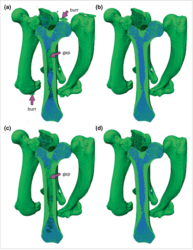
Computed tomography (CT) enables rapid imaging of large-scale studies of bone, but those datasets typically require manual segmentation, which is time-consuming and prone to error. Convolutional neural networks (CNNs) offer an automated solution, achieving superior performance on image data. In this methodology-focused paper, we used CNNs to train segmentation models from scratch on 2D and 3D patches from micro-CT scans of otter long bones. These new models, collectively called BONe (Bone One-shot Network), aimed to be fast and accurate, and we expected enhanced results from 3D training due to better spatial context. Contrary to expectations, 2D models performed slightly better than 3D models in labeling details such as thin trabecular bone. Although lacking in some detail, 3D models appeared to generalize better and predict smoother internal surfaces than 2D models. However, the massive computational costs of 3D models limit their scalability and practicality, leading us to recommend 2D models for bone segmentation. BONe models showed potential for broader applications with variation in performance across species and scan quality. Notably, BONe models demonstrated promising results on skull segmentation, suggesting their potential utility beyond long bones with further refinement and fine-tuning.