Welcome to the Amira-Avizo Software Use Case Gallery
Below you will find a collection of use cases of our 3D data visualization and analysis software. These use cases include scientific publications, articles, papers, posters, presentations or even videos that show how Amira-Avizo Software is used to address various scientific and industrial research topics.
Use the Domain selector to filter by main application area, and use the Search box to enter keywords related to specific topics you are interested in.
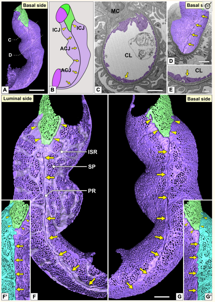
Three-Dimensional Architecture of Glomerular Endothelial Cells Revealed by FIB-SEM Tomography
Focused-ion beam-scanning electron microscopic (FIB-SEM) tomography enables easier acquisition of a series of ultrastructural, sectional images directly from resin-embedded biological samples. In this study, to clarify the three-dimensional (3D) architecture of glomerular endothelial cells (GEnCs) in adult rats, we manually extracted GEnCs from serial FIB-SEM images and reconstructed them on an Amira reconstruction software. The luminal and basal surface structures were clearly visualized in ... Read more
Yuto Kawasaki, Yasue Hosoyamada, Takayuki Miyaki, Junji Yamaguchi, Soichiro Kakuta, Tatsuo Sakai, and Koichiro Ichimura
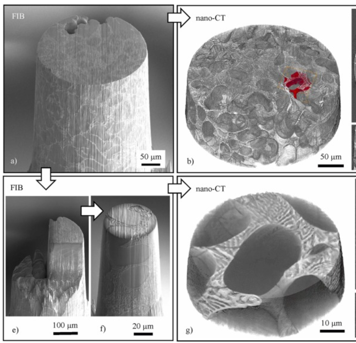
Three-dimensional imaging of microstructural evolution in SEM-based nano-CT
Scanning electron microscopy (SEM) is a powerful and versatile technique for materials characterization and present in many laboratories. The integration of an X-ray target holder and detector allows expanding the modalities of SEM by X-ray imaging. These little hardware adaptations enable radiography ... Read more
Jonas Fell, Christoph Pauly, Michael Maisl, Simon Zabler, Frank Mücklich, Hans-Georg Herrmann
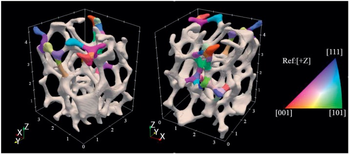
The complex mechanical response of open-cell foams depends strongly on the hierarchy of length scales inherent in them, from engineering-part scale to the ligament scale through the grain scale down to the crystal-lattice scale. A first step toward understanding and predicting the coordinated mechanical response across le... Read more
Jayden C. Plumb, Jonathan F. Lind, Joseph C. Tucker, Ron Kelley, Ashley D. Spear
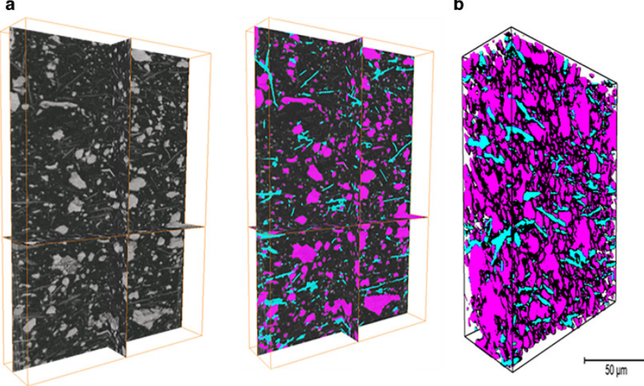
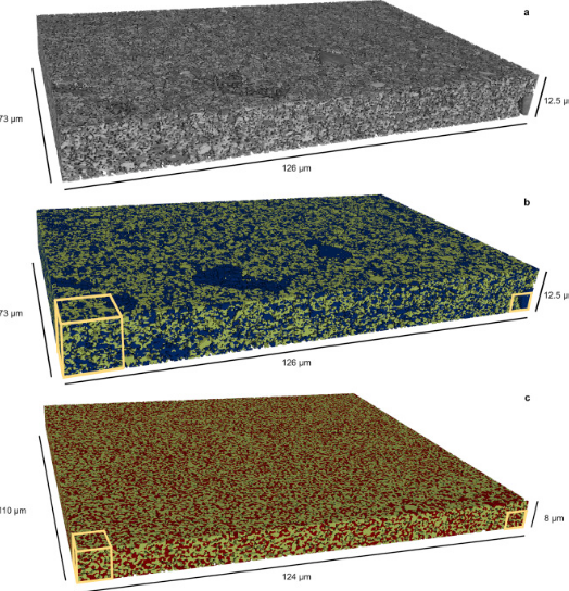
Multi-modal plasma focused ion beam serial section tomography of an organic paint coating
Pigment distributions have a critical role in the corrosion protection properties of organic paint coatings, but they are difficult to image in 3D over statistically significant volumes and at sufficiently high spatial resolutions required for detailed analysis. Here we report, for the first time, large volume analytical serial sectioning tomography of an organic composite coating using a xenon Plasma Focused Ion Beam (PFIB) combined with secondary electron imaging, energy dispersive X-ray (E... Read more
Zhong Xiangli, M. Grace Burke, Philip J. Withers, Zhang Xun, Zhou Xiaorong, Timothy L. Burnett, Liu Yanwen, Stuart B. Lyon, Simon R.Gibbon
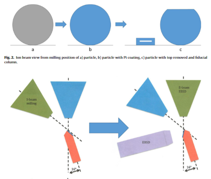
Automated 3D EBSD for metallic powders
Metallic powders are commonly used in additive manufacturing processes. While their post-process consolidated properties are widely studied, there is little research on the properties of the powders prior to consolidation. Understanding the powder characteristics before use in additive manufacturing processes could lead to fine-tuning properties of additively manufactured materials. The three-dimensional grain structure of metals can be useful in predicting their properties and ... Read more
Caitlin Walde, Roger Ristau, Danielle Cote
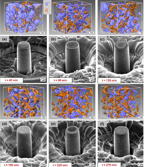
A unique approach to correlating an evolving 3D microstructure in an Al-Cu alloyand its micro-scale mechanical properties has been introduced. Using these nanoscale three-dimensional microstructures derived from Transmission X-rayMicroscopy (TXM), individual contributions from different strengthening mechanisms were quantified. The spatial distribution and morphology of the individual θ′ and θ phases were seen to play an important role in influencing dislocation storage. Uniaxi... Read more
C. Shashank Kaira, Christopher Kantzos, Jason J. Williams, Vincent De Andrade, Francesco De Carlo, Nikhilesh Chawlaa
Mesoscale characterization of local property distributions in heterogeneous electrodes
The performance of electrochemical devices depends on the three-dimensional (3D) distributions of microstructural features in their electrodes. Several mature methods exist to characterize 3D microstructures over the microscale (tens of microns), which are useful in understanding homogeneous electrodes. However, methods that capture mesoscale (hundreds of microns) volumes at appropriate resolution (tens of nm) are lacking, though they are needed to understand more common, less ideal electrode... Read more
Tim Hsu, William K. Epting, Rubayyat Mahbub, Noel T. Nuhfer, Sudip Bhattachary, Yinkai Lei, Herbert M. Miller, Paul R. Ohodnicki, Kirk R. Gerdes, Harry W. Abernathy, Gregory A. Hackett, Anthony D. Rollett, Marc De Graef, Shawn Litster, Paul A. Salvador
Laser processing of metal surfaces by ultrafast Read more
Edwin Peng, Alexander Roth, Craig A. Zuhlke, Soodabeh Azadehranjbar, Dennis R. Alexander, George Gogos, Jeffrey E. Shield
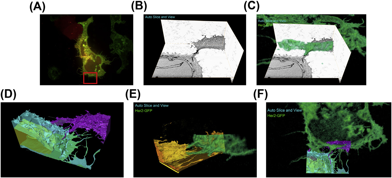
A fully integrated, three-dimensional fluorescence to electron microscopy correlative workflow
While fluorescence microscopy provides tools for highly specific labeling and sensitive detection, its resolution limit and lack of general contrast has hindered studies of cellular structure and protein localization. Recent advances in correlative light and electron microscopy (CLEM), including the fully integrated CLEM workflow instrument, the Thermo Scientific CorrSight with MAPS, have allowed for a more reliable, reproducible, and quicker approach to correlate three-dimensional time-lapse... Read more
Claudia S. Lopez, Cedric Bouchet-Marquis, Christopher P. Arthur, Jessica L. Riesterer, Gregor Heiss, Guillaume Thibault, Lee Pullan, Sunjong Kwon, Joe W. Gray

Synergistic role of nucleotides and lipids for the self-assembly of Shs1 septin oligomers
Amira capacities for membranes and filaments segmentation in cryo-TEM images are featured on the front cover of Biochemical Journal, July 2020.
Budding yeast septins are essential for cell division and polarity. (…) [The authors] have dissected, here, for the first time, the behavior of the Shs1 protomer bound to membranes at nanometer resolution, in complex with the other septins. Using electron microscopy, [the authors] have shown that on membranes, Shs1 protomers self-assembl... Read more
Cyntia Taveneau, Rémi Blanc, Gerard Pehau-Arnaudet, Aurélie Cicco, Aurélie Bertin
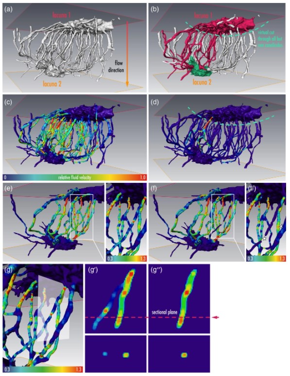
Osteocytes are the most frequent bone cells connected with each other through cell processes within tiny tubular-shaped canaliculi. The so-called osteocyte lacunar-canalicular network (LCN) plays a crucial role in bone remodeling and mineral homeostasis. Given the critical nature of these functions, it is herein hypothesized that the LCN must be structurally “overengineered” to provide network resilience.
This hypothesis is tested by characterizing canalicular networks in human bon... Read more
Emely Bortel, Liam M Grover, Neil Eisenstein, Christian Seim, Heikki Suhonen, Alexandra Pacureanu, Peter Westenberger, Kay Raum, Max Langer, Francoise Peyrin, Owen Addison, Bernhard Hesse
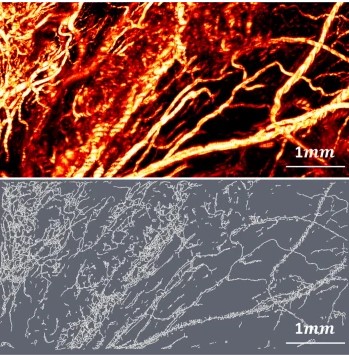
Optical Resolution Photoacoustic Microscopy of Ovary and Fallopian Tube
Ovarian cancer is the leading cause of death among gynecological cancers, but is poorly amenable to preoperative diagnosis. In this study, we investigate the feasibility of “optical biopsy,” using high-optical-resolution photoacoustic microscopy (OR-PAM) to quantify the microvasculature of ovarian and fallopian tube tissue. The technique is demonstrated using excised human ovary and fallopian tube specimens imaged immediately after surgery.
This report describes the first applicatio... Read more
Bin Rao, Xiandong Leng, Yifeng Zeng, Yixiao Lin, Ruimin Chen, Qifa Zhou, Andrea R. Hagemann, Lindsay M. Kuroki, Carolyn K. McCourt, David G. Mutch, Matthew A. Powell, Ian S. Hagemann & Quing Zhu
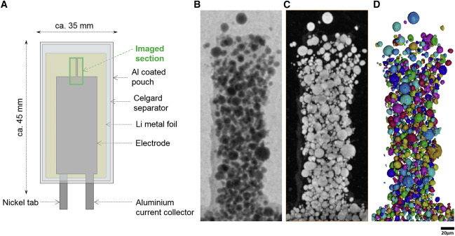
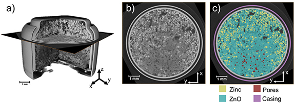
Nickel-rich transition metal oxide materials […] are of great interest for achieving immediate improvements in the energy density of Li-ion batteries and for risk reduction within the Li-ion battery supply chain.
[…] An increase in Ni content in NMC materials leads to accelerated degradation […]. This potentially complicates their adoption in applications requiring extended cycle life such as in electric vehicles.
Recent developments in X-ray characterization to... Read more
ChunTan, Andrew S.Leach, Thomas M.M. Heenan, Huw Parks, Rhodri Jervis, Johanna Nelson Weker, Daniel J.L. Brett, Paul R.Shearing
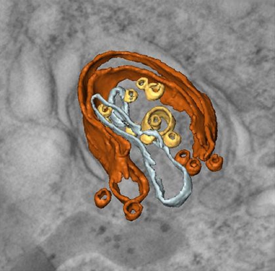
Given the urgent need to move to low-carbon technologies, batteries are being increasingly used in a range of applications. Lithium-ion batteries are the most widely used chemistry, but to meet the growing demand, there is a need to move beyond lithium towards alternative battery chemistries. Metal–air batteries are a group of such battery alternatives that hold promise, especially for stationary power and flexible electronics applications.
However, barriers to their widespread adopt... Read more
Jennifer Hack, Drasti Patel, Josh J Bailey, Francesco Iacoviello, Paul R Shearing, and Dan J L Brett
Macroautophagy is morphologically characterized by autophagosome formation. Autophagosomes are double-membraned vesicles that sequester cytoplasmic components for further degradation in the lysosome. Basal autophagy is paramount for intracellular quality control in post-mitotic cells but, surprisingly, the number of autophagosomes in post-mitotic neurons is very low, suggesting that alternative degradative structures could exist in neurons…
Read more
Maria Rosario Fernandez-Fernandez, Desire Ruiz-Garcia, Eva Martin-Solana, Francisco Javier Chichon, Jose L. Carrascosa, Jose-Jesus Fernandez
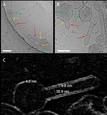
Influenza A matrix protein M1 is sufficient to induce lipid membrane deformation
The matrix protein M1 of the Influenza A virus is considered to mediate viral assembly and budding at the plasma membrane (PM) of infected cells. In order for a new viral particle to form, the PM lipid bilayer has to bend into a vesicle towards the extracellular side. Studies in cellular models have proposed that different viral proteins might be responsible for inducing membrane curvature in this context (including M1), but a clear consensus has not been reached. In this study, we use a comb... Read more
Ismail Dahmani, Kai Ludwig, Salvatore Chiantia
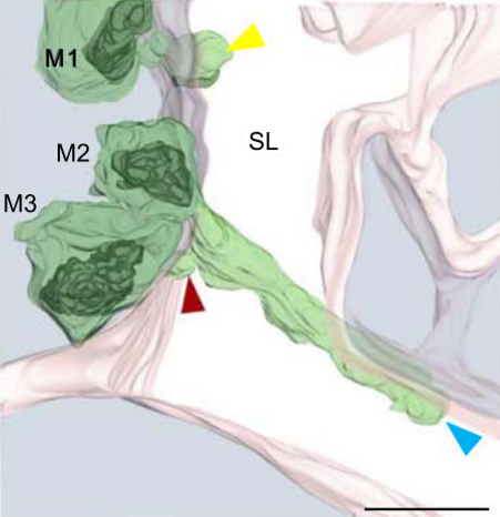
Multiple membrane extrusion sites drive megakaryocyte migration into bone marrow blood vessels
Platelets, cells central to hemostasis and thrombosis, are formed from parent cell megakaryocytes. Although the process is highly efficient in vivo, our ability to generate them in vitro is still remarkably inefficient. We proposed that greater understanding of the process in vivo is needed and used an imaging approach, intravital correlative light electron microscopy, to visualize platelet generation in bone marrow in the living mouse. In contrast to current understanding, we found that most... Read more
Edward Brown, Leo M Carlin, Claus Nerlov, Cristina Lo Celso, Alastair W Poole
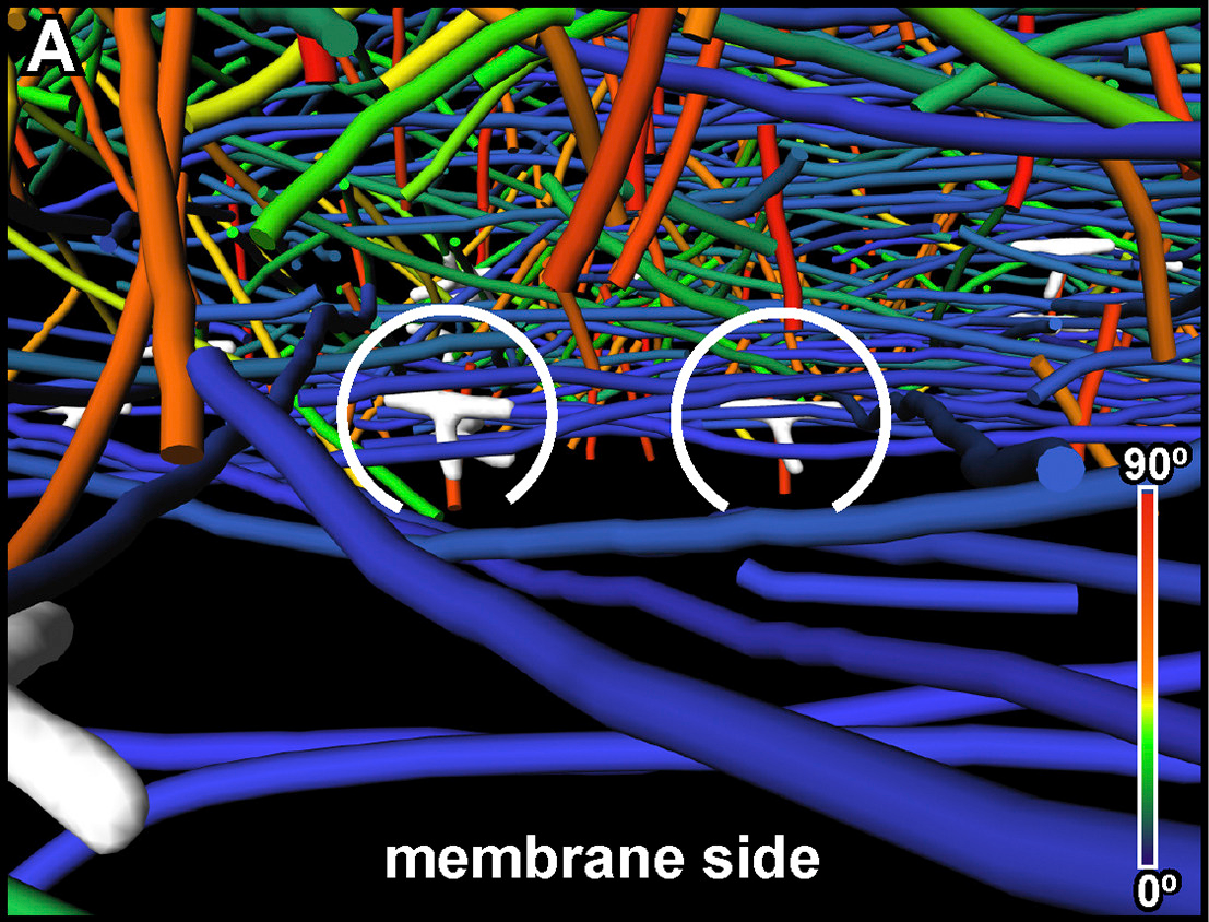
The Architecture of Traveling Actin Waves Revealed by Cryo-Electron Tomography
Actin waves are dynamic supramolecular structures involved in cell migration, cytokinesis, adhesion, and neurogenesis. Although wave-like propagation of actin networks is a widespread phenomenon, the actin architecture underlying wave propagation remained unknown. In situ cryo-electron tomography of Dictyostelium cells unveils the wave architecture and provides evidence for wave progression by de novo actin nucleation. Subtomogram averaging reveals the structu... Read more
Marion Jasnin, Florian Beck, Mary Ecke, Yoshiyuki Fukuda, Antonio Martinez-Sanchez, Wolfgang Baumeister, Günther Gerisch
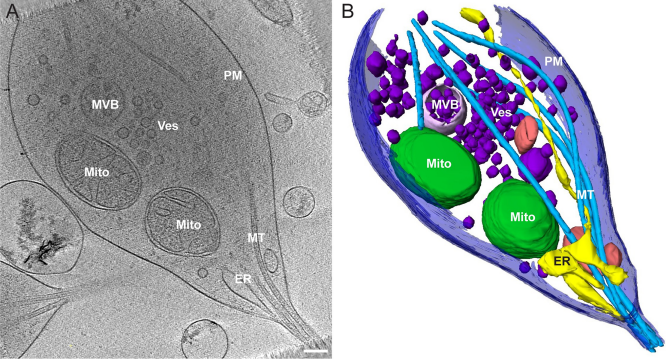
Morphology of mitochondria in spatially restricted axons revealed by cryo-electron tomography
Neurons project axons to local and distal sites and can display heterogeneous morphologies with limited physical dimensions that may influence the structure of large organelles such as mitochondria. Using cryo-electron tomography (cryo-ET), we characterized native environments within axons and presynaptic varicosities to examine whether spatial restrictions within these compartments influence the morphology of mitochondria. Segmented tomographic reconstructions revealed distinctive morphologi... Read more
Tara D. Fischer, Pramod K. Dash, Jun Liu, M. Neal Waxham
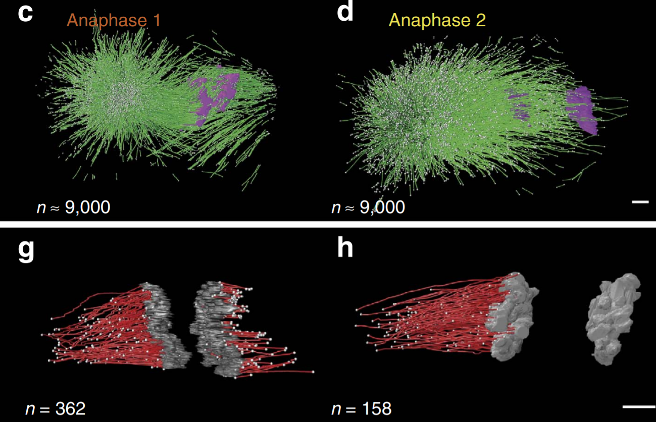
C. elegans chromosomes connect to centrosomes by anchoring into the spindle network
The mitotic spindle ensures the faithful segregation of chromosomes. Here we combine the first large-scale serial electron tomography of whole mitotic spindles in early C. elegans embryos with live-cell imaging to reconstruct all microtubules in 3D and identify their plus- and minus-ends. We classify them as kinetochore (KMTs), spindle (SMTs) or astral microtubules (AMTs) according to their positions, and quantify distinct properties of each class. While our light microscopy and muta... Read more
Stefanie Redemann, Johannes Baumgart, Norbert Lindow, Michael Shelley, Ehssan Nazockdast, Andrea Kratz, Steffen Prohaska, Jan Brugués, Sebastian Fürthauer & Thomas Müller-Reichert
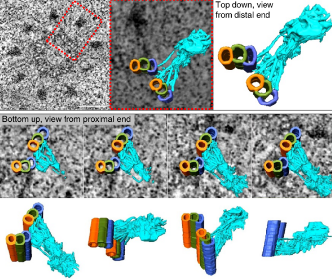
Centrioles are vital cellular structures that form centrosomes and cilia. The formation and function of cilia depends on a set of centriole’s distal appendages. In this study, we use correlative super resolution and electron microscopy to precisely determine where distal appendage proteins localize in relation to the centriole microtubules and appendage electron densities. Here we characterize a novel distal appendage protein ANKRD26 and detail, in high resolution, the initial steps of dist... Read more
Mathew Bowler, Dong Kong, Shufeng Sun, Rashmi Nanjundappa, Lauren Evans, Veronica Farmer, Andrew Holland, Moe R. Mahjoub, Haixin Sui & Jadranka Loncarek