Welcome to the Amira-Avizo Software Use Case Gallery
Below you will find a collection of use cases of our 3D data visualization and analysis software. These use cases include scientific publications, articles, papers, posters, presentations or even videos that show how Amira-Avizo Software is used to address various scientific and industrial research topics.
Use the Domain selector to filter by main application area, and use the Search box to enter keywords related to specific topics you are interested in.
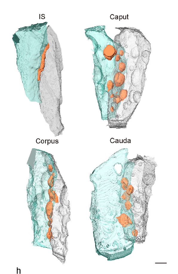
Mammalian epididymal epithelial cells are crucial for sperm maturation. Historically,
vacuole-like ultrastructures in epididymal epithelial cells were
observed via transmission electron microscopy but were undefined. Here, we
utilize volume electron microscopy (vEM) to generate 3D reconstructions of
epididymal epithelial cells and identify these vacuoles as intercellular organelle
reservoirs (IORs) in the lateral intercellular space (LIS), which contains
pr... Read more
Xia Li, Feng Qiao, Jiansheng Guo, Ting Jiang, Huifang Lou, Huixia Li, Gangcai Xie, Hangjun Wu, Weizhen Wang, Ruoyu Pei, Sha Liu, Mei Ye, Jin Li, Shiqin Huang, Mengya Zhang, Chaoye Ma, Yiwen Huang, Shushu Xu, Xiaofeng Li, Xiao Sun, Jun Yu, Kin Lam Fok, Shumin Duan & Hao Chen
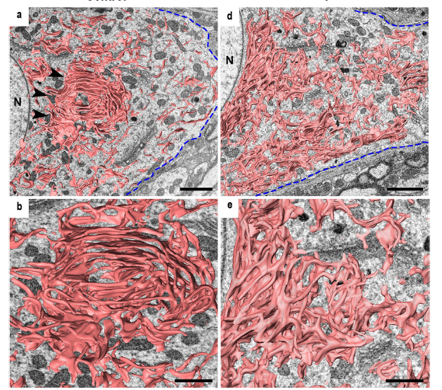
The endoplasmic reticulum (ER) extends throughout a cell and plays a critical role in maintaining cellular homeostasis.
Changes in ER shape could provide a clue to explore the mechanisms that underlie the fate determination of neurons after
axon injury because the ER drastically changes its morphology under neuronal stress to maintain cellular homeostasis and
recover from damage. Because of their tiny structures and richness in the soma, the detailed morphology of the ER and... Read more
Mahmoud Elgendy,Hiromi Tamada, Takaya Taira, Yuma Iio, Akinobu Kawamura, Ayusa Kunogi, Yuka Mizutani, Hiroshi Kiyama
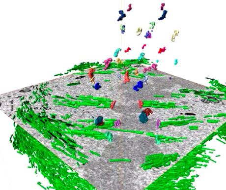
Serial block face scanning electron microscopy (SBF-SEM) is a powerful method to analyze cells in 3D. Here, working at the resolution limit of the method, we describe a correlative light–SBF-SEM workflow to resolve microtubules of the mitotic spindle in human cells. We present four examples of uses for this workflow that are not practical by light microscopy and/or transmission electron microscopy. First, distinguishing closely associated microtubules within K-fibers; second, resolving brid... Read more
Faye M. Nixon, Thomas R. Honnor, Nicholas I. Clarke, Georgina P. Starling, Alison J. Beckett, Adam M. Johansen, Julia A. Brettschneider, Ian A. Prior, Stephen J. Royle