Thermo Fisher Scientific › Electron Microscopy › Electron Microscopes › 3D Visualization, Analysis and EM Software › Use Case Gallery
A detailed understanding of scaphoid anatomy helps anatomic fracture reduction and optimal screw position. Therefore, we analyzed the size and shape variations of the cartilage and osseous surface, the distribution of volumetric bone mineral density (vBMD), and if the vBMD values differ between a peripheral and a central screw pathway?
Forty-three fresh frozen hand specimens (17 females, 26 males) were analysed with high-resolution peripheral quantitative computed tomography (HR-pQCT) and dissected to compute a 3D-statistical osseous and cartilage surface model and a 3D-averaged vBMD model of the scaphoid. 3D patterns were analysed using principal component analysis (PCA). vBMD was analysed via averaging HR-pQCT grey values and virtual bone probing along a central and peripheral pathway. High anatomical variations regarding the osseous and cartilage surfaces were associated with three distinct concentrically arranged zones with notable different vBMD. The complex scaphoid anatomy with its waist might alter the strategy of fracture fixation, education and research.
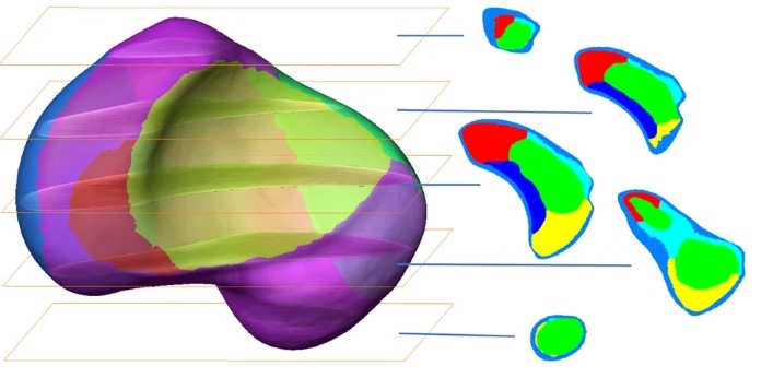
In Amira, the HU of non-osseous tissue with HU less than 0 were set to 0. All images of the left hand were mirrored to the right. Standard threshold-based, semi-automated image segmentation was performed to create 3D computer models of each scaphoid. Nutrient foramina which could be identified due to the high scan resolution were closed manually to facilitate the subsequent data processing. HR-pQCT post-processing resulted in two types of 3D statistical models of the scaphoid: a 3D statistical surface model that demonstrated 3D size and shape and variation patterns of the bony and cartilage surfaces of the scaphoid (external configuration), and a 3D averaged bone density model which provided information about the scaphoid’s internal configuration.
For Research Use Only. Not for use in diagnostic procedures.