Thermo Fisher Scientific › Electron Microscopy › Electron Microscopes › 3D Visualization, Analysis and EM Software › Use Case Gallery
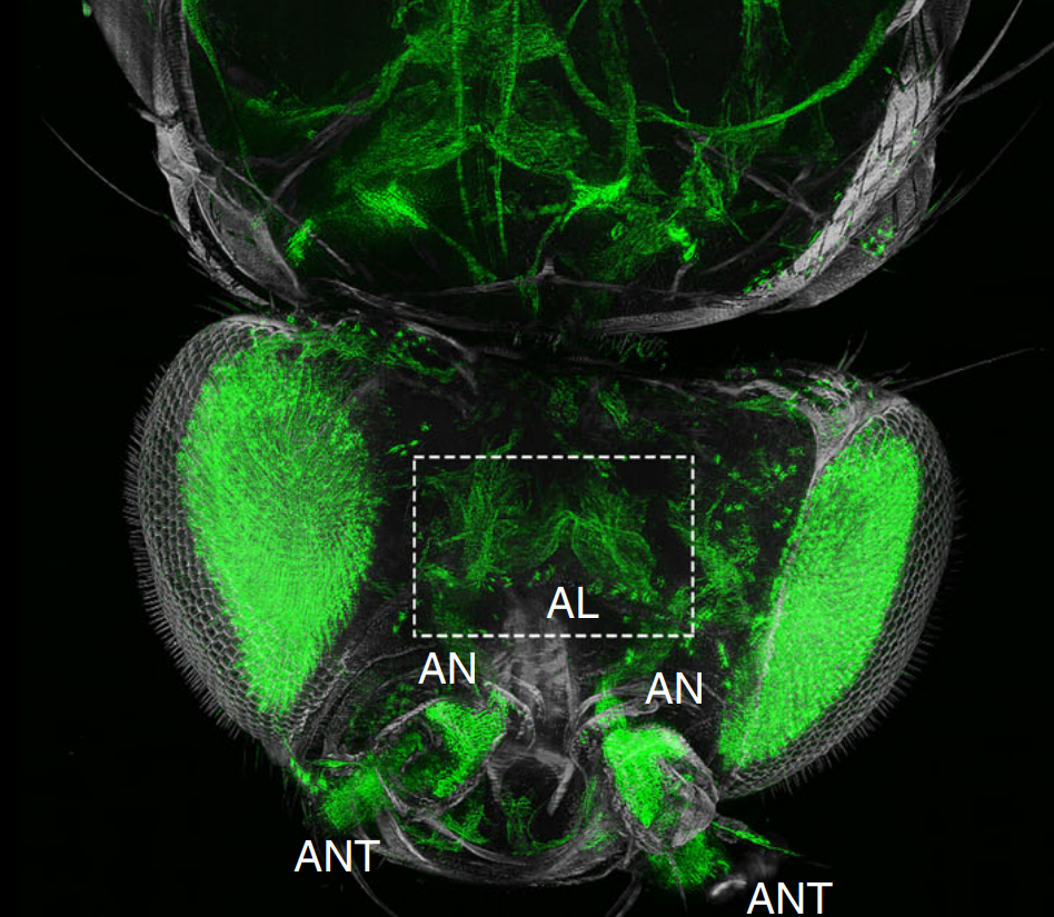
The fruit fly, Drosophila melanogaster, is an important experimental model to address central questions in neuroscience at an organismic level. However, imaging of neural circuits in intact fruit flies is limited due to structural properties of the cuticle. Here we present a novel approach combining tissue clearing, ultramicroscopy, and data analysis that enables the visualisation of neuronal networks with single-cell resolution from the larval stage up to the adult Drosophila. (…) This methodological integration of novel chemical, optical, and computational techniques allows a major advance in the analysis of global neural circuit organisation.
The recorded image stacks were 3D reconstructed and spatially registered using Amira software. The recorded image stacks were 3D reconstructed and spatially registered using Amira software.
For Research Use Only. Not for use in diagnostic procedures.