Thermo Fisher Scientific › Electron Microscopy › Electron Microscopes › 3D Visualization, Analysis and EM Software › Use Case Gallery
Ligaments serve as compliant connectors between hard tissues. In that role, they function under various load regimes and directions. The 3D structure of ligaments is considered to form as a uniform entity that changes due to function. The periodontal ligament (PDL) connects the tooth to the bone and sustains different types of loads in various directions. Using the PDL as a model, employing a fabricated motorized setup in a microCT, we demonstrate that the fibrous network structure within the PDL is not uniform, even before the tooth becomes functional. Utilizing morphological automated segmentation methods, directionality analysis, as well as second harmonic generation imaging, we find high correlation between blood vessel distribution and fiber density. We also show a structural feature in a form of a dense collar around the neck of the tooth as well as a preferred direction of the fibrous network. Finally, we show that the PDL develops as a nonuniform structure, with an architecture designed to sustain specific types of load in designated areas. Based on these findings, we propose that ligaments in general should be regarded as nonuniform entities, structured already at developmental stages for optimal functioning under variable load regimes.
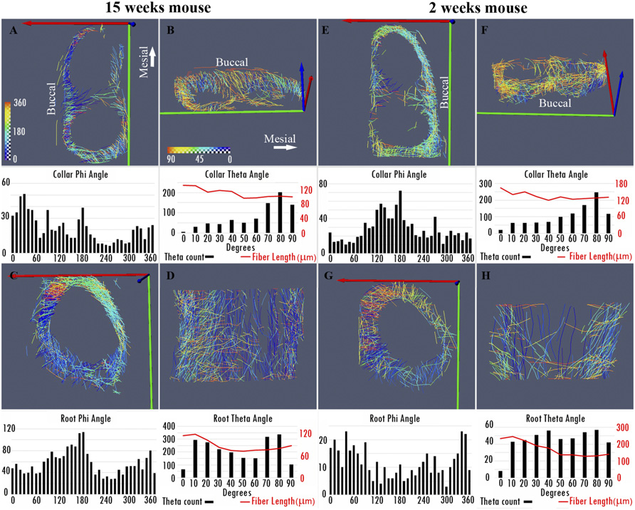
Directionality Analysis. Visualizing the entire PDL fibrous component in 3D revealed that the longitudinal backbone of the fibers displays a certain directionality in the mesial–distal direction. We therefore carried out a 3D directionality analysis comparing the phi and theta angles for fully functioning PDL and preeruptive state PDL. For the analysis, we used the cylinder correlation and trace correlation line modules in Avizo software (Xfiber Extension version 9.4, Thermo Scientific).
For Research Use Only. Not for use in diagnostic procedures.