Thermo Fisher Scientific › Electron Microscopy › Electron Microscopes › 3D Visualization, Analysis and EM Software › Use Case Gallery
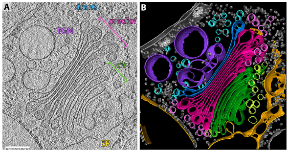
COPI-coated vesicles mediate trafficking within the Golgi apparatus and from the Golgi to the endoplasmic reticulum. Here, we applied cryo-focused ion beam milling, cryo-electron tomography and subtomogram averaging to determine the native structure of the COPI coat within vitrified Chlamydomonas reinhardtii cells. The native algal structure resembles the in vitro mammalian structure, but additionally reveals cargo bound beneath beta’–COP. We find that all coat components disassemble simultaneously and relatively rapidly after budding. Structural analysis in situ, maintaining Golgi topology, shows that vesicles change their size, membrane thickness, and cargo content as they progress from cis to trans, but the structure of the coat machinery remains constant.
Tomograms were visualized in Amira software. We localized 267 COPI vesicles and buds, measured their membrane-to-membrane diameters and determined their central coordinates.
For Research Use Only. Not for use in diagnostic procedures.