Thermo Fisher Scientific › Electron Microscopy › Electron Microscopes › 3D Visualization, Analysis and EM Software › Use Case Gallery
In biomedical research, a huge variety of different techniques is currently available for the structural examination of small specimens, including conventional light microscopy (LM), transmission electron microscopy (TEM), confocal laser scanning microscopy (CLSM), microscopic X-ray computed tomography (microCT), and many others. Since every imaging method is physically limited by certain parameters, a correlative use of complementary methods often yields a significant broader range of information. Here we demonstrate the advantages of the correlative use of microCT, light microscopy, and transmission electron microscopy for the analysis of small biological samples.
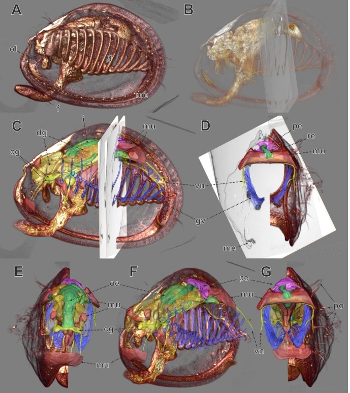
We used a small juvenile bivalve mollusc (Mytilus galloprovincialis, approximately 0.8 mm length) to demonstrate the workflow of a correlative examination by microCT, LM serial section analysis, and TEM-re-sectioning. Initially these three datasets were analyzed separately, and subsequently they were fused in one 3D scene. This workflow is very straightforward. The specimen was processed as usual for transmission electron microscopy including post-fixation in osmium tetroxide and embedding in epoxy resin. Subsequently it was imaged with microCT. Post-fixation in osmium tetroxide yielded sufficient X-ray contrast for microCT imaging, since the X-ray absorption of epoxy resin is low. Thereafter, the same specimen was serially sectioned for LM investigation. The serial section images were aligned and specific organ systems were reconstructed based on manual segmentation and surface rendering. According to the region of interest (ROI), specific LM sections were detached from the slides, re-mounted on resin blocks and re-sectioned (ultrathin) for TEM. For analysis, image data from the three different modalities was co-registered into a single 3D scene using the software AMIRA®. We were able to register both the LM section series volume and TEM slices neatly to the microCT dataset, with small geometric deviations occurring only in the peripheral areas of the specimen. Based on co-registered datasets the excretory organs, which were chosen as ROI for this study, could be investigated regarding both their ultrastructure as well as their position in the organism and their spatial relationship to adjacent tissues. We found structures typical for mollusc excretory systems, including ultrafiltration sites at the pericardial wall, and ducts leading from the pericardium towards the kidneys, which exhibit a typical basal infolding system.
The datasets gained from microCT (volume dataset), LM (volume dataset, surface model) and TEM (individual sections) were co-registered and combined to a single visualization using AMIRA Software.
For Research Use Only. Not for use in diagnostic procedures.