Thermo Fisher Scientific › Electron Microscopy › Electron Microscopes › 3D Visualization, Analysis and EM Software › Use Case Gallery
We investigated the synaptic innervation of apical tufts of cortical pyramidal cells in a region between layers 1 and 2 using 3-D electron microscopy (3D-EM) applied to four cortical regions in mouse. Across all cortices, we found the relative inhibitory input at the apical dendrite’s main bifurcation to be more than 3-fold stronger for layer 2 pyramidal cells than for all other pyramidal cells. Towards the distal tuft dendrites in upper layer 1, however, the relative inhibitory input was about 2-fold stronger for L5 pyramidal cells than for all others. Only L3 pyramidal cells showed homogeneous inhibitory input density. The inhibitory to excitatory synaptic balance is thus specific for the types of pyramidal cells. Inhibitory axons preferentially innervated either layer 2 or L3/5 apical dendrites, but not both. These findings describe connectomic principles for the control of pyramidal cells at their apical dendrites in the upper layers of the cerebral cortex and point to differential computational properties of layer 2, layer 3 and layer 5 pyramidal cells in cortex.
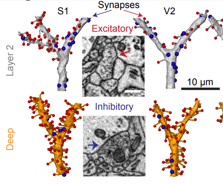
The surface of each main bifurcation was overlaid with skeleton reconstruction of spine necks and spheres demonstrating synapses. The visualizations were generated using Amira (Thermo Fisher Scientific, USA).
For Research Use Only. Not for use in diagnostic procedures.