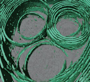
Membrane architecture of pulmonary lamellar bodies revealed by post-correlation on-lamella cryo-CLEM

Lamellar bodies (LBs) are surfactant rich organelles in alveolar type 2 cells. LBs disassemble into a lipid-protein network that reduces surface tension and facilitates gas exchange at the air-water interface in the alveolar cavity. Current knowledge of LB architecture is predominantly based on electron microscopy studies using disruptive sample preparation methods. We established a post-correlation on-lamella cryo-correlative light and electron microscopy approach for cryo-FIB milled lung cells to structurally characterize and validate LB identity in their unperturbed state using the well-established ABCA3-eGFP marker. In situ cryo-electron tomography revealed that LBs are composed of lipidic structures unique in organelle biology. We report open-ended membrane sheets frequently attached to the limiting membrane of LBs and so far undescribed dome-shaped protein complexes. We propose that LB biogenesis is driven by parallel membrane sheet import and the curvature of the limiting membrane to maximize lipid storage capacity.