Thermo Fisher Scientific › Electron Microscopy › Electron Microscopes › 3D Visualization, Analysis and EM Software › Use Case Gallery
Nickel-rich transition metal oxide materials […] are of great interest for achieving immediate improvements in the energy density of Li-ion batteries and for risk reduction within the Li-ion battery supply chain. […] An increase in Ni content in NMC materials leads to accelerated degradation […]. This potentially complicates their adoption in applications requiring extended cycle life such as in electric vehicles. Recent developments in X-ray characterization tools have provided unprecedented insight into the structural, chemical, and electronic states of materials, and these tools have been harnessed to probe Li-ion batteries both in the lab and in large-scale synchrotron facilities. […] Here, we present a proof-of-concept correlative in situ 3D micro-CT and 2D spectroscopic TXM study on a polycrystalline NMC811 electrode in a half-cell arrangement within our specialized tab pouch cell during its initial delithiation (charge).
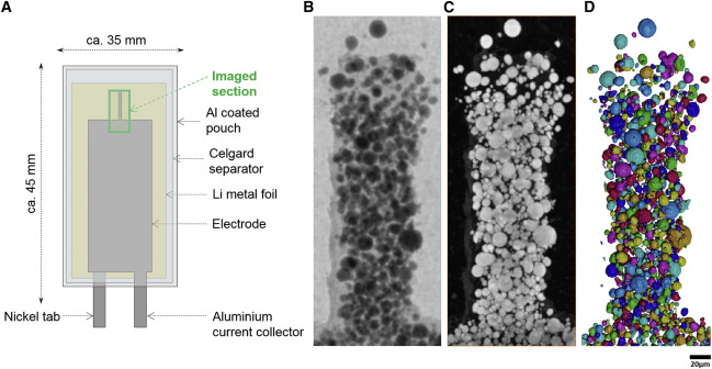
Image processing was performed in Avizo (V2020.2, Thermo Fisher Scientific) on the reconstructed micro-CT volume containing 975 × 975 × 975 voxels and the edge energy maps extracted from the output of TXM-Wizard described earlier. Segmentation of the NMC particles was carried out via a simple thresholding algorithm derived from Otsu’s method25 and available in the Auto-Threshold module in Avizo. Small features <5 voxels wide were removed, and the Separate Objects Avizo module was applied to separate neighboring particles that were in contact with one another. The separated particles were individually labeled using the Label Analysis Avizo module, which assigns different label values to each particle. A Z projection of the micro-CT grayscale dataset was obtained using the Image Stack Projection module in Avizo, to emulate the TXM mosaics. Another intensity-maxima projection of the individually numbered particle label field was also obtained using the Image Stack Projection module in Avizo to show any stacking of particles in the direction orthogonal to the current collector. All of the datasets were manually aligned using the Transform Editor tool in Avizo.
For Research Use Only. Not for use in diagnostic procedures.