Thermo Fisher Scientific › Electron Microscopy › Electron Microscopes › 3D Visualization, Analysis and EM Software › Use Case Gallery

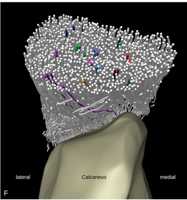
Tendon insertions to bone are heavily loaded transitions between soft and hard tissues. The fiber courses in the tendon have profound effects on the distribution of stress along and across the insertion. We tracked fibers of the Achilles tendon in mice in micro-computed tomographies and extracted virtual transversal sections. The fiber tracks and shapes were analyzed from a position in the free tendon to the insertion with regard to their mechanical consequences. The fiber number was found to stay about constant along the tendon. But the fiber cross-sectional areas decrease towards the insertion. The fibers mainly interact due to tendon twist, while branching only creates small branching clusters with low levels of divergence along the tendon. The highest fiber curvatures were found within the unmineralized entheseal fibrocartilage. The fibers inserting at a protrusion of the insertion area form a distinct portion within the tendon. Tendon twist is expected to contribute to a homogeneous distribution of stress among the fibers. According to the low cross-sectional areas and the high fiber curvatures, tensile and compressive stress are expected to peak at the insertion. These findings raise the question whether the insertion is reinforced in terms of fiber strength or by other load-bearing components besides the fibers.
The volume images were segmented manually in a 3D image analysis software (Amira Software 2019.1 by Thermo Fisher Scientific Inc., Waltham, Massachusetts, USA). Tendon, bone, the insertion area and the protrusion of the insertion area were marked as segments.
The fibers were tracked within the tendon segments with a template-based fiber tracking procedure (Amira Xtracing Extension 2019.1). The extraction of transversal tendon sections from the datasets was implemented as an Amira script.
For Research Use Only. Not for use in diagnostic procedures.