Thermo Fisher Scientific › Electron Microscopy › Electron Microscopes › 3D Visualization, Analysis and EM Software › Use Case Gallery
The Asian citrus psyllid (ACP), Diaphorina citri, is a harmful pest of citrus trees that transmits Candidatus Liberibacter spp. which causes Huanglongbing (HLB) (citrus greening disease); this is considered to be the most serious bacterial disease of citrus plants.
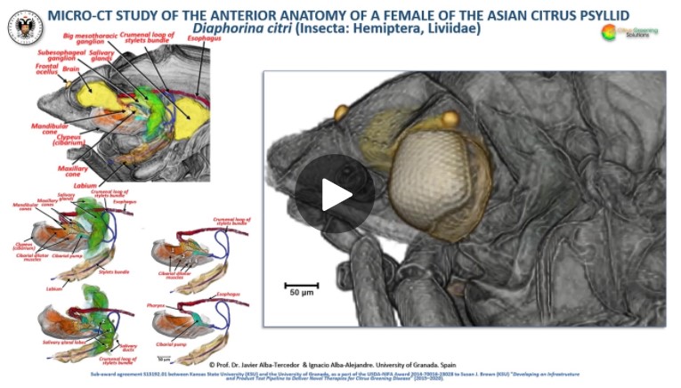
Here we detail an anatomical study of the external and internal anatomy (excluding the reproductive system) using micro-computed tomography (micro-CT). This is the first complete 3D micro-CT reconstruction of the anatomy of a psylloid insect and includes a 3D reconstruction of an adult feeding on a citrus leaf that can be used on mobile devices. Detailed rendered images and videos support first descriptions of coxal and scapus antennal glands and sexual differences in the internal anatomy (hindgut rectum, mesothoracic ganglion and brain). This represents a significant advance in our knowledge of ACP anatomy, and of psyllids in general. Together the images, videos and 3D model constitute a unique anatomical atlas and are useful tools for future research and as teaching aids.
Amira software with the built-in ‘volrenRed.col’ colour filter, was used to obtain the volume-rendered images seen in Figs. 3, 4, 5, 9, 10, 11, 12, 13, 14, 15, 16, 17, 18, 19 and Supplementary Videos S1–S11; the built-in ‘volrenGreen.col’ colour filter was used for Figs. 8, 16b,d–i. Different anatomical parts were independently segmented to obtain the final rendered colorized images seen in Figs. 11, 12, 13, 15, 16b,d, and Supplementary Videos S5–S8 and S9. To observe the actual texture of structures after segmentation, and in desired colours, each structure was subjected to the following arithmetic operation: A*(B > 0), where A represents the whole animal and B the segmented structure.
For Research Use Only. Not for use in diagnostic procedures.