Cellular in vivo 3D imaging of the cornea by confocal laser scanning microscopy

Sebastian Bohn, Karsten Sperlich, Stephan Allgeier, Andreas Bartschat, Ruby Prakasam, Klaus-martin Reichert, Heinrich Stolz, Rudolf Guthoff, Ralf Mikut, Bernd Köhler, and Oliver Stachs - Department of Ophthalmology, University Medical Center Rostock, Germany ; Institute for Automation and Applied Informatics, Karlsruhe Institute of Technology, Germany ; Institute of Physics, University of Rostock, Germany

We present an in vivo confocal laser scanning microscopy based method for large 3D reconstruction of the cornea on a cellular level with cropped volume sizes up to 266 x 286x 396 µm3.
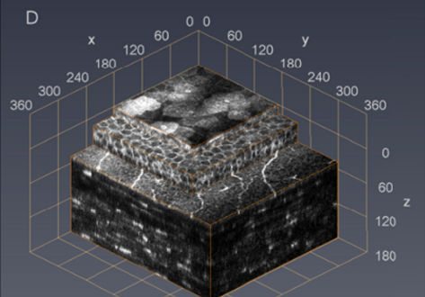
The microscope objective used is equipped with a piezo actuator for automated, fast and precise closed-loop focal plane control. Furthermore, we present a novel concave surface contact cap, which significantly reduces eye movements by up to 87%, hence increasing the overlapping image area of the whole stack. This increases the cuboid volume
of the generated 3D reconstruction significantly. The possibility to generate oblique sections using isotropic volume stacks opens the window to slit lamp microscopy on a cellular level.

How Amira-Avizo Software is used

Amira 3D, was used for 3D volume reconstruction of image stacks and for the imaging of sectional views. To align each image relative to the next one, we used both a rigid image registration method from Amira 3D and a specially adapted image registration algorithm developed by the Karlsruhe Institute of Technology (KIT) [7], which we term in the following as KIT-alignment. The registration method from Amira 3D uses rigid transformations (translations and rotations) to align consecutive images.