Thermo Fisher Scientific › Electron Microscopy › Electron Microscopes › 3D Visualization, Analysis and EM Software › Use Case Gallery
The aim of this study was to evaluate the influence of additional hydroxyapatite (HA) in collagen-based matrices (CM) and membrane placement on bone formation in calvarial defects.
Critical size defects in the calvaria of 16 New Zealand White Rabbits were randomly treated with CM or mineralized collagen-based matrices (mCM). Half of the sites were covered with a collagen membrane. Animals were euthanized after 12 weeks of healing. The samples were studied by micro-CT and histology. Newly formed lamellar bone was observed in all samples at the periphery of the defect. In the central areas, however, new bone composed of both woven and lamellar bone was embedded in the soft tissue. Samples treated with mCM showed more residual biomaterial and induced more small bony islands in the central areas of the defects than samples with CM. Nevertheless, a complete defect closure was not observed in any of the samples at 12 weeks. Membrane placement resulted in a decrease in bone density and height. Significant differences between the groups were revealed only between CM groups with and without membrane coverage for bone height in the central area of the defect. Neither mineralization of CM nor membrane placement improved the osteogenic capacity in this particular defect. Nevertheless, mineralisation influenced bone density without a membrane placement and bone volume underneath a membrane. CM may be used as a scaffold in bone regeneration procedures, without the need of a membrane coverage. Further preclinical studies are warrant to optimise the potential of mCM.
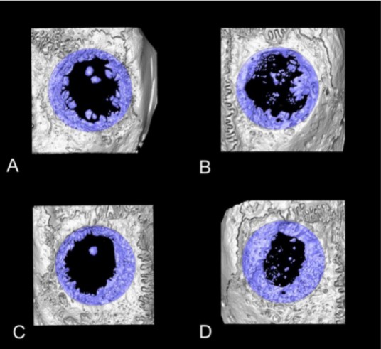
The micro-CT images were then analyzed and reconstructed by using 3D structural analysis software (Amira, Visualization Sciences Group, Düsseldorf, Germany). The volume of interest (VOI) was a 10-mm diameter, full thickness cylinders, selected corresponding to the dimensions of the defect sites. 2D parameters included defect closure measured on the horizontal plane (DC, relative % to whole surface at the defect site) and bone height (BH) measured on the sagittal plane, at the middle 5 mm (BH_M) and at the lateral part of the defect (BH_L). Bone volume (BV, mm3), BV fraction (BV/TV, ratio of the segmented BV to the total VOI) and relative BD (BD, relative % to initial bone at the defect site) were calculated. The micro-CT data of the harvested bone disc was used as reference threshold values for the BD analyses of tested groups.
For Research Use Only. Not for use in diagnostic procedures.