Welcome to the Amira-Avizo Software Use Case Gallery
Below you will find a collection of use cases of our 3D data visualization and analysis software. These use cases include scientific publications, articles, papers, posters, presentations or even videos that show how Amira-Avizo Software is used to address various scientific and industrial research topics.
Use the Domain selector to filter by main application area, and use the Search box to enter keywords related to specific topics you are interested in.
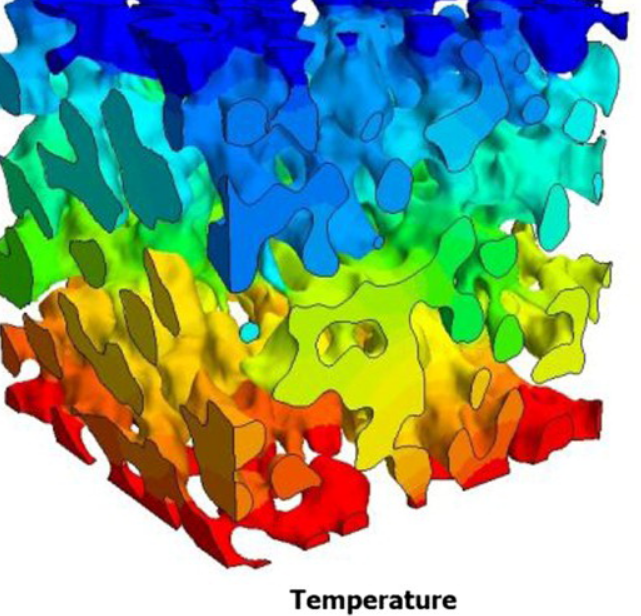
Tortuosity in electrochemical devices: a review of calculation approaches
Here, a review of tortuosity calculation procedures applied in the field of electrochemical devices is presented to better understand the resulting values presented in the literature. Visible differences between calculation methods are observed, especially when using porosity–tortuosity relationships and when comparing geometric and flux-based tortuosity calculation approaches.
Read more
Bernhard Tjaden, Dan J. L. Brett, Paul R. Shearing
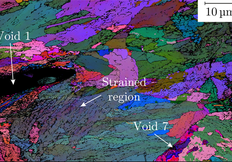
A multi-scale correlative investigation of ductile fracture
The use of novel multi-scale correlative methods, which involve the coordinated characterization of matter across a range of length scales, are becoming of increasing value to materials scientists. Here, we describe for the first time how a multi-scale correlative approach can be used to investigate the nature of ductile fracture in metals. Specimens of a nuclear pressure vessel steel, SA508 Grade 3, are examined following ductile fracture using medium and high-resolution 3D X-ray computed to... Read more
School of Materials, University of Manchester; National Nuclear Laboratory; BAM Federal Institute for Materials Research and Testing; Thermo Fischer Scientific
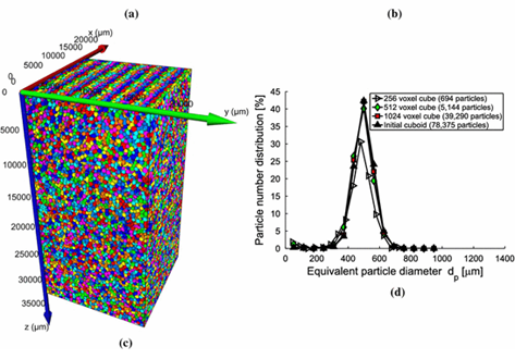
Hydraulic properties of porous sintered glass bead systems
In this paper, porous sintered glass bead packings are studied, using X-ray Computed Tomography (XRCT) images at 16μm16μm voxel resolution, to obtain not only the porosity field, but also other properties like particle sizes, pore throats and the permeability. The influence of the sintering procedure and the original particle size distributions on the microstructure, and thus on the hydraulic properties, is analyzed in detail. The XRCT data are visualized and studied by advanced image fil... Read more
University of Twente, Enschede | Ruhr-University Bochum; Eindhoven University of Technology | Helmholtz Institute Erlangen-Nürnberg for Renewable Energy | University of Stuttgart
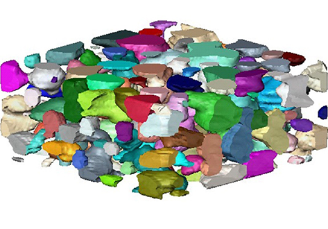
Physical characteristics critical to chromatography including geometric porosity and tortuosity within the packed column were analysed based upon three dimensional reconstructions of bed structure in-situ. Image acquisition was performed using two X-ray computed tomography systems, with optimisation of column imaging performed for each sample in order to produce three dimensional representations of packed beds at 3 μm resolution.
Read more
Department of Biochemical Engineering, University College London | Pall Life Sciences, Portsmouth | Electrochemical Innovation Lab, Department of Chemical Engineering, University College London
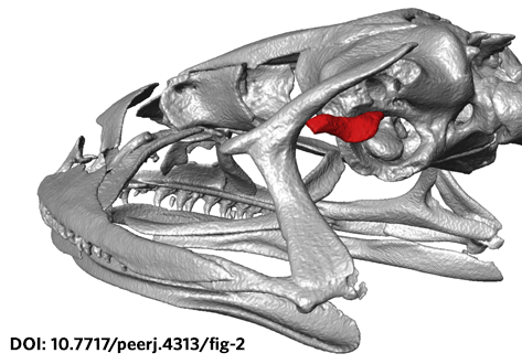
The loss of hearing structures and loss of advertisement calls in many terrestrial breeding frogs (Strabomantidae) living at high elevations in South America are common and intriguing phenomena. The Andean frog genus Phrynopus Peters, 1873 has undergone an evolutionary radiation in which most species lack the tympanic membrane and tympanic annulus, yet the phylogenetic relationships among species in this group remain largely unknown. Here, we present an expanded molecular phylogeny o... Read more
May R, Lehr E, Rabosky DL. PeerJ 6:e4313
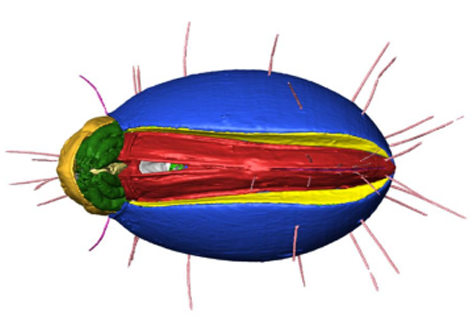
The NOVA project: maximizing beam time efficiency through synergistic analyses of SRµCT data
Beamtime and resulting SRμCT data are a valuable resource for researchers of a broad scientific community in life sciences. Most research groups, however, are only interested in a specific organ and use only a fraction of their data. The rest of the data usually remains untapped. By using a new collaborative approach, the NOVA project (Network for Online Visualization and synergistic Analysis of tomographic data) aims to demonstrate, that more efficient use of the valuable beam time is possi... Read more
The international society for optics and photonics (SPIE)
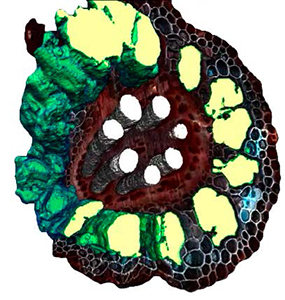
L4iS uses Avizo software to analyze 3D images from laser ablation tomography
Lasers for Innovative Solutions, LLC (L4iS) is developing a new class of tomography technology with the aim of allowing material characterization in three dimensions with sub-micron resolution. The method uses a nanosecond, Q-switched, pulsed ultraviolet laser coupled with high-resolution imaging to generate highly detailed specimen models. Using this system, sequential images similar to light-sheet fluorescence microscopy are used to digitally reconstruct the specimen.
Read more
Brian Reinhardt and Benjamin Hall, L4iS (USA)
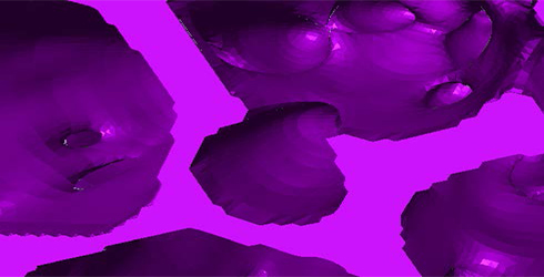
FAMU uses Avizo software to visualize and understand heat transfer and fluid flow
The CHEFF (Computational Heat Fluid Flow) Research group at Florida Agricultural & Mechanical University is using computational fluid dynamics to model flow and heat transfer in various engineering applications for industry, government and the private sector. The primary goal of this research is to first examine and then enhance the thermal performance of current and future low-density reticulated porous media, and explore their use as heat sinks in high power electronics (computer chips... Read more
CHEFF Research Group at Florida Agricultural & Mechanical University: Dr. G.D. Wesson, Professor of Chemical Engineering/Biological Agricultural Systems Engineering, Shawn Austin (Graduate student), Shari Briggs (Graduate student), Mellissa McCole (Graduate student), David Mosley (Graduate student)
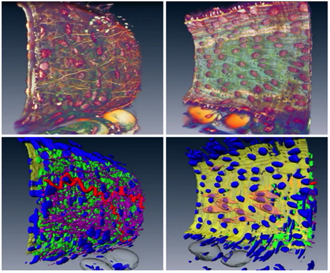
University of Glasgow uses Amira software to analyze vascular structures
The vascular (arterial or venous) wall is a fascinating structure. At the simplest level, we can think of the wall as being composed of three distinct, but interacting, layers . The vascular wall changes its structure in conditions such as hypertension which can cause a thickening of the wall. Unfortunately, the details of this ‘remodeling’ process are poorly understood.
Therefore, studying the 3D architecture may provide vital clues for future therapeutic targets.
R... Read more
Dr. Craig J Daly, School of Life Sciences, College of Medical Veterinary & Life Sciences, University of Glasgow
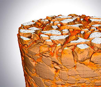
The Division of Highway and Railway Engineering, Royal Institute of Technology (KTH) promotes advances in computational and experimental science in order to develop new materials, tools and systems for improved mobility, transportation safety and infrastructure durability. The group works on analysis and performance-based design of roads and tracks, management as well as operation and maintenance of roads.
Read more
Denis Jelagin, Alvaro Guarin, Ibrahim Onifade, Nicole Kringos, and Bjorn Birgisson (KTH)
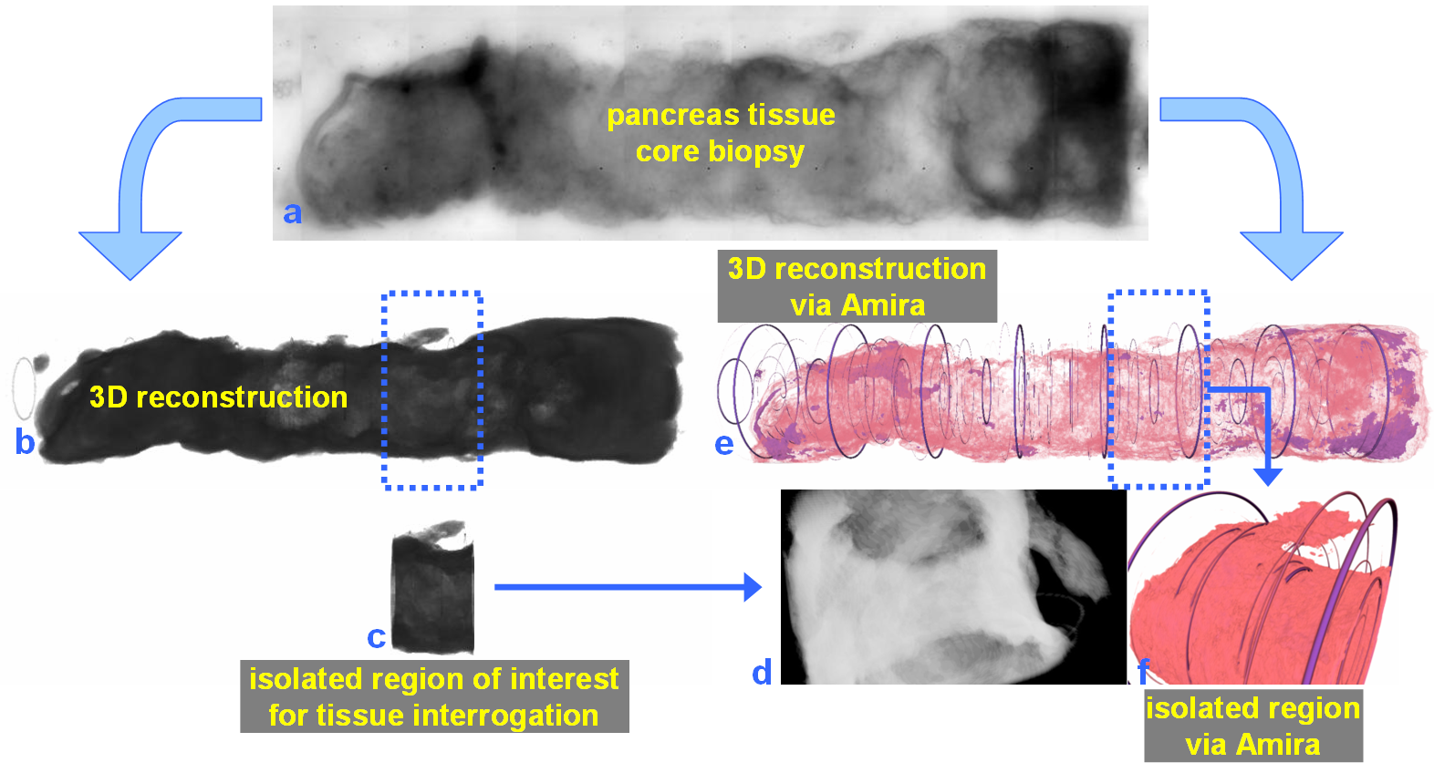
For nearly 100 years, pathology for cancer diagnosis has involved a standard, but complex series of steps to process tissue biopsies procured from a patient in the clinic. Many procedures are a direct result of the fact that observation and evaluation of specimens by pathologists occur using a standard microscope (in 2D).
In 2014, the Human Photonics Laboratory at the University of Washington demonstrated that the rudimentary operations of a pathology laboratory may be replicated on wh... Read more
Ronnie Das, PhD and Eric J. Seibel, PhD
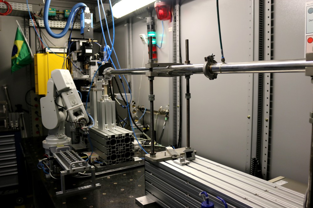
The imaging beamline, IMX, at LNLS extracts synchrotron radiation from bending magnet D6 with magnetic field of 1.67 T and bending radius of 2.736 m. It has an electron source size of 391 Read more
Brazilian Synchotron Light Laboratory
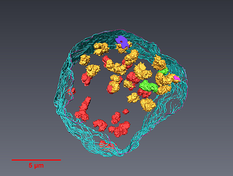
A team led by London Centre for Nanotechnology researchers, Prof. Ian Robinson and Dr. Bo Chen (now a professor at the Tongji University, Shanghai) used newly-developed serial block-face scanning electron microscopy (SBFSEM) and Thermo Scientific™ Avizo® Software, one dominant tool in 3D reconstructed image processing, to reveal the spatial structure of human chromosomes and nucleus quantitatively at high resolution of approximately 50 nm in three dimensions.
Read more
Prof. Ian Robinson, Dr. Bo Chen, London Centre for Nechnology, UCL and Tongji University
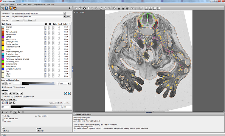
The Academic Medical Center (AMC) uses Amira to build a 3D Atlas for Human Embryology
The 3D Atlas of Human Embryology project was funded by the Academic Medical Center (AMC) in Amsterdam, the Netherlands, in 2009. Since then, over 75 students, under the supervision of embryologists of the Department of Anatomy, Embryology & Physiology, have contributed to this labor-int... Read more
Academic Medical Center
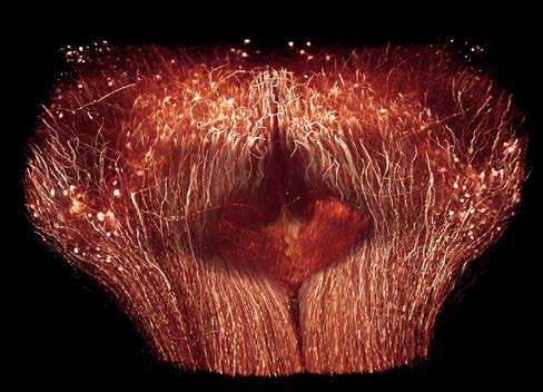
The Max Planck Institute of Neurobiology uses Amira software for axon tracing from head to toe
Ali Ertürk and his collaborators described a novel histo-chemical technique to clear spinal cord tissue of adult mice for fluorescence microscopy imaging of the intact spinal cord.
Previously, the high lipid content of the spinal cord tissue in adult mice allowed imaging of the spinal cord only through destructive histological methods, making the tracing of complete axons impossible. As applied in this study, the improved histo-chemistry enabled Ertürk and his colleagues to show that... Read more
Ali Ertürk, Christoph P Mauch, Farida Hellal, Friedrich Förstner, Tara Keck, Klaus Becker, Nina Jährling, Heinz Steffens, Melanie Richter, Mark Hübener, Edgar Kramer, Frank Kirchhoff, Hans Ulrich Dodt & Frank Bradke