Welcome to the Amira-Avizo Software Use Case Gallery
Below you will find a collection of use cases of our 3D data visualization and analysis software. These use cases include scientific publications, articles, papers, posters, presentations or even videos that show how Amira-Avizo Software is used to address various scientific and industrial research topics.
Use the Domain selector to filter by main application area, and use the Search box to enter keywords related to specific topics you are interested in.
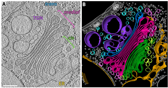
The structure of the COPI coat determined within the cell
COPI-coated vesicles mediate trafficking within the Golgi apparatus and from the Golgi to the endoplasmic reticulum. Here, we applied cryo-focused ion beam milling, cryo-electron tomography and subtomogram averaging to determine the native structure of the COPI coat within vitrified Chlamydomonas reinhardtii cells. The native algal structure resembles the in vitro mammalian structure, but additionally reveals cargo bound beneath beta’–COP. We find that all coat components disassemble... Read more
Yury S Bykov, Miroslava Schaffer, Svetlana O Dodonova, Sahradha Albert, Jurgen M Plitzko, Wolfgang Baumeister, Benjamin D Engel, John AG Briggs
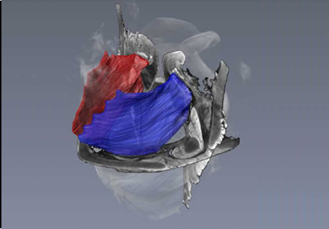
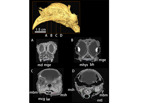
Juvenile Ovine Ex Vivo Larynges: Phonatory, Histologic, and Micro CT Based Anatomic Analyses
It is well known that the phonatory process changes during the life span. However, detailed investigations on potential factors concerned are rare. To deal with this issue, we performed extended biomechanical, macro anatomical, and histological analyses of the contributing laryngeal structures in ex vivo juvenile sheep models. Altogether twelve juvenile sheep larynges were analyzed within the phonatory experiments. Three different elongation levels and 16 different flow levels were applied to... Read more
Michael Döllinger, Olaf Wendler, Claus Gerstenberger, Tanja Grossmann, Marion Semmler, Hossein Sadeghi, and Markus Gugatschka
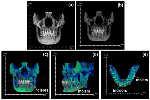
During the acts of biting and chewing, the muscles of the jaw (consisting of the masseter, temporalis, and medial pterygoid in the elevator group, and lateral pterygoid as the main depressor) generate forces that dictate jaw kinematics . The movement of jaws hinges about the temporomandibular joint and are brought together by the muscles attached to respective bones through bone-tendon interfaces known as entheses . Thus, chewing forces affect aspects of craniofacial structure as well as bo... Read more
Kyle H.-Y. Chan, fourth-year undergraduate student in Molecular and Cell Biology, and Public Health at UC Berkeley, FeiFei Yang, Ph.D., postdoctoral scholar in the Laboratory of Multiscale Biomechanics and Biomineralization, School of Dentistry, UCSF, and Sunita P. Ho, Ph.D., Division of Biomaterials and Bioengineering, Department of Preventive and Restorative Dental Sciences, School of Dentistry, University of California San Francisco
Aetosauria is a clade of heavily armored, quadrupedal omnivorous to herbivorous archosaurs known from the Late Triassic across what was the supercontinent of Pangea. Their abundance in many deposits relative to the paucity of other Triassic herbivores indicates that they were key components of Late Triassic ecosystems. However, their evolutionary relationships remain contentious due, in large part, to their extensive dermal armor, which often obstructs observation of internal skeletal anatomy... Read more
Devin K. Hoffman, Andrew B. Heckert, Lindsay E. Zanno
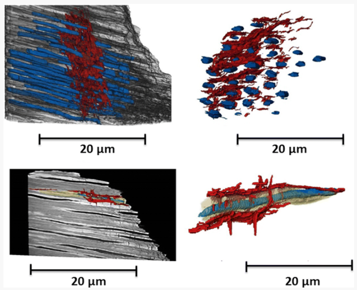
Microbial-tubeworm associations in a 440 million year old hydrothermal vent community
Microorganisms are the chief primary producers within present-day deep-sea hydrothermal vent ecosystems, and play a fundamental role in shaping the ecology of these environments. (…) The oldest known hydrothermal vent community that includes metazoans is preserved within the Ordovician to early Silurian Yaman Kasy massive sulfide deposit, Ural Mountains, Russia. (…) A re-examination of these fossils using a range of microscopy, chemical analysis and nano-tomography techniques re... Read more
Magdalena N. Georgieva , Crispin T. S. Little , Russell J. Bailey , Alexander D. Ball and Adrian G. Glover
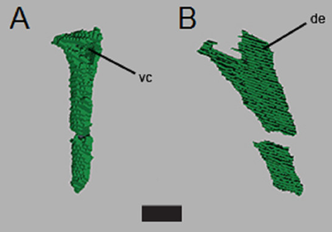
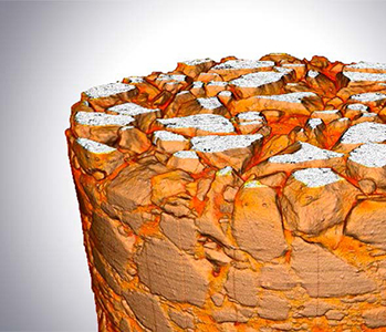
Micron-scale crack propagation in laser-irradiated enamel and dentine studied with nano-CT
The aim of this study was to see the effect of Er:YAG laser irradiation in dentine and compare this with its effect in enamel. The mechanism of crack propagation in dentine was emphasised and its clinical implications were discussed. A possible mechanism is that laser radiation is transmitted down the dentinal tubules causing micro-cracks to form in the dentinal tubule walls that tend to be limited to this region. Crack might be a source of fracture as it represents a weak point and subsequen... Read more
Abtesam Aljdaimi, Hugh Devlin, Mark Dickinson, Timothy Burnett, Thomas J. A. Slater
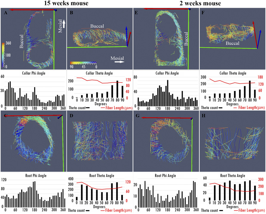
Nonuniformity in ligaments is a structural strategy for optimizing functionality
Ligaments serve as compliant connectors between hard tissues. In that role, they function under various load regimes and directions. The 3D structure of ligaments is considered to form as a uniform entity that changes due to function. The periodontal ligament (PDL) connects the tooth to the bone and sustains different types of loads in various directions. Using the PDL as a model, employing a fabricated motorized setup in a microCT, we demonstrate that the fibrous network structure with... Read more
Gili R. S. Naveh, Jonathan E. Foster, Tomas M. Silva Santisteban, Xianrui Yang, and Bjorn R. Olsen
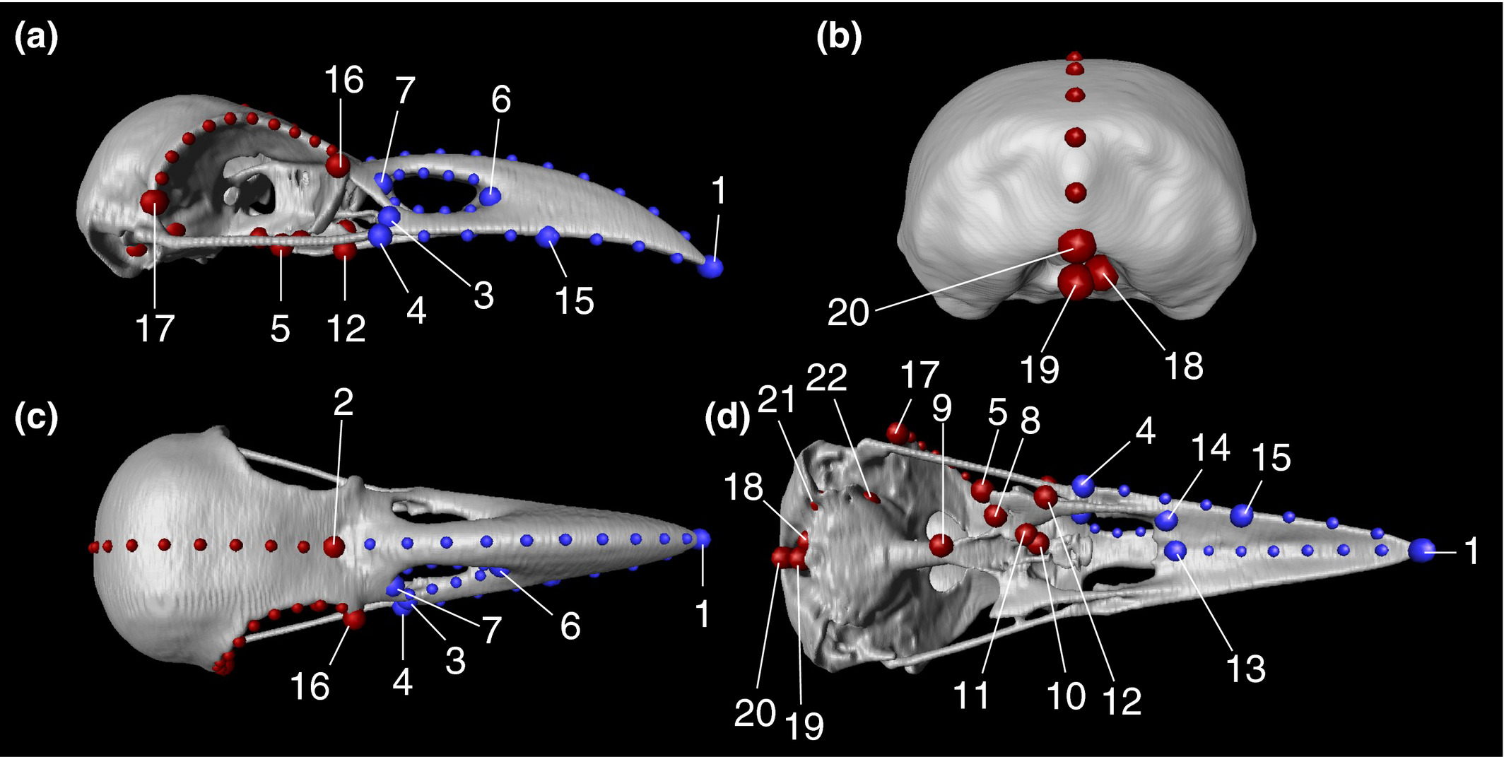
Here, to evaluate the intensity of evolutionary constraints on avian beak shape more appropriately, we selected large billed (Corvus macrorhynchos) and carrion crows (Corvus corone) as study objects. These landbird species seem to experience selection pressures favoring a departure from an allometric trajectory. A landmark based geometric morphometric approach using three dimensional reconstructions of CT scan images revealed that only 45.4% of the total shape variation was explained by allom... Read more
Takeshi Yamasaki, Sou Aoki, Masayoshi Tokita
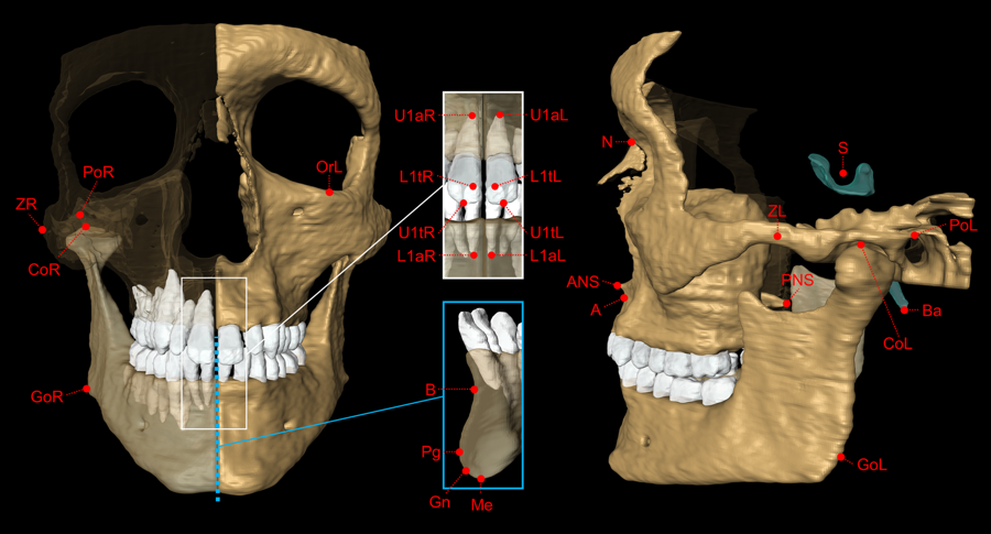
The aim of this study was to validate geometric accuracy and in vivo reproducibility of landmark-based cephalometric measurements using high-resolution 3D Magnetic Resonance Imaging (MRI) at 3 Tesla. (…) In conclusion, this study demonstrates that accurate and reproducible 3D cephalometric analysis can be performed without exposure to ionizing radiation using MRI.
Read more
Alexander Juerchott, Muhammad Abdullah Saleem, Tim Hilgenfeld, Christian Freudlsperger, Sebastian Zingler, Christopher J. Lux, Martin Bendszus & Sabine Heiland
Convergent evolution of a mobile bony tongue in flighted dinosaurs and pterosaurs
The tongue, with fleshy, muscular, and bony components, is an innovation of the earliest land-dwelling vertebrates with key functions in both feeding and respiration. Here, we bring together evidence from preserved hyoid elements from dinosaurs and outgroup archosaurs, including pterosaurs, with enhanced contrast x-ray computed tomography data from extant taxa. Midline ossification is a key component of the origin of an avian hyoid. The elaboration of the avian tongue includes the evolution o... Read more
Zhiheng Li, Zhonghe Zhou, Julia A. Clarke
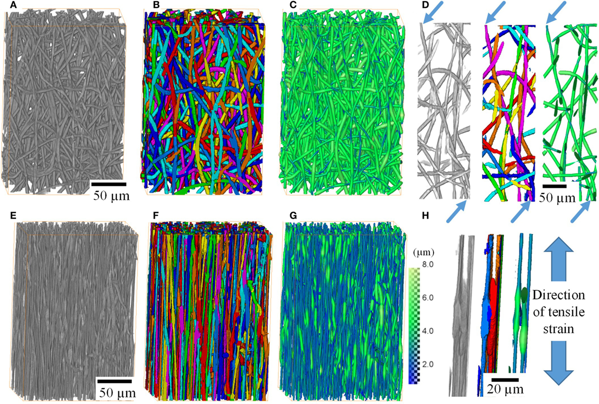
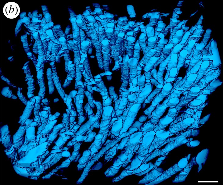
X-ray Tomographic Imaging of Tensile Deformation Modes of Electrospun Biodegradable Polyester Fibers
Electrospun constructs for the repair of load-bearing tissues are required to have adequate mechanical properties. However, the failure mechanisms of electrospun fibrous materials are not well understood. Existing literature focuses on failure modes of individual fibers and/or on bulk mechanical properties of whole fiber mats.
Electrospinning allows the production of fibrous networks for tissue engineering, drug delivery, and wound healing in health care. It enables the production of c... Read more
Jekaterina Maksimcuka; Akiko Obata ; William W. Sampson ; Remi Blanc ; Chunxia Gao ; Philip J. Withers ; Olga Tsigkou ; Toshihiro Kasuga ; Peter D. Lee ; Gowsihan Poologasundarampillai
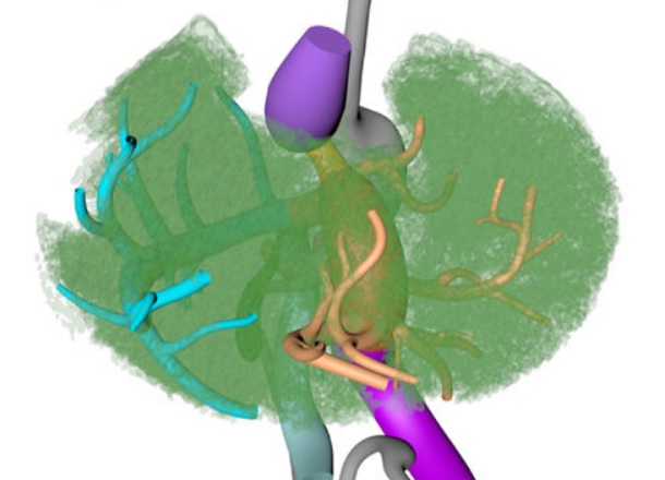
Couinaud based his well-known subdivision of the liver into (surgical) segments on the branching order of portal veins and the location of hepatic veins. However, both segment boundaries and number remain controversial due to an incomplete understanding of the role of liver lobes and vascular physiology on hepatic venous development. Human embryonic livers (5–10 weeks of development) were visualized with Amira 3D-reconstruction and Cinema 4D-remodeling software.
Read more
Jill P. J. M. Hikspoors, Mathijs M. J. P. Peeters, Nutmethee Kruepunga, Hayelom K. Mekonen, Greet M. C. Mommen, S. Eleonore Köhler & Wouter H. Lamers
The Division of Highway and Railway Engineering, Royal Institute of Technology (KTH) promotes advances in computational and experimental science in order to develop new materials, tools and systems for improved mobility, transportation safety and infrastructure durability. The group works on analysis and performance-based design of roads and tracks, management as well as operation and maintenance of roads.
Read more
Denis Jelagin, Alvaro Guarin, Ibrahim Onifade, Nicole Kringos, and Bjorn Birgisson (KTH)
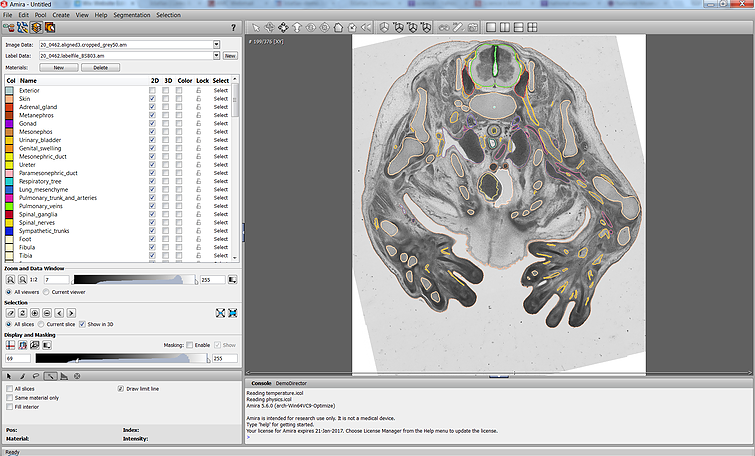
The Academic Medical Center (AMC) uses Amira to build a 3D Atlas for Human Embryology
The 3D Atlas of Human Embryology project was funded by the Academic Medical Center (AMC) in Amsterdam, the Netherlands, in 2009. Since then, over 75 students, under the supervision of embryologists of the Department of Anatomy, Embryology & Physiology, have contributed to this labor-int... Read more
Academic Medical Center