Welcome to the Amira-Avizo Software Use Case Gallery
Below you will find a collection of use cases of our 3D data visualization and analysis software. These use cases include scientific publications, articles, papers, posters, presentations or even videos that show how Amira-Avizo Software is used to address various scientific and industrial research topics.
Use the Domain selector to filter by main application area, and use the Search box to enter keywords related to specific topics you are interested in.
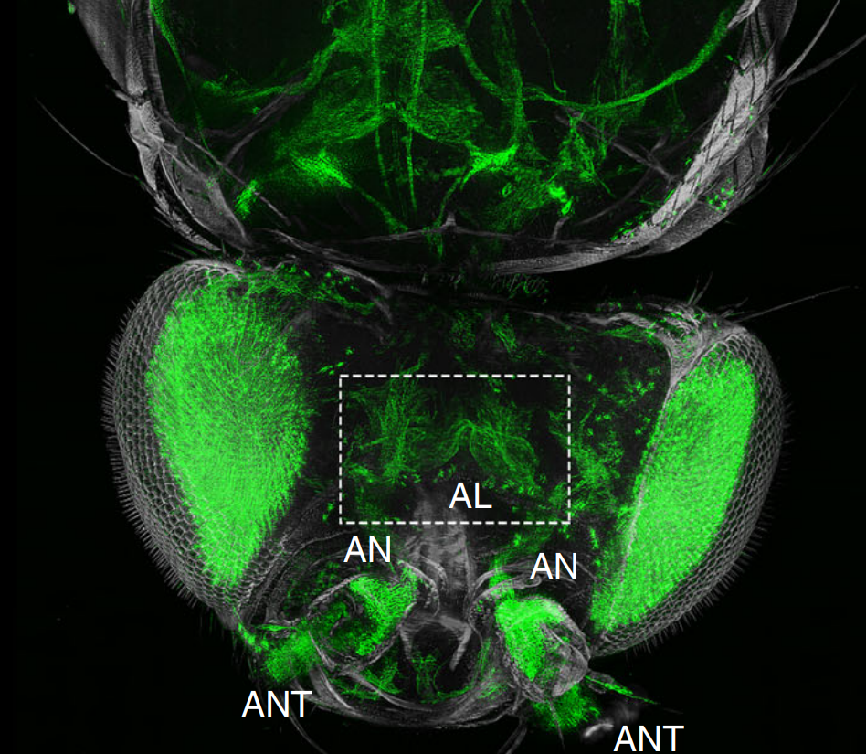
The fruit fly, Drosophila melanogaster, is an important experimental model to address central questions in neuroscience at an organismic level. However, imaging of neural circuits in intact fruit flies is limited due to structural properties of the cuticle. Here we present a novel approach combining tissue clearing, ultramicroscopy, and data analysis that enables the visualisation of neuronal networks with single-cell resolution from the larval stage up to the adult Drosophila. (…) This... Read more
Marko Pende, Klaus Becker, Martina Wanis, Saiedeh Saghafi, Rashmit Kaur, Christian Hahn, Nika Pende, Massih Foroughipour, Thomas Hummel & Hans-Ulrich Dodt
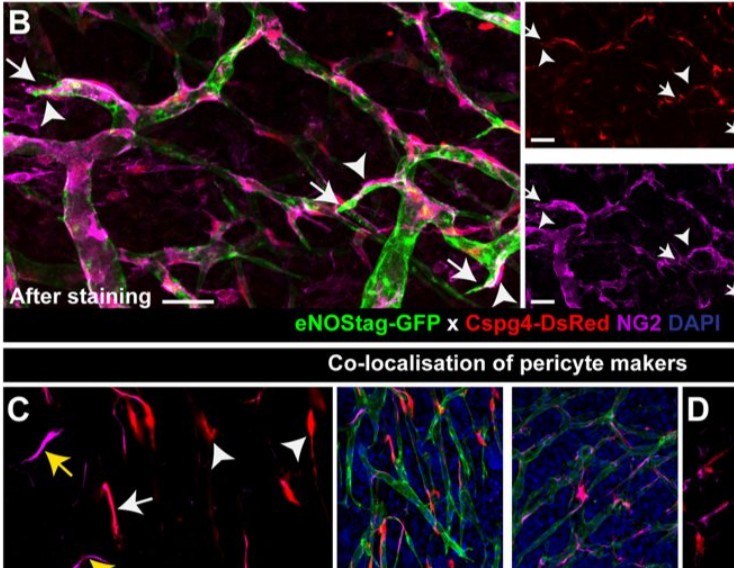
Endothelial cells and pericytes are integral cellular components of the vasculature with distinct interactive functionalities. To study dynamic interactions between these two cells we created two transgenic animal lines. A truncated eNOS (endothelial nitric oxide synthase) construct was used as a GFP tag for endothelial cell evaluation and an inducible Cre-lox recombination, under control of the Pdgfrb (platelet derived growth factor receptor beta) promoter, was created for pericyte assessmen... Read more
Ann L. B. Seynhaeve, Douwe Oostinga, Rien van Haperen, Hanna M. Eilken, Susanne Adams, Ralf H. Adams & Timo L. M. ten Hagen
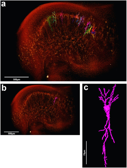
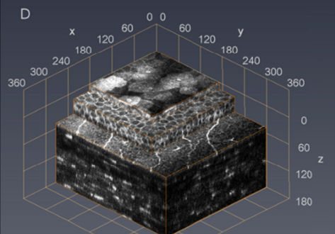
The assessment of neuronal number, spatial organization and connectivity is fundamental for a complete understanding of brain function. However, the evaluation of the three-dimensional (3D) brain cytoarchitecture at cellular resolution persists as a great challenge in the field of neuroscience. In this context, X-ray microtomography has shown to be a valuable non-destructive tool for imaging a broad range of samples, from dense materials to soft biological specimens, arisen as a new method fo... Read more
Matheus de Castro Fonseca, Bruno Henrique Silva Araujo, Carlos Sato Baraldi Dias, Nathaly Lopes Archilha, Dionísio Pedro Amorim Neto, Esper Cavalheiro, Harry Westfahl Jr, Antônio José Roque da Silva, Kleber Gomes Franchini
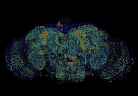
Combined expansion and lattice light sheet microscopy enables high
speed, nanoscale molecular imaging of neural circuits over large volumes.
Optical and electron microscopy have made tremendous inroads in understanding the complexity of the brain, but the former offers insufficient resolution to reveal subcellular details and the latter lacks the throughput and molecular contrast to visualize specific molecular constituents over mm-scale or larger dimensions. We combined expansio... Read more
Ruixuan Gao, Shoh M Asano, Srigokul Upadhyayula, Igor Pisarev, Daniel E Milkie, Tsung-Li Liu, Ved Singh, Austin Graves, Grace H Huynh, Yongxin Zhao, John Bogovic, Jennifer Colonell, Carolyn M Ott, Christopher Zugates, Susan Tappan, Alfredo Rodriguez, Kishore R Mosaliganti, Sean G Megason, Jennifer Lippincott-Schwartz, Adam Hantman, Gerald M Rubin, Tom Kirchhausen, Stephan Saalfeld, Yoshinori Aso, Edward S Boyden, Eric Betzig
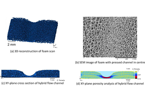
The flow distribution behaviour of open-cell metallic foam fuel cell flow-fields are evaluated using ex-situ optical analysis and X-ray computed tomography (X-ray CT). Five different manifold designs are evaluated and flow distribution and pressure drop ... Read more
A.Fly, D.Butcher, Q.Meyer, M.Whiteley, A.Spencer, C.Kim, P.R.Shearing, D.J.L.Brett, R.Chen
Cellular in vivo 3D imaging of the cornea by confocal laser scanning microscopy
We present an in vivo confocal laser scanning microscopy based method for large 3D reconstruction of the cornea on a cellular level with cropped volume sizes up to 266 x 286x 396 µm3.
The microscope objective used is equipped with a piezo actuator for automated, fast and precise closed-loop focal plane control. Furthermore, we present a novel concave surface contact cap, which significantly reduces eye movements by up to 87%, hence increasing the overlapping image area of the whole st... Read more
Sebastian Bohn, Karsten Sperlich, Stephan Allgeier, Andreas Bartschat, Ruby Prakasam, Klaus-martin Reichert, Heinrich Stolz, Rudolf Guthoff, Ralf Mikut, Bernd Köhler, and Oliver Stachs
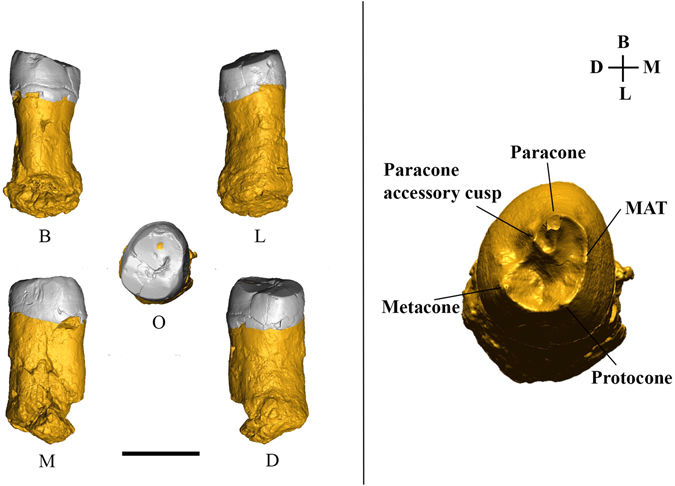
The first Neanderthal remains from an open-air Middle Palaeolithic site in the Levant
The late Middle Palaeolithic (MP) settlement patterns in the Levant included the repeated use of caves and open landscape sites. The fossil record shows that two types of hominins occupied the region during this period—Neandertals and Homo sapiens. Until recently, diagnostic fossil remains were found only at cave sites. Because the two populations in this region left similar material cultural remains, it was impossible to attribute any open-air site to either species. In this study... Read more
Ella Been, Erella Hovers, Ravid Ekshtain, Ariel Malinski-Buller, Nuha Agha, Alon Barash, Daniella E. Bar-Yosef Mayer, Stefano Benazzi, Jean-Jacques Hublin, Lihi Levin, Noam Greenbaum, Netta Mitki, Gregorio Oxilia, Naomi Porat, Joel Roskin, Michalle Soudack, Reuven Yeshurun, Ruth Shahack-Gross, Nadav Nir, Mareike C. Stahlschmidt, Yoel Rak & Omry Barzilai
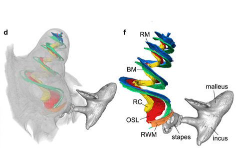
Propagation-based phase-contrast x-ray tomography of cochlea using a compact synchrotron source
We demonstrate that phase retrieval and tomographic imaging at the organ level of small animals can be advantageously carried out using the monochromatic radiation emitted by a compact x-ray light source, without further optical elements apart from source and detector. This approach allows to carry out microtomography experiments which – due to the large performance gap with respect to conventional laboratory instruments – so far were usually limited to synchrotron sources. We dem... Read more
Mareike Töpperwien, Regine Gradl, Daniel Keppeler, Malte Vassholz, Alexander Meyer, Roland Hessler, Klaus Achterhold, Bernhard Gleich, Martin Dierolf, Franz Pfeiffer, Tobias Moser & Tim Salditt
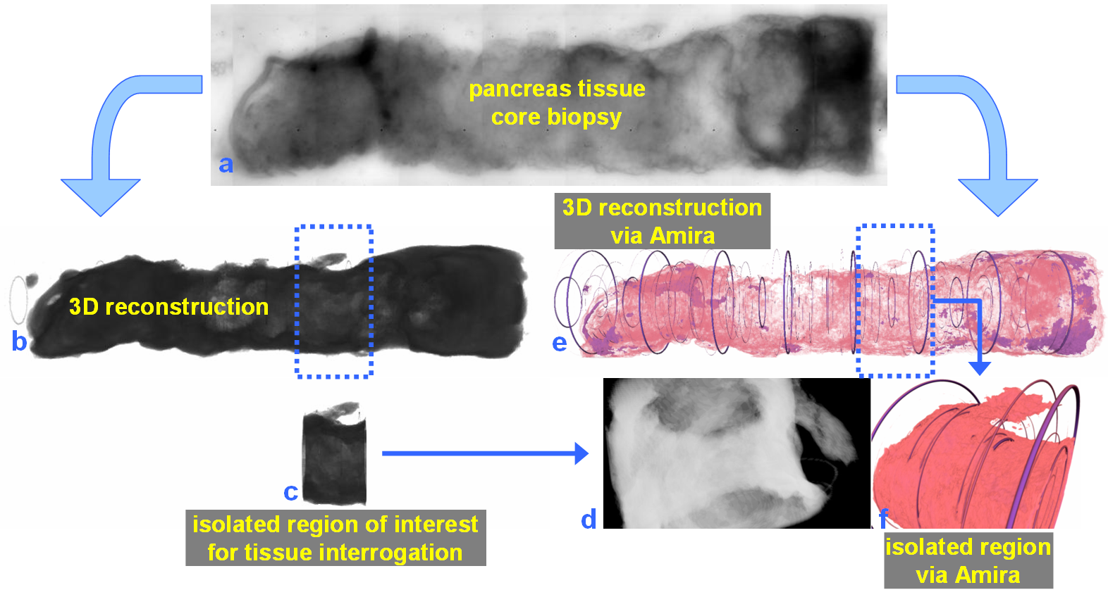
For nearly 100 years, pathology for cancer diagnosis has involved a standard, but complex series of steps to process tissue biopsies procured from a patient in the clinic. Many procedures are a direct result of the fact that observation and evaluation of specimens by pathologists occur using a standard microscope (in 2D).
In 2014, the Human Photonics Laboratory at the University of Washington demonstrated that the rudimentary operations of a pathology laboratory may be replicated on wh... Read more
Ronnie Das, PhD and Eric J. Seibel, PhD