Welcome to the Amira-Avizo Software Use Case Gallery

Below you will find a collection of use cases of our 3D data visualization and analysis software. These use cases include scientific publications, articles, papers, posters, presentations or even videos that show how Amira-Avizo Software is used to address various scientific and industrial research topics.

Use the Domain selector to filter by main application area, and use the Search box to enter keywords related to specific topics you are interested in.

Testing hypotheses for the function of the carnivoran baculum using finite-element analysis

Testing hypotheses for the function of the carnivoran baculum using finite-element analysis

The baculum (os penis) is a mineralized bone within the glans of the mammalian penis and is one of the most morphologically diverse structures in the mammal skeleton. (…) For the first time, to our knowledge, we apply a computational simulation approach (finite-element analysis; FEA) to quantify the three-dimensional biomechanical performance of carnivoran bacula (n = 74) based upon high-resolution micro-computed tomography scans. (…) a highly significant negative relation... Read more

Charlotte A. Brassey, James D. Gardiner, and Andrew C. Kitchener

Read full paper
Micron-scale crack propagation in laser-irradiated enamel and dentine studied with nano-CT

Micron-scale crack propagation in laser-irradiated enamel and dentine studied with nano-CT

The aim of this study was to see the effect of Er:YAG laser irradiation in dentine and compare this with its effect in enamel. The mechanism of crack propagation in dentine was emphasised and its clinical implications were discussed. A possible mechanism is that laser radiation is transmitted down the dentinal tubules causing micro-cracks to form in the dentinal tubule walls that tend to be limited to this region. Crack might be a source of fracture as it represents a weak point and subsequen... Read more

Abtesam Aljdaimi, Hugh Devlin, Mark Dickinson, Timothy Burnett, Thomas J. A. Slater

Read full paper
Vascular structure of the earliest shark teeth

Vascular structure of the earliest shark teeth

Here we use synchrotron tomography to characterise dental vasculature in the oldest known tooth-bearing sharks, Leonodus carlsi Mader, 1986 and Celtiberina maderi Wang, 1993. Three dimensional reconstruction of the vascular system and microstructure of both taxa revealed a complex and dense network of canals, including horizontal, ascending and secondary bifurcated canals, as well as histological features consistent with an osteodont histotype. However, L. carlsi and C. maderi also exhibit si... Read more

Carlos Martinez-Perez, Alba Martin-Lazaro, Humberto G Ferron, Martina Kirstein, Philip C.J. Donoghue, Hector Botella

Read full paper
Branching morphogenesis of the urinary collecting system in the human embryonic metanephros

Branching morphogenesis of the urinary collecting system in the human embryonic metanephros

An elaborate system of ducts collects urine from all nephrons, and this structure is known as the urinary collecting system (UCS). This study focused on how the UCS is formed during human embryogenesis. Fifty human embryos between the Carnegie stage (CS) 14 and CS23 were selected from the Kyoto Collection at the Congenital Anomaly Research Center of Kyoto University, Japan. Metanephroses, including the UCS, were segmented on serial digital virtual histological sections. Three-dimensional imag... Read more

Hana Ishiyama, Aoi Ishikawa, Haruka Kitazawa, Sena Fujii, Jun Matsubayashi, Shigehito Yamada, Tetsuya Takakuwa

Read full paper
Nonuniformity in ligaments is a structural strategy for optimizing functionality

Nonuniformity in ligaments is a structural strategy for optimizing functionality

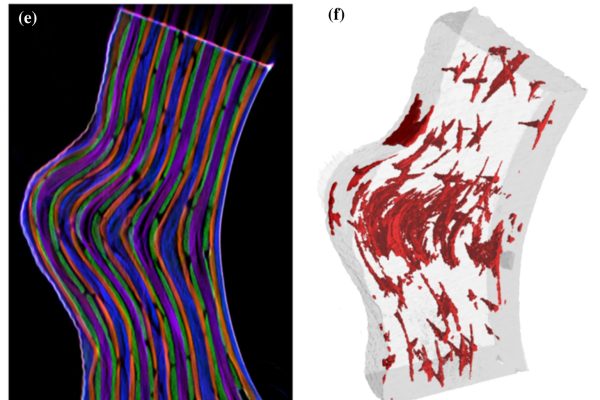
Ligaments serve as compliant connectors between hard tissues. In that role, they function under various load regimes and directions. The 3D structure of ligaments is considered to form as a uniform entity that changes due to function. The periodontal ligament (PDL) connects the tooth to the bone and sustains different types of loads in various directions. Using the PDL as a model, employing a fabricated motorized setup in a microCT, we demonstrate that the fibrous network structure with... Read more

Gili R. S. Naveh, Jonathan E. Foster, Tomas M. Silva Santisteban, Xianrui Yang, and Bjorn R. Olsen

Read full paper
Functional Electrical Stimulation Leads to Increased Volume of the Aged Thyroarytenoid Muscle

Functional Electrical Stimulation Leads to Increased Volume of the Aged Thyroarytenoid Muscle

A stimulation electrode was placed unilaterally near the terminal adduction branch of the recurrent laryngeal nerve (RLN) adjacent to the right cricothyroid joint. The electrode was connected to an implant located subcutaneously at the neck region. Predesigned training patterns were automatically delivered by a bidirectional radio frequency link using a programming device and were repeated automatically by the implant every other day over 11 weeks in the awake animal. Outcome parameters compr... Read more

Markus Gugatschka, MD, DMSci, Jonathan C. Jarvis, PhD, Justin D. Perkins, MSc, Vladimir Bubalo, PhD, Iris Wiederstein-Grasser, PhD, Hermann Lanmüller, PhD, Claus Gerstenberger, MSc and Michael Karbiener, PhD

Read full paper
3D cephalometric analysis using Magnetic Resonance Imaging: validation of accuracy and reproducibility

3D cephalometric analysis using Magnetic Resonance Imaging: validation of accuracy and reproducibility

The aim of this study was to validate geometric accuracy and in vivo reproducibility of landmark-based cephalometric measurements using high-resolution 3D Magnetic Resonance Imaging (MRI) at 3 Tesla. (…) In conclusion, this study demonstrates that accurate and reproducible 3D cephalometric analysis can be performed without exposure to ionizing radiation using MRI.

Read more

Alexander Juerchott, Muhammad Abdullah Saleem, Tim Hilgenfeld, Christian Freudlsperger, Sebastian Zingler, Christopher J. Lux, Martin Bendszus & Sabine Heiland

Read full paper
Spatiotemporal endothelial cell – pericyte association in tumors as shown by high resolution 4D intravital imaging

Spatiotemporal endothelial cell – pericyte association in tumors as shown by high resolution 4D intravital imaging

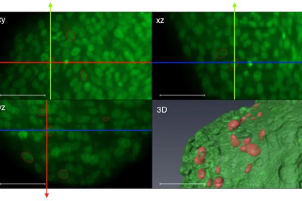
Endothelial cells and pericytes are integral cellular components of the vasculature with distinct interactive functionalities. To study dynamic interactions between these two cells we created two transgenic animal lines. A truncated eNOS (endothelial nitric oxide synthase) construct was used as a GFP tag for endothelial cell evaluation and an inducible Cre-lox recombination, under control of the Pdgfrb (platelet derived growth factor receptor beta) promoter, was created for pericyte assessmen... Read more

Ann L. B. Seynhaeve, Douwe Oostinga, Rien van Haperen, Hanna M. Eilken, Susanne Adams, Ralf H. Adams & Timo L. M. ten Hagen

Read full paper
STIM1 promotes migration, phagosomal maturation and antigen cross-presentation in dendritic cells

STIM1 promotes migration, phagosomal maturation and antigen cross-presentation in dendritic cells

Antigen cross-presentation by dendritic cells (DC) stimulates cytotoxic T cell activation to promote immunity to intracellular pathogens, viruses and cancer. Phagocytosed antigens generate potent T cell responses, but the signalling and trafficking pathways regulating their cross-presentation are unclear. Here, we show that ablation of the store-operated-Ca2+-entry regulator STIM1 in mouse myeloid cells impairs cross-presentation and DC migration in vivo and in vitro. Stim1... Read more

Paula Nunes-Hasler, Sophia Maschalidi, Carla Lippens, Cyril Castelbou, Samuel Bouvet, Daniele Guido, Flavien Bermont, Esen Y. Bassoy, Nicolas Page, Doron Merkler, Stéphanie Hugues, Denis Martinvalet, Bénédicte Manoury & Nicolas Demaurex

Read full paper
High-resolution synchrotron-based X-ray microtomography as a tool to unveil the three-dimensional neuronal architecture of the brain

High-resolution synchrotron-based X-ray microtomography as a tool to unveil the three-dimensional neuronal architecture of the brain

The assessment of neuronal number, spatial organization and connectivity is fundamental for a complete understanding of brain function. However, the evaluation of the three-dimensional (3D) brain cytoarchitecture at cellular resolution persists as a great challenge in the field of neuroscience. In this context, X-ray microtomography has shown to be a valuable non-destructive tool for imaging a broad range of samples, from dense materials to soft biological specimens, arisen as a new method fo... Read more

Matheus de Castro Fonseca, Bruno Henrique Silva Araujo, Carlos Sato Baraldi Dias, Nathaly Lopes Archilha, Dionísio Pedro Amorim Neto, Esper Cavalheiro, Harry Westfahl Jr, Antônio José Roque da Silva, Kleber Gomes Franchini

Read full paper
Ptychographic X-ray CT characterization of the osteocyte lacuno-canalicular network in a male rat's glucocorticoid induced osteoporosis model

Ptychographic X-ray CT characterization of the osteocyte lacuno-canalicular network in a male rat's glucocorticoid induced osteoporosis model

Ptychographic X-ray computed tomography (PXCT) is a quantitative imaging modality that non-destructively maps the 3D electron density inside an object with tens of nanometers spatial resolution. This method provides unique access to the morphology and structure of the osteocyte lacuno-canalicular network (LCN) and nanoscale density of the tissue in the vicinity of an osteocyte lacuna.  Our findings indicate that PXCT can non-destructively provide detailed, nanoscale information on the 3D org... Read more

Antonia Ciani, Hechmi Toumi, Stéphane Pallu, Esther H.R.Tsai, Ana Diaz, Manuel Guizar-Sicairos, Mirko Holler, Eric Lespessailles, Cameron M.Kewish | Synchrotron Soleil, France

Read full paper
Cortical Column and Whole Brain Imaging of Neural Circuits with Molecular Contrast and Nanoscale Resolution

Cortical Column and Whole Brain Imaging of Neural Circuits with Molecular Contrast and Nanoscale Resolution

Combined expansion and lattice light sheet microscopy enables high
speed, nanoscale molecular imaging of neural circuits over large volumes.

Optical and electron microscopy have made tremendous inroads in understanding the complexity of the brain, but the former offers insufficient resolution to reveal subcellular details and the latter lacks the throughput and molecular contrast to visualize specific molecular constituents over mm-scale or larger dimensions. We combined expansio... Read more

Ruixuan Gao, Shoh M Asano, Srigokul Upadhyayula, Igor Pisarev, Daniel E Milkie, Tsung-Li Liu, Ved Singh, Austin Graves, Grace H Huynh, Yongxin Zhao, John Bogovic, Jennifer Colonell, Carolyn M Ott, Christopher Zugates, Susan Tappan, Alfredo Rodriguez, Kishore R Mosaliganti, Sean G Megason, Jennifer Lippincott-Schwartz, Adam Hantman, Gerald M Rubin, Tom Kirchhausen, Stephan Saalfeld, Yoshinori Aso, Edward S Boyden, Eric Betzig

Read full paper
Morphology of the bryozoan Cinctipora elegans (Cyclostomata, Cinctiporidae) with first data on its sexual reproduction and the cyclostome neuro-muscular system

Morphology of the bryozoan Cinctipora elegans (Cyclostomata, Cinctiporidae) with first data on its sexual reproduction and the cyclostome neuro-muscular system

Cyclostome bryozoans are an ancient group of marine colonial suspension-feeders comprising approximately 700 extant species. Previous morphological studies are mainly restricted to skeletal characters whereas data on soft tissues obtained by state-of-the-art methods are still lacking. In order to contribute to issues related to cyclostome ground pattern reconstruction, we analyzed the morphology of the neuromuscular system Cinctipora elegans by means of immunocytochemical staining,... Read more

Thomas F. Schwaha, Stephan Handschuh, Andrew N. Ostrovsky, Andreas Wanninger

Read full paper
Secure corridor for infraacetabular screws in acetabular fracture fixation—a 3-D radiomorphometric analysis of 124 pelvic CT datasets

Secure corridor for infraacetabular screws in acetabular fracture fixation—a 3-D radiomorphometric analysis of 124 pelvic CT datasets

Acetabular fracture surgery is directed toward anatomical reduction and stable fixation to allow for the early functional rehabilitation of an injured hip joint. Recent biomechanical investigations have shown the superiority of using an additional screw in the infraacetabular (IA) region, thereby transfixing the separated columns to strengthen the construct by closing the periacetabular fixation frame. However, the inter-individual existence and variance concerning secure IA screw corridors a... Read more

Stephan Arlt, Hansrudi Noser, Andreas Wienke, Florian Radetzki, Gunther Olaf Hofmann, Thomas Mendel

Read full paper
Three-dimensional virtual histology of human cerebellum by X-ray phase-contrast tomography

Three-dimensional virtual histology of human cerebellum by X-ray phase-contrast tomography

To quantitatively evaluate brain tissue and its corresponding function, knowledge of the 3D cellular distribution is essential. The gold standard to obtain this information is histology, a destructive and labor-intensive technique where the specimen is sliced and examined under a light microscope, providing 3D information at nonisotropic resolution. To overcome the limitations of conventional histology, we use phase-contrast X-ray tomography with optimized optics, reconstruction, and image an... Read more

Mareike Töpperwien, Franziska van der Meer, Christine Stadelmann, and Tim Salditt

Read full paper
Resurrecting Darwin’s Niata - anatomical, biomechanical, genetic, and morphometric studies of morphological novelty in cattle

Resurrecting Darwin’s Niata - anatomical, biomechanical, genetic, and morphometric studies of morphological novelty in cattle

The Niata was a cattle variety from South America that figured prominently in writings on evolution by Charles Darwin. Its shortened head and other aspects of its unusual morphology have been subject of unsettled discussions since Darwin’s time. Here, we examine the anatomy, cranial shape, skull biomechanics, and population genetics of the Niata. Our results show that the Niata was a viable variety of cattle and exhibited anatomical differences to known chondrodysplastic forms. In cranial s... Read more

Kristof Veitschegger, Laura A. B. Wilson, Beatrice Nussberger, Glauco Camenisch, Lukas F. Keller, Stephen Wroe, Marcelo R. Sánchez-Villagra

Read full paper
Mechanical adaptation of trabecular bone morphology in the mammalian mandible

Mechanical adaptation of trabecular bone morphology in the mammalian mandible

Alveolar bone, together with the underlying trabecular bone, fulfils an important role in providing structural support against masticatory forces. Diseases such as osteoporosis or periodontitis cause alveolar bone resorption which weakens this structural support and is a major cause of tooth loss. However, the functional relationship between alveolar bone remodelling within the molar region and masticatory forces is not well understood. This study investigated this relationship by comparing m... Read more

Peter J. Watson, Laura C. Fitton, Carlo Meloro, Michael J. Fagan, Flora Gröning

Read full paper
3D Reconstruction of Lipid Droplets in the Seed of Brassica napus

3D Reconstruction of Lipid Droplets in the Seed of Brassica napus

Rapeseed is one of the most important and widely cultured oilseed crops for food and nonfood purposes worldwide. Neutral lipids are stored in lipid droplets (LDs) as fuel for germination and subsequent seedling growth. Most of the LD detection in seeds was still in 2D levels, and some of the details might have been lost in previous studies. In the present work, the configuration of LDs in seeds was obtained by confocal imaging combined with 3D reconstruction technology in Brassica napus<... Read more

Yongtai Yin, Liangxing Guo, Kang Chen, Zhenyi Guo, Hongbo Chao, Baoshan Wang, and Maoteng Licorresponding author

Read full paper
The morphological diversity of Garra barreimiae

The morphological diversity of Garra barreimiae

Garra barreimiae (Fowler and Steinitz, Bull Res Counc Isr 5B:262–289, 1956) is a freshwater fish that lives throughout the Hajar Mountains of Oman and the United Arab Emirates. Previously, four different genetic clades (West clade, C... Read more

Arthur Pichler; Harald Ahnelt; Sandra Kirchner; Helmut Sattmann; Elisabeth Haring; Stephan Handschuh; Jörg Freyhof; Reginald Victor; Luise Kruckenhauser

Read full paper
Morphology and ontogeny of Lophopus crystallinus lophophore support the epistome as ancestral character of phylactolaemate bryozoans

Morphology and ontogeny of Lophopus crystallinus lophophore support the epistome as ancestral character of phylactolaemate bryozoans

Phylactolaemate bryozoans are the sister-group to all remaining bryozoan taxa. Consequently, their study is essential to reveal and analyze ancestral traits of Phylactolaemata and Bryozoa in general.

They are the only bryozoans to possess an epistome which traditionally has been regarded as shared with phoronids and brachiopods. Contrary to older observations, an epistome was recently reported to be missing in the early branching phylactolaemate Lophopus crystallinus. In this... Read more

Thomas Schwaha

Read full paper
Cellular in vivo 3D imaging of the cornea by confocal laser scanning microscopy

Cellular in vivo 3D imaging of the cornea by confocal laser scanning microscopy

We present an in vivo confocal laser scanning microscopy based method for large 3D reconstruction of the cornea on a cellular level with cropped volume sizes up to 266 x 286x 396 µm3.

The microscope objective used is equipped with a piezo actuator for automated, fast and precise closed-loop focal plane control. Furthermore, we present a novel concave surface contact cap, which significantly reduces eye movements by up to 87%, hence increasing the overlapping image area of the whole st... Read more

Sebastian Bohn, Karsten Sperlich, Stephan Allgeier, Andreas Bartschat, Ruby Prakasam, Klaus-martin Reichert, Heinrich Stolz, Rudolf Guthoff, Ralf Mikut, Bernd Köhler, and Oliver Stachs

Read full paper