Welcome to the Amira-Avizo Software Use Case Gallery
Below you will find a collection of use cases of our 3D data visualization and analysis software. These use cases include scientific publications, articles, papers, posters, presentations or even videos that show how Amira-Avizo Software is used to address various scientific and industrial research topics.
Use the Domain selector to filter by main application area, and use the Search box to enter keywords related to specific topics you are interested in.
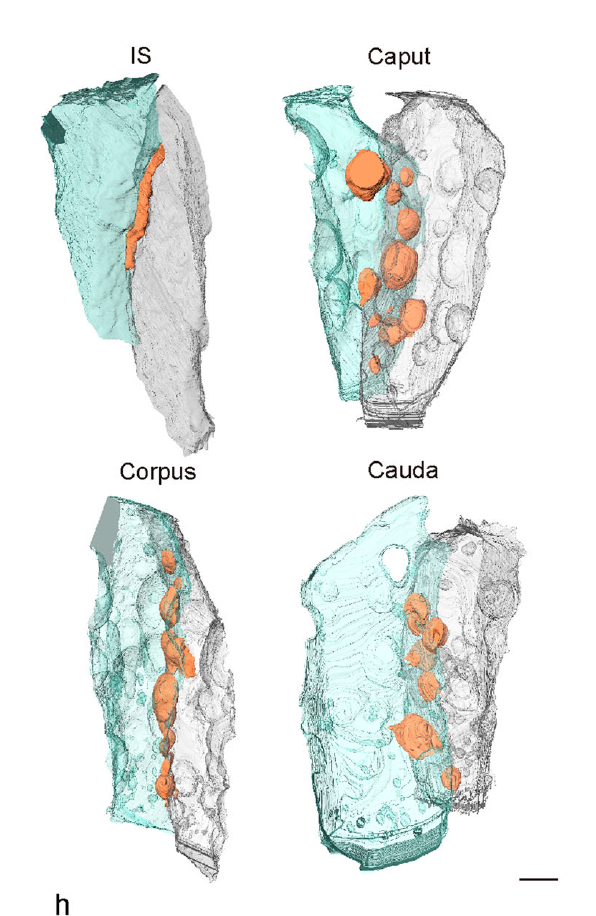
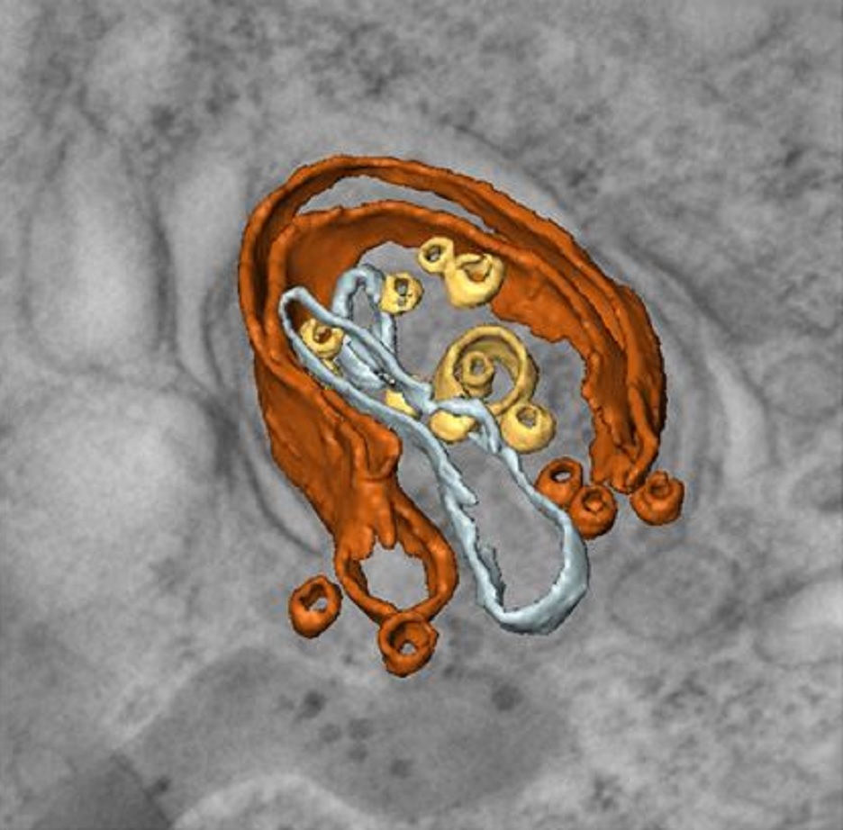
Mammalian epididymal epithelial cells are crucial for sperm maturation. Historically,
vacuole-like ultrastructures in epididymal epithelial cells were
observed via transmission electron microscopy but were undefined. Here, we
utilize volume electron microscopy (vEM) to generate 3D reconstructions of
epididymal epithelial cells and identify these vacuoles as intercellular organelle
reservoirs (IORs) in the lateral intercellular space (LIS), which contains
pr... Read more
Xia Li, Feng Qiao, Jiansheng Guo, Ting Jiang, Huifang Lou, Huixia Li, Gangcai Xie, Hangjun Wu, Weizhen Wang, Ruoyu Pei, Sha Liu, Mei Ye, Jin Li, Shiqin Huang, Mengya Zhang, Chaoye Ma, Yiwen Huang, Shushu Xu, Xiaofeng Li, Xiao Sun, Jun Yu, Kin Lam Fok, Shumin Duan & Hao Chen
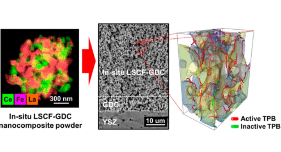
Composite cathodes comprising nanoscale
powders are expected to impart with high specific surface
area and triple phase boundary (TPB) density, which will lead
to better performance.
However, uniformly mixing nanosized heterophase powders remains a challenge due to their high surface energy and thus ease with which they agglomerate into their individual phases during the mixing and sintering
processes. In this study, we successfully synthesized La0.6Sr0.4Co0.2Fe... Read more
Dong Woo Joh, Areum Cha, Jeong Hwa Park, Kyeong Joon Kim, Kyung Taek Bae, Doyeub Kim, Young Ki Choi, Hyegsoon An, Ji Su Shin, Kyung Joong Yoon, and Kang Taek Lee
Cryo-STEM mapping of solid–liquid interfaces and dendrites in lithium-metal batteries
Solid–liquid interfaces are important in a range of chemical, physical and biological processes but are often not fully understood owing to the lack of high-resolution characterization methods that are compatible with both solid and liquid components. For example, the related processes of dendritic deposition of lithium metal and the formation of solid–electrolyte interphase layers are known to be key determinants of battery safety and performance in high-energy-density lithium-metal bat... Read more
Michael J. Zachman, Zhengyuan Tu, Snehashis Choudhury, Lynden A. Archer & Lena F. Kourkoutis
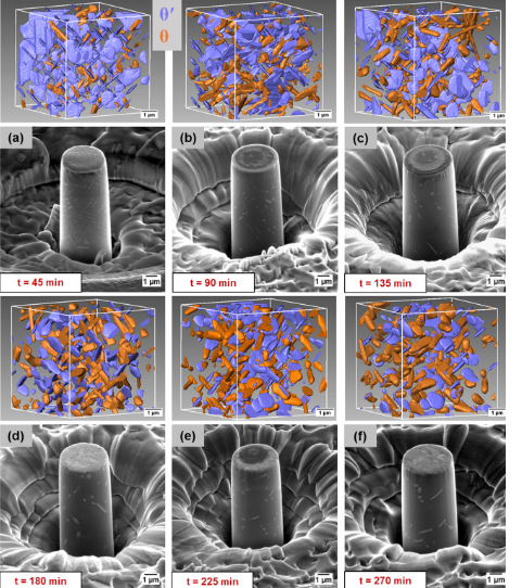
A unique approach to correlating an evolving 3D microstructure in an Al-Cu alloyand its micro-scale mechanical properties has been introduced. Using these nanoscale three-dimensional microstructures derived from Transmission X-rayMicroscopy (TXM), individual contributions from different strengthening mechanisms were quantified. The spatial distribution and morphology of the individual θ′ and θ phases were seen to play an important role in influencing dislocation storage. Uniaxi... Read more
C. Shashank Kaira, Christopher Kantzos, Jason J. Williams, Vincent De Andrade, Francesco De Carlo, Nikhilesh Chawlaa
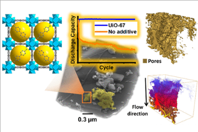
Porous Metal–Organic Frameworks for Enhanced Performance Silicon Anodes in Lithium-Ion Batteries
Maintaining the physical integrity of electrode microstructures in Li-ion batteries is critical to significantly extend their cycle life. This is especially important for high-capacity anode materials such as silicon, whose operational volume expansion exerts huge internal stress within the anode, resulting in electrode destruction and capacity fade. In this study, we demonstrate that by incorporating metal–organic frameworks (MOFs) with carboxylate organic linkers into Si-based anodes, a s... Read more
Romeo Malik, Melanie. J. Loveridge, Luke J. Williams, Qianye Huang, Geoff West, Paul R. Shearing, Rohit Bhagat, Richard I. Walton

Protocols for Generating Surfaces and Measuring 3D Organelle Morphology Using Amira
High-resolution 3D images of organelles are of paramount importance in cellular biology. Although light microscopy and transmission electron microscopy (TEM) have provided the standard for imaging cellular structures, they cannot provide 3D images.
However, recent technological advances such as serial block-face scanning electron microscopy (SBF-SEM) and focused ion beam scanning electron microscopy (FIB-SEM) provide the tools to create 3D images for the ultrastructural analysis of org... Read more
Edgar Garza-Lopez, Zer Vue, Prasanna Katti, Kit Neikirk, Michelle Biete, Jacob Lam, Heather K. Beasley, Andrea G. Marshall, Taylor A. Rodman, Trace A. Christensen, Jeffrey L. Salisbury, Larry Vang, Margaret Mungai, Salma Ash Shareef, Sandra A. Murray, Jianqiang Shao, Jennifer Streeter, Brian Glancy, Renata O. Pereira1, E. Dale Abel, and Antentor Hinton, Jr.
Macroautophagy is morphologically characterized by autophagosome formation. Autophagosomes are double-membraned vesicles that sequester cytoplasmic components for further degradation in the lysosome. Basal autophagy is paramount for intracellular quality control in post-mitotic cells but, surprisingly, the number of autophagosomes in post-mitotic neurons is very low, suggesting that alternative degradative structures could exist in neurons…
Read more
Maria Rosario Fernandez-Fernandez, Desire Ruiz-Garcia, Eva Martin-Solana, Francisco Javier Chichon, Jose L. Carrascosa, Jose-Jesus Fernandez
Influenza A matrix protein M1 is sufficient to induce lipid membrane deformation
The matrix protein M1 of the Influenza A virus is considered to mediate viral assembly and budding at the plasma membrane (PM) of infected cells. In order for a new viral particle to form, the PM lipid bilayer has to bend into a vesicle towards the extracellular side. Studies in cellular models have proposed that different viral proteins might be responsible for inducing membrane curvature in this context (including M1), but a clear consensus has not been reached. In this study, we use a comb... Read more
Ismail Dahmani, Kai Ludwig, Salvatore Chiantia
Membrane architecture of pulmonary lamellar bodies revealed by post-correlation on-lamella cryo-CLEM
Lamellar bodies (LBs) are surfactant rich organelles in alveolar type 2 cells. LBs disassemble into a lipid-protein network that reduces surface tension and facilitates gas exchange at the air-water interface in the alveolar cavity. Current knowledge of LB architecture is predominantly based on electron microscopy studies using disruptive sample preparation methods. We established a post-correlation on-lamella cryo-correlative light and electron microscopy approach for cryo-FIB milled lung ce... Read more
Steffen Klein, Benedikt H. Wimmer, Sophie L. Winter, Androniki Kolovou, Vibor Laketa, Petr Chlanda
Serial block face scanning electron microscopy (SBF-SEM) is a powerful method to analyze cells in 3D. Here, working at the resolution limit of the method, we describe a correlative light–SBF-SEM workflow to resolve microtubules of the mitotic spindle in human cells. We present four examples of uses for this workflow that are not practical by light microscopy and/or transmission electron microscopy. First, distinguishing closely associated microtubules within K-fibers; second, resolving brid... Read more
Faye M. Nixon, Thomas R. Honnor, Nicholas I. Clarke, Georgina P. Starling, Alison J. Beckett, Adam M. Johansen, Julia A. Brettschneider, Ian A. Prior, Stephen J. Royle
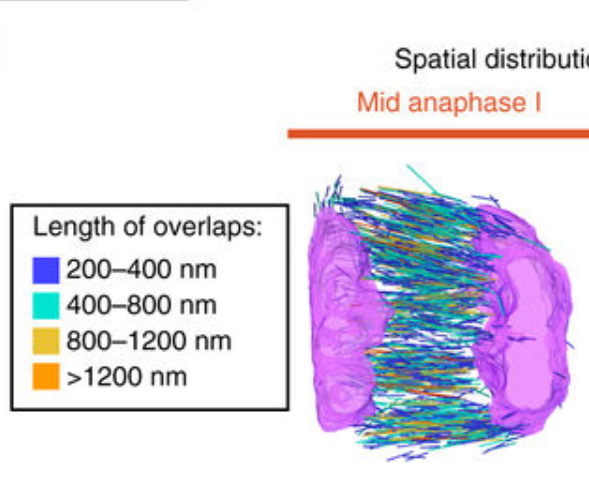
Chromosome segregation occurs by microtubule pushing in oocytes
During cell division, spindle microtubules ensure an equal repartition of chromosomes between the two daughter cells. While the kinetochore-dependent mechanisms that drive mitotic chromosome segregation are well understood, in oocytes of most species atypical spindles assembled in absence of centrosomes entail poorly understood mechanisms of chromosome segregation. In particular, the structure(s) responsible for force generation during meiotic chromosome separation in oocytes is unclear. Usin... Read more
Kimberley Laband, Rémi Le Borgne, Frances Edwards, Marine Stefanutti, Julie C. Canman, Jean-Marc Verbavatz, Julien Dumont
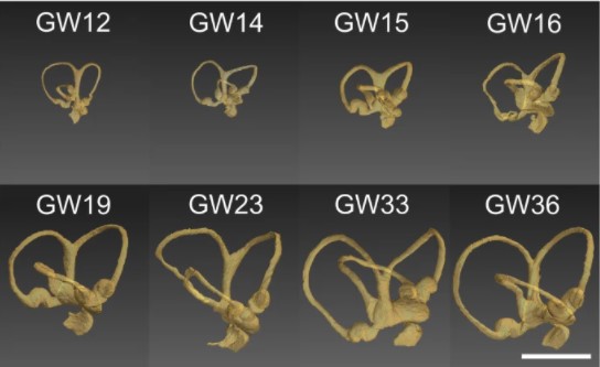
Progressive transformation of the otic placode into the functional inner ear during gestational development in humans leads to the acquisition of hearing perception via the cochlea and balance and spatial orientation via the vestibular organ.
Using a correlative approach involving micro-computerized tomography (micro-CT), transmission electron microscopy and histological techniques we were able to examine both the morphological and cellular changes associated with human inner ear devel... Read more
Lejo Johnson Chacko, David Wertjanz, Consolato Sergi, Jozsef Dudas, Natalie Fischer, Theresa Eberharter, Romed Hoermann, Rudolf Glueckert, Helga Fritsch, Helge Rask-Andersen, Anneliese Schrott-Fischer & Stephan Handschuh
In biomedical research, a huge variety of different techniques is currently available for the structural examination of small specimens, including conventional light microscopy (LM), transmission electron microscopy (TEM), confocal laser scanning microscopy (CLSM), microscopic X-ray computed tomography (microCT), and many others. Since every imaging method is physically limited by certain parameters, a correlative use of complementary methods often yields a significant broader range of inform... Read more
Stephan Handschuh, Natalie Baeumler, Thomas Schwaha & Bernhard Ruthensteiner
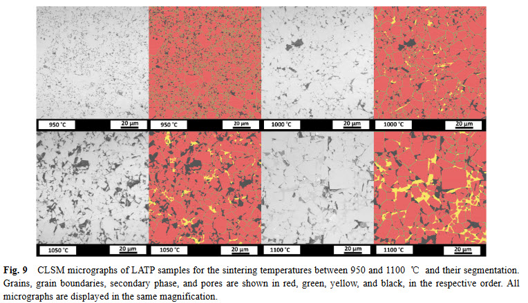
Lithium aluminum titanium phosphate (LATP) is one of the materials under consideration as an electrolyte in future all-solid-state lithium-ion batteries. In ceramic processing, the presence of secondary phases and porosity play an important role. In a presence of more than one secondary phase and pores, image analysis must tackle the difficulties about distinguishing between these microstructural features. In this study, we study the phase evolution of LATP ceramics sintered at temperatures b... Read more
Deniz Cihan GUNDUZ, Roland SCHIERHOLZ, Shicheng YUa, Hermann TEMPEL, Hans KUNGL, Rüdiger-A. EICHEL
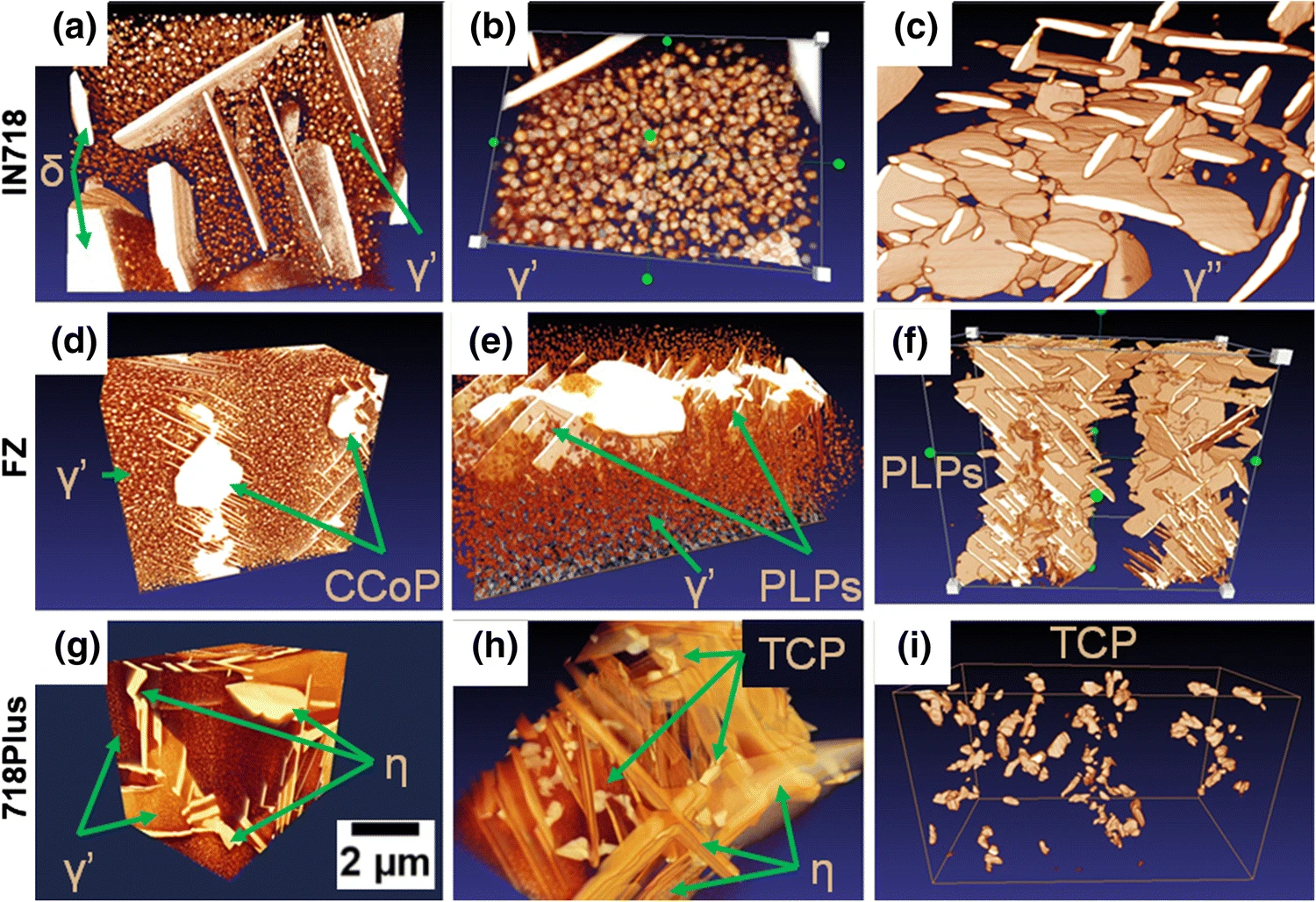
Inconel 718 (IN718) is the most popular precipitation-strengthened nickel-based superalloy introduced by the Huntington Alloys Division of INCO in 1959 (Ref Read more
Oskar Dziuba, Grzegorz Cempura, Agnieszka Wusatowska-Sarnek & Adam Kruk
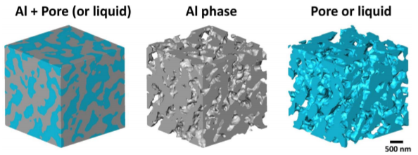
Nanoporous Aluminum by Galvanic Replacement: Dealloying and Inward-Growth Plating
In aqueous solutions, electro/chemically deposited metals usually grow outward into electrolyte. Here we report that the reduced Al grows inward into the sample, surprisingly, while Mg (in pure Mg and Al2Mg3 alloy) is galvanically replaced with Al in an ionic liquid. The galvanic replacement reaction (GRR) of Al2Mg3 involves a dealloying process that generates a nanoporous Al skeleton, and simultaneously the inward-growth plating of Al that thicke... Read more
Wei Yang, Xian-Gui Zheng, Shao-Gang Wang, Hai-Jun Jin
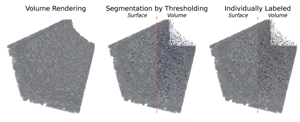
Aging of a Pt/Al2O3 exhaust gas catalyst monitored by quasi in situ X-ray micro computed tomography
Catalyst aging effects were analyzed using X-ray absorption micro-computed tomography in combination with conventional characterization methods on various length scales ranging from nm to μm to gain insight into deactivation mechanisms.
For this purpose, a 4 wt% Pt/Al2O3 model exhaust gas catalyst was coated on a cordierite honeycomb and subjected to sequential thermal aging in static air at 950 °C for 4, 8, 12 and 24 hours. The ag... Read more
Georg Hofmann, Amélie Rochet, Elen Ogel, Maria Casapu, Stephan Ritter, Malte Ogurreck and Jan-Dierk Grunwaldt
Despite the importance of studying the instability of delithiated cathode materials, it remains difficult to underpin the degradation mechanism of lithium-rich cathode materials due to the complication of combined chemical and structural evolutions. Herein, we use state-of-the-art electron microscopy tools, in conjunction with synchrotron X-ray techniques and first-principle calculations to study a 4d-element-containing compound, Li2Ru0.5Mn0.5O3. We find surprisingly, after cycling, ruthenium... Read more
Lin, Ruoqian AU - Hu, Enyuan AU - Liu, Mingjie AU - Wang, Yi AU - Cheng, Hao AU - Wu, Jinpeng AU - Zheng, Jin-Cheng AU - Wu, Qin AU - Bak, Seongmin AU - Tong, Xiao AU - Zhang, Rui AU - Yang, Wanli AU - Persson, Kristin A. AU - Yu, Xiqian AU - Yang, Xiao-Qing AU - Xin, Huolin L. PY
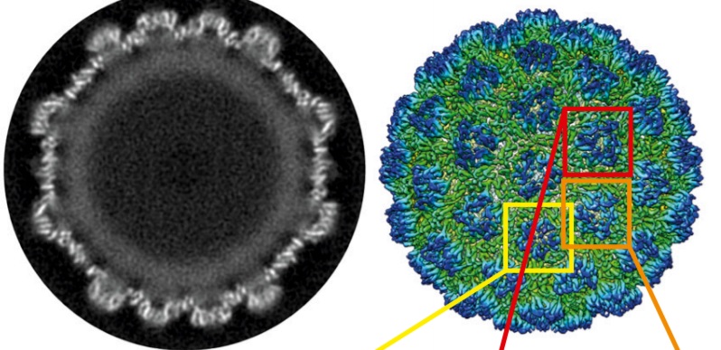
Structure of the Ty3/Gypsy retrotransposon capsid and the evolution of retroviruses
Retroviruses evolved from long terminal repeat (LTR) retrotransposons by acquisition of envelope functions, and subsequently reinvaded host genomes. Together, endogenous retroviruses and LTR retrotransposons represent major components of animal, plant, and fungal genomes. Sequences from these elements have been exapted to perform essential host functions, including placental development, synaptic communication, and transcriptional regulation. They encode a Gag polypeptide, the capsid domains ... Read more
Svetlana O. Dodonova, Simone Prinz, Virginia Bilanchone, Suzanne Sandmeyer, and John A. G. Briggs
As key functional units in neural circuits, different types of neuronal synapses play distinct roles in brain information processing, learning, and memory. Synaptic abnormalities are believed to underlie various neurological and psychiatric disorders. Here, by combining cryo-electron tomography and cryo-correlative light and electron microscopy, we distinguished intact excitatory and inhibitory synapses of cultured hippocampal neurons, and visualized the in situ 3D organization of ... Read more
Chang-Lu Tao, Yun-Tao Liu, Rong Sun, Bin Zhang, Lei Qi, Sakar Shivakoti, Chong-Li Tian, Peijun Zhang, Pak-Ming Lau, Z. Hong Zhou and Guo-Qiang Bi
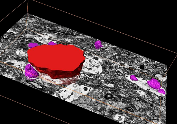
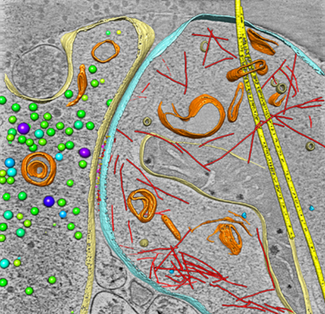
Corpora amylacea are cell-derived structures that appear physiologically in the aged human brain. While their histological identification is straightforward, their ultrastructural composition and microenvironment at the nanoscale have remained unclear so far, as has their relevance to aging and certain disease states that involve the sequestration of toxic cellular metabolites. Here, we apply correlative serial block-face scanning electron microscopy and transmission electron tomograp... Read more
Paula P. Navarro, Christel Genoud, Daniel Castaño-Díez, Alexandra Graff-Meyer, Amanda J. Lewis, Yvonne de Gier, Matthias E. Lauer, Markus Britschgi, Bernd Bohrmann, Stephan Frank, Jürgen Hench, Gabriel Schweighauser, Annemieke J. M. Rozemuller, Wilma D. J. van de Berg, Henning Stahlberg & Sarah H. Shahmoradian