Welcome to the Amira-Avizo Software Use Case Gallery
Below you will find a collection of use cases of our 3D data visualization and analysis software. These use cases include scientific publications, articles, papers, posters, presentations or even videos that show how Amira-Avizo Software is used to address various scientific and industrial research topics.
Use the Domain selector to filter by main application area, and use the Search box to enter keywords related to specific topics you are interested in.

Macropinosomes are key players in early shigella invasion and vacuolar escape in epithelial cells
Intracellular pathogens include all viruses, many bacteria and parasites capable of invading and surviving within host cells. Key to survival is the subversion of host cell pathways by the pathogen for the purpose of propagation and evading the immune system. The intracellular bacterium Shigella flexneri, the causative agent of bacillary dysentery, invades host cells in a vacuole that is subsequently ruptured to allow growth of the pathogen within the host cytoplasm…
Read more
Allon Weiner , Nora Mellouk , Noelia Lopez-Montero , Yuen-Yan Chang, Célia Souque, Christine Schmitt, Jost Enninga
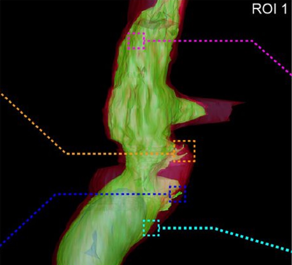
Fast and precise targeting of single tumor cells in vivo by multimodal correlative microscopy
Intravital microscopy provides dynamic understanding of multiple cell biological processes, but its limited resolution has so far precluded structural analysis. Because it is difficult to capture rare and transient events, only a few attempts have been made to observe specific developmental and pathological processes in animal models using electron microscopy. The multimodal correlative approach that we propose here combines intravital microscopy, microscopic X-ray computed tomography and thr... Read more
Matthia A. Karreman, Luc Mercier, Nicole L. Schieber, Gergely Solecki, Guillaume Allio, Frank Winkler, Bernhard Ruthensteiner, Jacky G. Goetz, Yannick Schwab
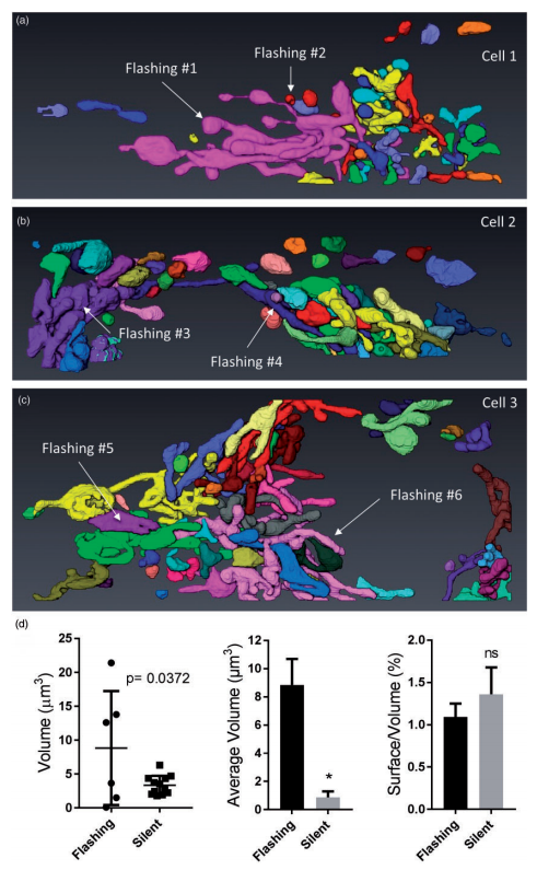
Ultrastructural Characterization of Flashing Mitochondria
Mitochondria undergo spontaneous transient elevations in matrix pH associated with drops in mitochondrial membrane potential. These mitopHlashes require a functional respiratory chain and the profusion protein optic atrophy 1, but their mechanistic basis is unclear. To gain insight on the origin of these dynamic events, we resolved the ultrastructure of flashing mitochondria by correlative light and electron microscopy. HeLa cells expressing the matrix-targeted pH probe mitoSypHer were screen... Read more
Manon Rosselin, Paula Nunes-Hasler, and Nicolas Demaurex
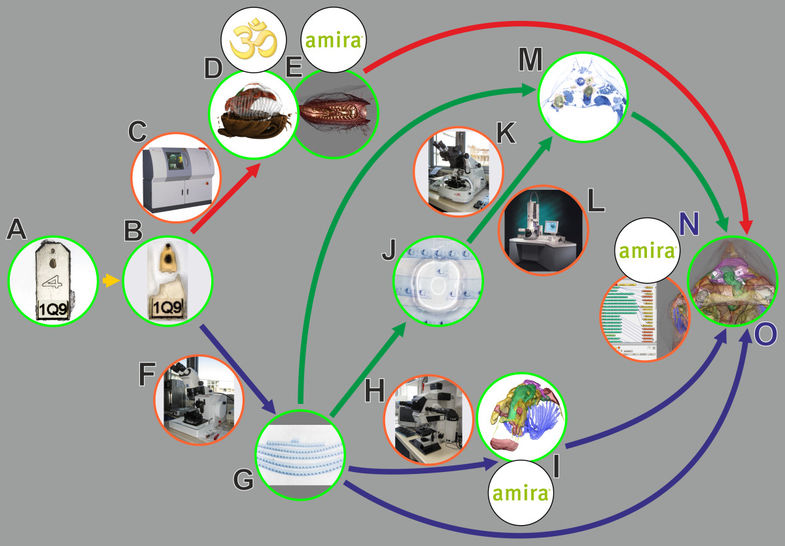
In biomedical research, a huge variety of different techniques is currently available for the structural examination of small specimens, including conventional light microscopy (LM), transmission electron microscopy (TEM), confocal laser scanning microscopy (CLSM), microscopic X-ray computed tomography (microCT), and many others. Since every imaging method is physically limited by certain parameters, a correlative use of complementary methods often yields a significant broader range of inform... Read more
Stephan Handschuh, Natalie Baeumler, Thomas Schwaha and Bernhard Ruthensteiner
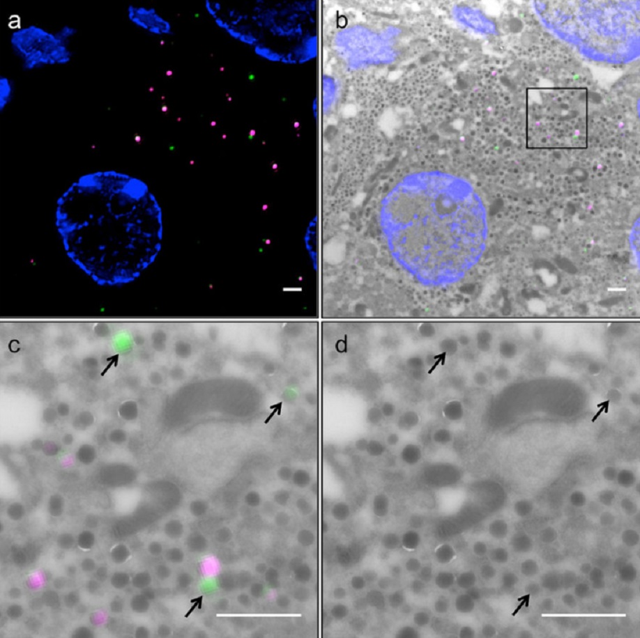
Correlative light and electron microscopy (CLEM) is a powerful approach to investigate the molecular ultrastructure of labeled cell compartments. However, quantitative CLEM studies are rare, mainly due to small sample sizes and the sensitivity of fluorescent proteins to strong fixatives and contrasting reagents for EM. Here, we show that fusion of…
Read more
Andreas Müller, Martin Neukam, Anna Ivanova, Anke Sönmez, Carla Münster, Susanne Kretschmar, Yannis Kalaidzidis, Thomas Kurth, Jean-Marc Verbavatz & Michele Solimena
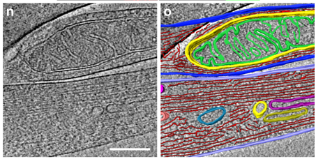
Correlative cryo-electron microscopy reveals the structure of TNTs in neuronal cells
The orchestration of intercellular communication is essential for multicellular organisms. One mechanism by which cells communicate is through long, actin-rich membranous protrusions called tunneling nanotubes (TNTs), which allow the intercellular transport of various cargoes, between the cytoplasm of distant cells in vitro and in vivo. Here, we use correlative FIB-SEM, light- and cryo-electron microscopy approaches to elucidate the structural organization of neuronal TNTs. Our data indicate ... Read more
Anna Sartori-Rupp, Diégo Cordero Cervantes, Anna Pepe, Karine Gousset, Elise Delage, Simon Corroyer-Dulmont, Christine Schmitt, Jacomina Krijnse-Locker & Chiara Zurzolo
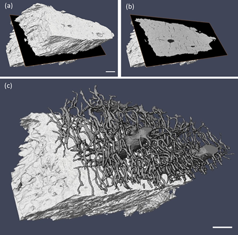
Ptychographic X-ray computed tomography (PXCT) is a quantitative imaging modality that non-destructively maps the 3D electron density inside an object with tens of nanometers spatial resolution. This method provides unique access to the morphology and structure of the osteocyte lacuno-canalicular network (LCN) and nanoscale density of the tissue in the vicinity of an osteocyte lacuna. Our findings indicate that PXCT can non-destructively provide detailed, nanoscale information on the 3D org... Read more
Antonia Ciani, Hechmi Toumi, Stéphane Pallu, Esther H.R.Tsai, Ana Diaz, Manuel Guizar-Sicairos, Mirko Holler, Eric Lespessailles, Cameron M.Kewish | Synchrotron Soleil, France
Volcanogenic Pseudo-Fossils from the ∼3.48 Ga
The ∼3.48 billion-year-old Dresser Formation, Pilbara Craton, Western Australia, is a key geological unit for the study of Earth’s earliest life and the habitats it occupied. Here, we describe a new suite of spheroidal to lenticular microstructures that morphologically resemble some previously reported Archean microfossils. Correlative microscopy shows that these objects have a size distribution, wall ultrastructure, and chemistry that are incompatible with a microfossil origin and in... Read more
Wacey David , Noffke Nora , Saunders Martin , Guagliardo Paul , and Pyle David M
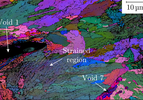
A multi-scale correlative investigation of ductile fracture
The use of novel multi-scale correlative methods, which involve the coordinated characterization of matter across a range of length scales, are becoming of increasing value to materials scientists. Here, we describe for the first time how a multi-scale correlative approach can be used to investigate the nature of ductile fracture in metals. Specimens of a nuclear pressure vessel steel, SA508 Grade 3, are examined following ductile fracture using medium and high-resolution 3D X-ray computed to... Read more
School of Materials, University of Manchester; National Nuclear Laboratory; BAM Federal Institute for Materials Research and Testing; Thermo Fischer Scientific