Welcome to the Amira-Avizo Software Use Case Gallery
Below you will find a collection of use cases of our 3D data visualization and analysis software. These use cases include scientific publications, articles, papers, posters, presentations or even videos that show how Amira-Avizo Software is used to address various scientific and industrial research topics.
Use the Domain selector to filter by main application area, and use the Search box to enter keywords related to specific topics you are interested in.
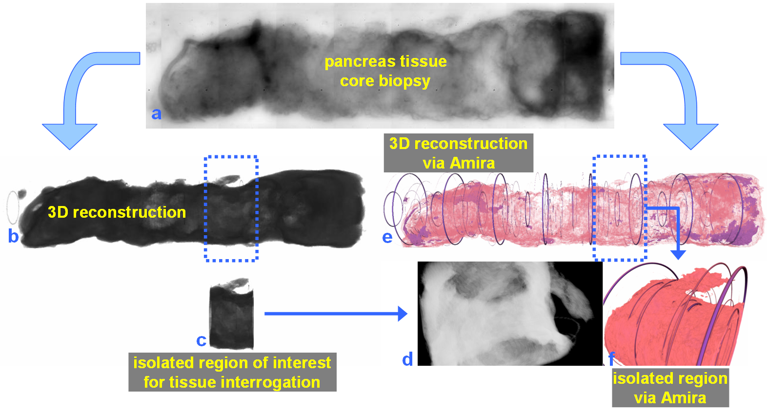
For nearly 100 years, pathology for cancer diagnosis has involved a standard, but complex series of steps to process tissue biopsies procured from a patient in the clinic. Many procedures are a direct result of the fact that observation and evaluation of specimens by pathologists occur using a standard microscope (in 2D).
In 2014, the Human Photonics Laboratory at the University of Washington demonstrated that the rudimentary operations of a pathology laboratory may be replicated on wh... Read more
Ronnie Das, PhD and Eric J. Seibel, PhD
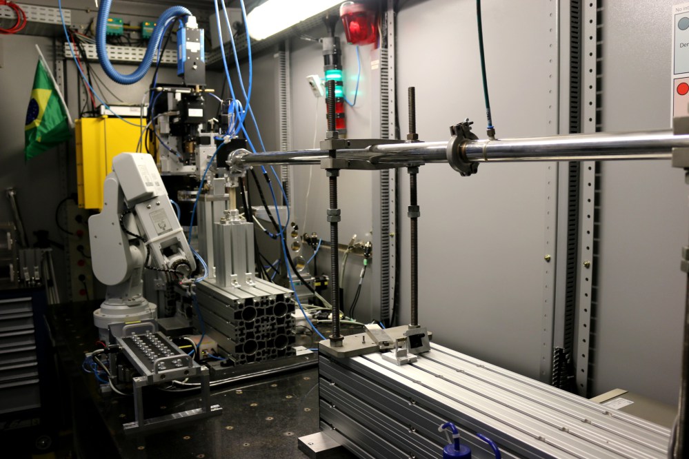
The imaging beamline, IMX, at LNLS extracts synchrotron radiation from bending magnet D6 with magnetic field of 1.67 T and bending radius of 2.736 m. It has an electron source size of 391 Read more
Brazilian Synchotron Light Laboratory
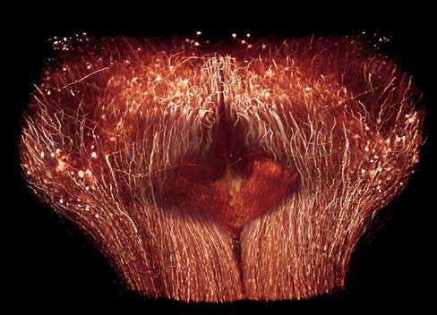
The Max Planck Institute of Neurobiology uses Amira software for axon tracing from head to toe
Ali Ertürk and his collaborators described a novel histo-chemical technique to clear spinal cord tissue of adult mice for fluorescence microscopy imaging of the intact spinal cord.
Previously, the high lipid content of the spinal cord tissue in adult mice allowed imaging of the spinal cord only through destructive histological methods, making the tracing of complete axons impossible. As applied in this study, the improved histo-chemistry enabled Ertürk and his colleagues to show that... Read more
Ali Ertürk, Christoph P Mauch, Farida Hellal, Friedrich Förstner, Tara Keck, Klaus Becker, Nina Jährling, Heinz Steffens, Melanie Richter, Mark Hübener, Edgar Kramer, Frank Kirchhoff, Hans Ulrich Dodt & Frank Bradke