Welcome to the Amira-Avizo Software Use Case Gallery
Below you will find a collection of use cases of our 3D data visualization and analysis software. These use cases include scientific publications, articles, papers, posters, presentations or even videos that show how Amira-Avizo Software is used to address various scientific and industrial research topics.
Use the Domain selector to filter by main application area, and use the Search box to enter keywords related to specific topics you are interested in.
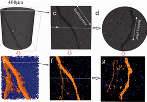
Experimental study on the cracking process of layered shale using X-ray microCT
The cracking process in Longmaxi formation shale was experimentally studied during uniaxial compressive loading. Both the evolution of the three-dimensional fracture network and the micromechanics of failure in the layered shale were examined as a function of the inclination angle of the bedding plane. To visualize the cracking process, the test devices presented here used an industrial X-ray CT scanner that enabled scanning during the uniaxial compressive loading. Scanning electron microscop... Read more
Institue of Geomechanic, Chinese Academy of Geological Sciences, Laboratory of Shale Oil & Gas, Beijing, China
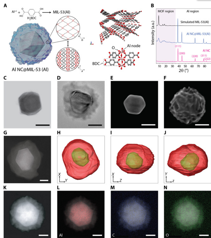
Metal-organic frameworks tailor the properties of aluminum nanocrystals
Metal-organic frameworks (MOFs) and metal nanoparticles are two classes of materials that have received considerable recent attention, each for controlling chemical reactivities, albeit in very different ways. Here, we report the growth of MOF shell layers surrounding aluminum nanocrystals (Al NCs), an Earth-abundant metal with energetic, plasmonic, and photocatalytic properties. The MOF shell growth proceeds by means of dissolution-and-growth chemistry that uses the intrinsic surface oxide o... Read more
Hossein Robatjazi, Daniel Weinberg, Dayne F. Swearer, Christian Jacobson, Ming Zhang, Shu Tian, Linan Zhou, Peter Nordlander, Naomi J. Halas
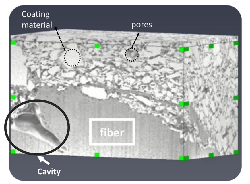
Characterization of the Interface Between Coating and Fibrous Layers of Paper
Coated paper is an example of a multi-layer porous medium, involving a coating layer along the two surfaces of the paper and a fibrous layer in the interior of the paper. The interface between these two media needs to be characterized in order to develop relevant modeling tools. After careful cutting of the paper, a cross section was imaged using focused ion beam scanning electron microscopy. The resulting image was analyzed to characterize the coating layer and its transition to the fibrous ... Read more
H. Aslannejad, S. M. Hassanizadeh, M. A. Celia
The development of focused ion beam-scanning electron microscopy (FIB-SEM) techniques has allowed high-resolution 3D imaging of nanometre-scale porous materials. These systems are of important interest to the oil and gas sector, as well as for the safe long-term storage of carbon and nuclear waste. This work focuses on validating the accurate representation of sample pore space in FIB-SEM-reconstructed volumes and the predicted permeability of these systems from subsequent single-phase flow s... Read more
Department of Chemical Engineering, Qatar Carbonates and Carbon Storage Research Centre, Imperial College London | Department of Applied Mathematics and Theoretical Physics, Cambridge University
Visualizing the Carbon Binder Phase of Battery Electrodes in Three Dimensions
This study presents a technique to directly characterize the carbon and binder domain (CBD) in lithium-ion (Li-ion) battery electrodes in three dimensions and use it to determine the effective transport properties of a LiNi0.33Mn0.33Co0.33O2 (NMC) electrode. X-ray nanocomputed tomography (nano-CT) is used to image an electrode composed solely of carbon and binder, whereas focused ion beam–scanning electron microscopy is used to analyze cross-sect... Read more
Sohrab R. Daemi, Chun Tan, Tobias Volkenandt, Samuel J. Cooper, Anna Palacios-Padros, James Cookson, Dan J. L. Brett, and Paul R. Shearing
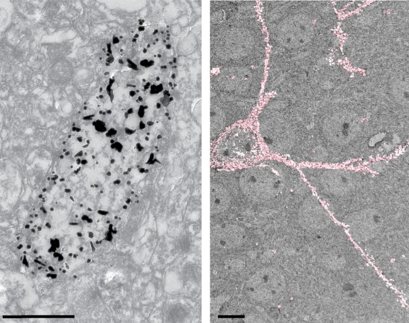
Corpora amylacea are cell-derived structures that appear physiologically in the aged human brain. While their histological identification is straightforward, their ultrastructural composition and microenvironment at the nanoscale have remained unclear so far, as has their relevance to aging and certain disease states that involve the sequestration of toxic cellular metabolites. Here, we apply correlative serial block-face scanning electron microscopy and transmission electron tomograp... Read more
Paula P. Navarro, Christel Genoud, Daniel Castaño-Díez, Alexandra Graff-Meyer, Amanda J. Lewis, Yvonne de Gier, Matthias E. Lauer, Markus Britschgi, Bernd Bohrmann, Stephan Frank, Jürgen Hench, Gabriel Schweighauser, Annemieke J. M. Rozemuller, Wilma D. J. van de Berg, Henning Stahlberg & Sarah H. Shahmoradian

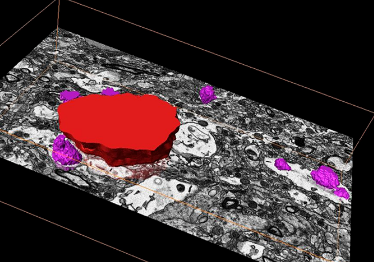
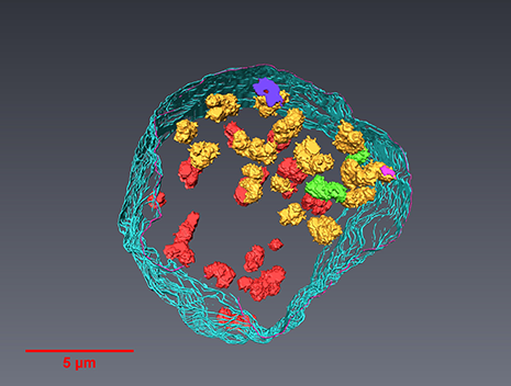
Macropinosomes are key players in early shigella invasion and vacuolar escape in epithelial cells
Intracellular pathogens include all viruses, many bacteria and parasites capable of invading and surviving within host cells. Key to survival is the subversion of host cell pathways by the pathogen for the purpose of propagation and evading the immune system. The intracellular bacterium Shigella flexneri, the causative agent of bacillary dysentery, invades host cells in a vacuole that is subsequently ruptured to allow growth of the pathogen within the host cytoplasm…
Read more
Allon Weiner , Nora Mellouk , Noelia Lopez-Montero , Yuen-Yan Chang, Célia Souque, Christine Schmitt, Jost Enninga
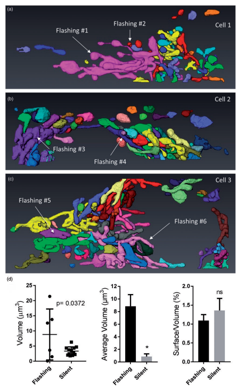
Ultrastructural Characterization of Flashing Mitochondria
Mitochondria undergo spontaneous transient elevations in matrix pH associated with drops in mitochondrial membrane potential. These mitopHlashes require a functional respiratory chain and the profusion protein optic atrophy 1, but their mechanistic basis is unclear. To gain insight on the origin of these dynamic events, we resolved the ultrastructure of flashing mitochondria by correlative light and electron microscopy. HeLa cells expressing the matrix-targeted pH probe mitoSypHer were screen... Read more
Manon Rosselin, Paula Nunes-Hasler, and Nicolas Demaurex
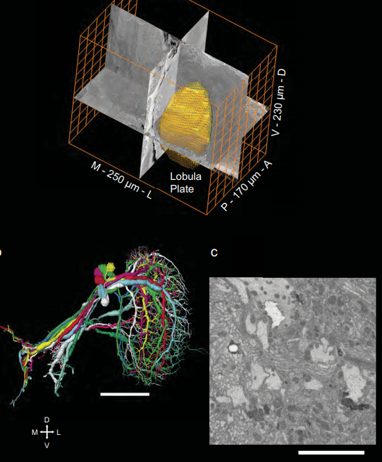
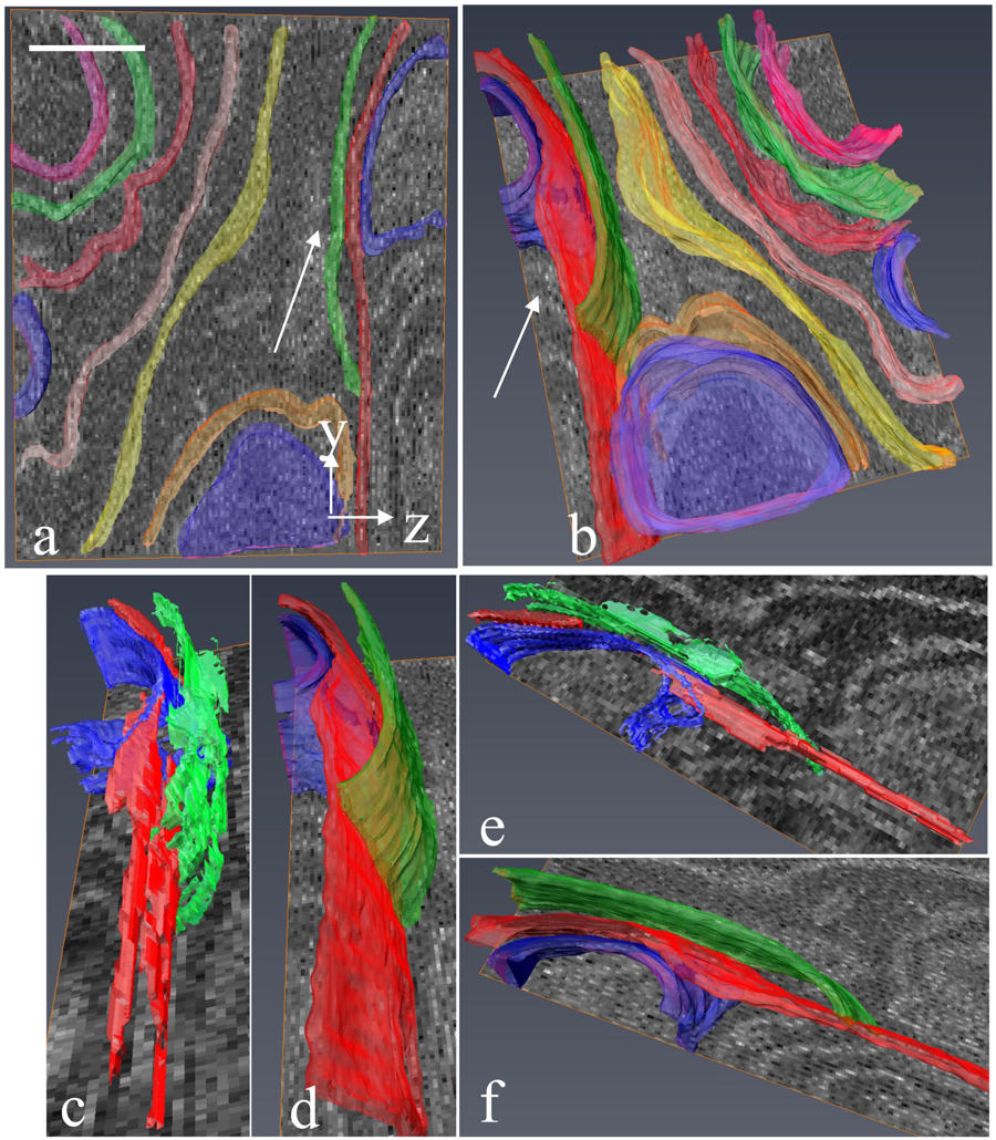
Full reconstruction of large lobula plate tangential cells in Drosophila from a 3D EM dataset
With the advent of neurogenetic methods, the neural basis of behavior is presently being analyzed in more and more detail. This is particularly true for visually driven behavior of Drosophila melanogaster where cell-specific driver lines exist that, depending on the combination with appropriate effector genes, allow for targeted recording, silencing and optogenetic stimulation of individual cell-types. Together with detailed connectomic data of large parts of the fly optic lobe, this has rece... Read more
Kevin M. Boergens , Christoph Kapfer, Moritz Helmstaedter, Winfried Denk, Alexander Borst
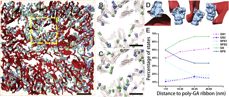
In Situ Structure of Neuronal C9orf72 Poly-GA Aggregates Reveals Proteasome Recruitment
Protein aggregation and dysfunction of the ubiquitin-proteasome system are hallmarks of many neurodegenerative diseases. Here, we address the elusive link between these phenomena by employing cryo-electron tomography to dissect the molecular architecture of protein aggregates within intact neurons at high resolution. We focus on the poly-Gly-Ala (poly-GA) aggregates resulting from aberrant translation of an expanded GGGGCC repeat in C9orf72, the most common genetic cause of amyotrophic latera... Read more
Qiang Guo, Carina Lehmer, Antonio Martinez-Sanchez, Till Rudack, Florian Beck, Hannelore Hartmann, Manuela Perez-Berlanga, Frederic Frottin, Mark S.Hipp, F. Ulrich Hartl, Dieter Edbauer, Wolfgang Baumeister, Ruben Fernandez-Busnadiego
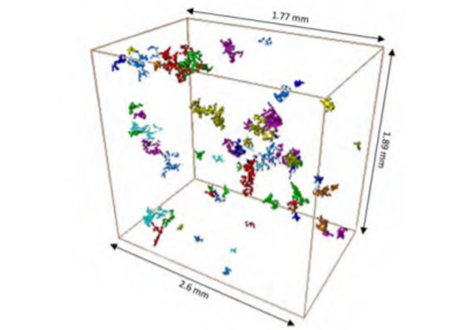
Analysis of neuronal arborization and connections is a powerful tool in fundamental and clinical neuroscience. Changes in neuronal morphology are central to brain development and plasticity and are associated with numerous diseases. Golgi staining is a classical technique based on a deposition of metal precipitate in a random set of neurons. Despite their versatility, Golgi methods have limitations that largely precluded their use in advanced microscopy. We combined Golgi staining with fluore... Read more
Katlijn Vints, Dorien Vandael, Pieter Baatsen, Benjamin Pavie, Frank Vernaillen, Nikky Corthout, Vasily Rybakin, Sebastian Munck & Natalia V. Gounko
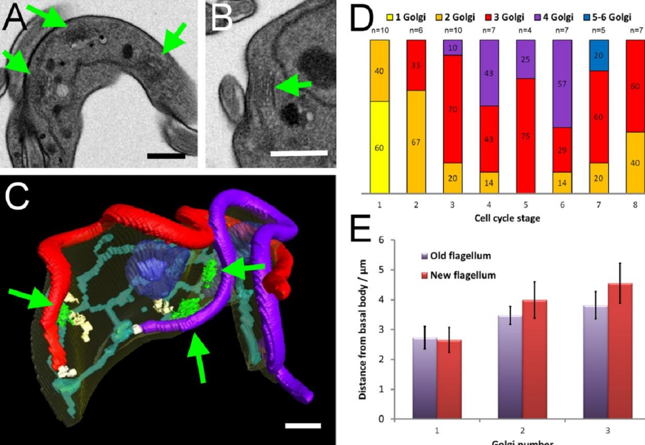
The major mammalian bloodstream form of the African sleeping sickness parasite Trypanosoma bruceimultiplies rapidly, and it is important to understand how these cells divide. Organelle inheritance involves complex spatiotemporal re-arrangements to ensure correct distribution to daughter cells…
Read more
Louise Hughes, Samantha Borrett, Katie Towers, Tobias Starborg, Sue Vaughan
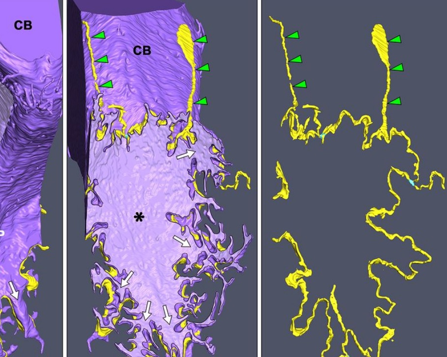
Morphological process of podocyte development revealed by block-face scanning electron microscopy
Podocytes present a unique 3D architecture specialized for glomerular filtration. However, several 3D morphological aspects on podocyte development remain partially understood because they are difficult to reveal using conventional scanning electron microscopy (SEM). Here, we adopted serial block-face SEM imaging…
Read more
Koichiro Ichimura, Soichiro Kakuta, Yuto Kawasaki, Takayuki Miyaki, Takahiro Nonami, Naoyuki Miyazaki, Tomoyo Nakao, Sakiko Enomoto, Shigeo Arai, Masato Koike, Kazuyoshi Murata, Tatsuo Sakai
Serial block-face electron microscopy (SBEM) provides nanoscale 3D ultrastructure of embedded and stained cells and tissues in volumes of up to 107 µm3. In SBEM, electrons with 1–3 keV energies are incident on a specimen block, from which backscattered electron (BSE) images are collected with x, y resolution of 5–10 nm in the block-face plane, and successive layers are removed by an in situ ultramicrotome. Sp... Read more
Q. He, M. Hsueh, G. Zhang, D. C. Joy & R. D. Leapman
3D characterization of the fracture mechanisms of a Fe-rich Al-Si-Cu alloy
The effect of the defect size and morphology on the fatigue damage evolution was analysed in a recycled Al-Si-Cu alloy by micro-computed tomography and scanning electron microscopy. Fatigue tests were performed and the different crack initiation scenarios were characterized and classified. The interaction between shrinkage and gas pores was the key crack initiation mechanism and the ß-Al5FeSi particles did not play any role in the crack initiation phase. However, crack path analysis indicate... Read more
Angelika Brueckner-Foit, Inigo Bacaicoa, Martin Luetje, Marcel Wicke, Andreas Geisert, Martin Fehlbier
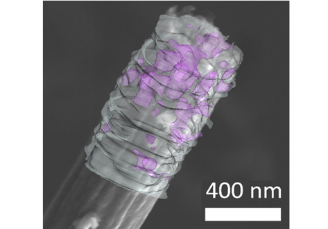
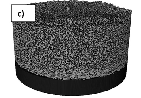
Flexible all-fiber electrospun supercapacitor
Electrospun all-fiber flexible supercapacitor with nanofiber electrodes/separator.
- Increased graphitic degree with the addition of MnACAC and thermal decomposition.
- Enhanced capacitive performance with the addition of MnO.
- Quantified nanofiber alignment and increased bias with MnO over undoped fibers.
- FIBSEM tomography of nanofibers showing MnO disitribution in carbon nanofibers.
We present an all-fiber flexible supercapacitor with compo... Read more
Xinhua Liu, Max Naylor Marlow, Samuel J. Cooper, Bowen Song, Xiaolong Chen, Nigel P. Brandon, Billy Wu
A team led by London Centre for Nanotechnology researchers, Prof. Ian Robinson and Dr. Bo Chen (now a professor at the Tongji University, Shanghai) used newly-developed serial block-face scanning electron microscopy (SBFSEM) and Thermo Scientific™ Avizo® Software, one dominant tool in 3D reconstructed image processing, to reveal the spatial structure of human chromosomes and nucleus quantitatively at high resolution of approximately 50 nm in three dimensions.
Read more
Prof. Ian Robinson, Dr. Bo Chen, London Centre for Nechnology, UCL and Tongji University