Welcome to the Amira-Avizo Software Use Case Gallery
Below you will find a collection of use cases of our 3D data visualization and analysis software. These use cases include scientific publications, articles, papers, posters, presentations or even videos that show how Amira-Avizo Software is used to address various scientific and industrial research topics.
Use the Domain selector to filter by main application area, and use the Search box to enter keywords related to specific topics you are interested in.
3D Reconstruction of Lipid Droplets in the Seed of Brassica napus
Rapeseed is one of the most important and widely cultured oilseed crops for food and nonfood purposes worldwide. Neutral lipids are stored in lipid droplets (LDs) as fuel for germination and subsequent seedling growth. Most of the LD detection in seeds was still in 2D levels, and some of the details might have been lost in previous studies. In the present work, the configuration of LDs in seeds was obtained by confocal imaging combined with 3D reconstruction technology in Brassica napus<... Read more
Yongtai Yin, Liangxing Guo, Kang Chen, Zhenyi Guo, Hongbo Chao, Baoshan Wang, and Maoteng Licorresponding author
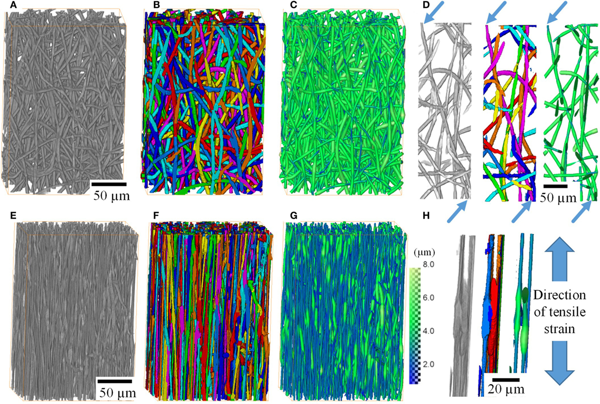
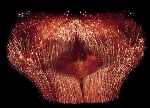
X-ray Tomographic Imaging of Tensile Deformation Modes of Electrospun Biodegradable Polyester Fibers
Electrospun constructs for the repair of load-bearing tissues are required to have adequate mechanical properties. However, the failure mechanisms of electrospun fibrous materials are not well understood. Existing literature focuses on failure modes of individual fibers and/or on bulk mechanical properties of whole fiber mats.
Electrospinning allows the production of fibrous networks for tissue engineering, drug delivery, and wound healing in health care. It enables the production of c... Read more
Jekaterina Maksimcuka; Akiko Obata ; William W. Sampson ; Remi Blanc ; Chunxia Gao ; Philip J. Withers ; Olga Tsigkou ; Toshihiro Kasuga ; Peter D. Lee ; Gowsihan Poologasundarampillai
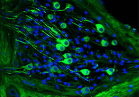
Chronic cigarette smoke exposure drives spiral ganglion neuron loss in mice
Tobacco use is associated with an increased risk of hearing loss in older individuals, suggesting cigarette smoke (CS) exposure may target the peripheral auditory organs. However, the effects of CS exposure on general cochlear anatomy have not previously been explored.
Here we compare control and chronic CS exposed cochleae from adult mice to assess changes in structure and cell survival. Two-photon imaging techniques, including the imaging of second harmonic generation (SHG) and two-p... Read more
Stephen T. Paquette, Ryan P. Dawes, Isaac K. Sundar, Irfan Rahman, Edward B. Brown & Patricia M. White
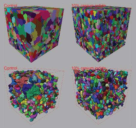
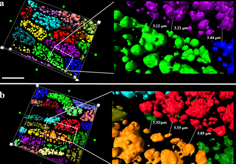
Contrast-enhanced 3D micro-CT of plant tissues using different impregnation techniques
X-ray micro-CT has increasingly been used for 3D imaging of plant structures. At the micrometer reso-lution however, limitations in X-ray contrast often lead to datasets with poor qualitative and quantitative measures, especially within dense cell clusters of plant tissue specimens. The current study developed protocols for delivering a cesium based contrast enhancing solution to varying plant tissue specimens for the purpose of improving 3D tissue structure characterization within plant spec... Read more
Zi Wang, Pieter Verboven and Bart Nicolai, Department of Biosystems KU Leuven – University of Leuven Willem de Croylaan, Leuven Belgium
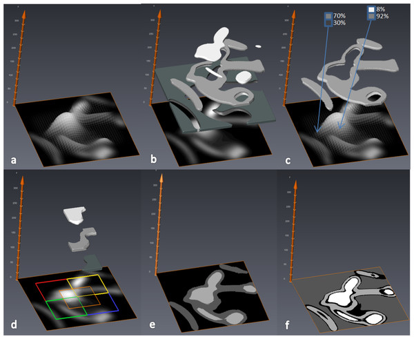
MIA-Clustering: a novel method for segmentation of paleontological material
Paleontological research increasingly uses high-resolution micro-computed tomography (μCT) to study the inner architecture of modern and fossil bone material to answer important questions regarding vertebrate evolution. This non-destructive method allows for the measurement of otherwise inaccessible morphology. Digital measurement is predicated on the accurate segmentation of modern or fossilized bone from other structures imaged in μCT scans, as errors in segmentation can result in inaccur... Read more
Christopher J. Dunmore, Gert Wollny, Matthew M. Skinner

The terrestrial Judith River Formation of northern Montana was deposited over an approximately 4 Myr interval during the Campanian (Late Cretaceous). Despite having been prospected and collected continuously by palaeontologists for over a century, few relatively complete dinosaur skeletons have been recovered from this unit to date. Here we describe a new genus and species of ankylosaurine dinosaur, Zuul crurivastator, from the Coal Ridge Member of the Judith River Formation, based ... Read more
Victoria M. Arbour, David C. Evans
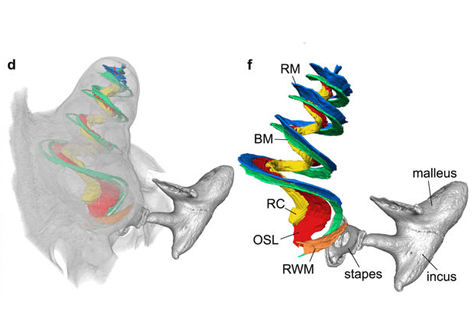
Propagation-based phase-contrast x-ray tomography of cochlea using a compact synchrotron source
We demonstrate that phase retrieval and tomographic imaging at the organ level of small animals can be advantageously carried out using the monochromatic radiation emitted by a compact x-ray light source, without further optical elements apart from source and detector. This approach allows to carry out microtomography experiments which – due to the large performance gap with respect to conventional laboratory instruments – so far were usually limited to synchrotron sources. We dem... Read more
Mareike Töpperwien, Regine Gradl, Daniel Keppeler, Malte Vassholz, Alexander Meyer, Roland Hessler, Klaus Achterhold, Bernhard Gleich, Martin Dierolf, Franz Pfeiffer, Tobias Moser & Tim Salditt
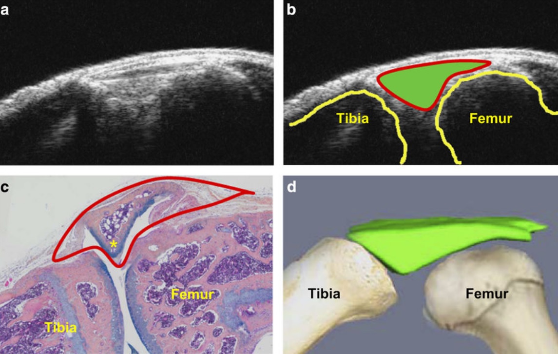
Ultrasound could be a fast and cost-effective means of assessing joint changes in mouse models of posttraumatic osteoarthritis (PTOA). Such models are essential for understanding the biology of this degenerative joint disease and developing new treatments, but noninvasive methods of evaluating disease activity are lacking. Because ultrasound can visualize both joint space volumes and blood flow in the joints, it could provide an alternative to microscopic examination of tissue, assuming it ac... Read more
Hao Xu, Echoe M Bouta, Ronald W Wood, Edward M Schwarz, Yongjun Wang & Lianping Xing
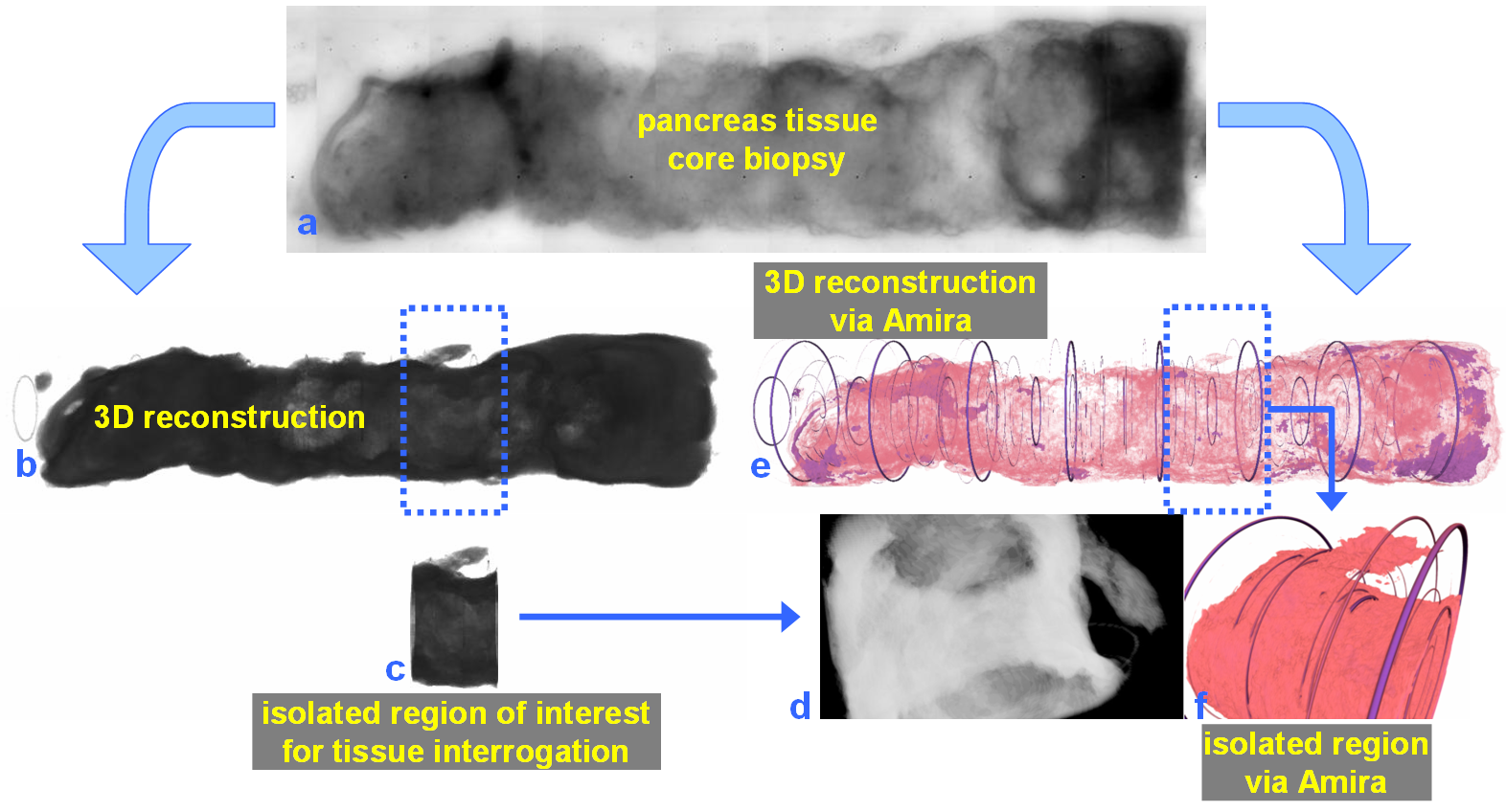
For nearly 100 years, pathology for cancer diagnosis has involved a standard, but complex series of steps to process tissue biopsies procured from a patient in the clinic. Many procedures are a direct result of the fact that observation and evaluation of specimens by pathologists occur using a standard microscope (in 2D).
In 2014, the Human Photonics Laboratory at the University of Washington demonstrated that the rudimentary operations of a pathology laboratory may be replicated on wh... Read more
Ronnie Das, PhD and Eric J. Seibel, PhD
The Max Planck Institute of Neurobiology uses Amira software for axon tracing from head to toe
Ali Ertürk and his collaborators described a novel histo-chemical technique to clear spinal cord tissue of adult mice for fluorescence microscopy imaging of the intact spinal cord.
Previously, the high lipid content of the spinal cord tissue in adult mice allowed imaging of the spinal cord only through destructive histological methods, making the tracing of complete axons impossible. As applied in this study, the improved histo-chemistry enabled Ertürk and his colleagues to show that... Read more
Ali Ertürk, Christoph P Mauch, Farida Hellal, Friedrich Förstner, Tara Keck, Klaus Becker, Nina Jährling, Heinz Steffens, Melanie Richter, Mark Hübener, Edgar Kramer, Frank Kirchhoff, Hans Ulrich Dodt & Frank Bradke