Welcome to the Amira-Avizo Software Use Case Gallery
Below you will find a collection of use cases of our 3D data visualization and analysis software. These use cases include scientific publications, articles, papers, posters, presentations or even videos that show how Amira-Avizo Software is used to address various scientific and industrial research topics.
Use the Domain selector to filter by main application area, and use the Search box to enter keywords related to specific topics you are interested in.
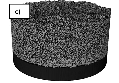
Lithium-ion (Li-ion) batteries operate via electrochemical reactions between positive and negative electrodes, formed by complex porous microstructures. An improved understanding of these materials can lead to a greater insight into the link between microscopic electrode morphology and macroscopic performance. The practice of calendering electrodes after manufacturing has been widely used to increase the volumetric energy density and improve the electrical contact between electrode... Read more
S. R. Daemi,X. Lu, D. Sykes, J. Behnsen, C. Tan, A. Palacios-Padros, J. Cookson, E. Petrucco, P. J. Withers, D. J. L. Brett and P. R. Shearing
Four-Dimensional Studies of Morphology Evolution in Lithium–Sulfur Batteries
Lithium sulfur (Li–S) batteries have great potential as a successor to Li-ion batteries, but their commercialization has been complicated by a multitude of issues stemming from their complex multiphase chemistry. In situ X-ray tomography investigations enable direct observations to be made about a battery, providing unprecedented insight into the microstructural evolution of the sulfur cathode and shedding light on the reaction kinetics of the sulfur phase. Here, for the first time, the mor... Read more
Chun Tan, Thomas M. M. Heenan, Ralf F. Ziesche, Sohrab R. Daemi, Jennifer Hack, Maximilian Maier, Shashidhara Marathe, Christoph Rau, Daniel J. L. Brett, Paul R. Shearing
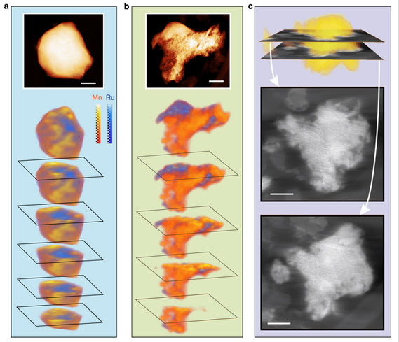
Despite the importance of studying the instability of delithiated cathode materials, it remains difficult to underpin the degradation mechanism of lithium-rich cathode materials due to the complication of combined chemical and structural evolutions. Herein, we use state-of-the-art electron microscopy tools, in conjunction with synchrotron X-ray techniques and first-principle calculations to study a 4d-element-containing compound, Li2Ru0.5Mn0.5O3. We find surprisingly, after cycling, ruthenium... Read more
Lin, Ruoqian AU - Hu, Enyuan AU - Liu, Mingjie AU - Wang, Yi AU - Cheng, Hao AU - Wu, Jinpeng AU - Zheng, Jin-Cheng AU - Wu, Qin AU - Bak, Seongmin AU - Tong, Xiao AU - Zhang, Rui AU - Yang, Wanli AU - Persson, Kristin A. AU - Yu, Xiqian AU - Yang, Xiao-Qing AU - Xin, Huolin L. PY
Visualizing the Carbon Binder Phase of Battery Electrodes in Three Dimensions
This study presents a technique to directly characterize the carbon and binder domain (CBD) in lithium-ion (Li-ion) battery electrodes in three dimensions and use it to determine the effective transport properties of a LiNi0.33Mn0.33Co0.33O2 (NMC) electrode. X-ray nanocomputed tomography (nano-CT) is used to image an electrode composed solely of carbon and binder, whereas focused ion beam–scanning electron microscopy is used to analyze cross-sect... Read more
Sohrab R. Daemi, Chun Tan, Tobias Volkenandt, Samuel J. Cooper, Anna Palacios-Padros, James Cookson, Dan J. L. Brett, and Paul R. Shearing
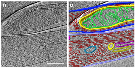
As key functional units in neural circuits, different types of neuronal synapses play distinct roles in brain information processing, learning, and memory. Synaptic abnormalities are believed to underlie various neurological and psychiatric disorders. Here, by combining cryo-electron tomography and cryo-correlative light and electron microscopy, we distinguished intact excitatory and inhibitory synapses of cultured hippocampal neurons, and visualized the in situ 3D organization of ... Read more
Chang-Lu Tao, Yun-Tao Liu, Rong Sun, Bin Zhang, Lei Qi, Sakar Shivakoti, Chong-Li Tian, Peijun Zhang, Pak-Ming Lau, Z. Hong Zhou and Guo-Qiang Bi

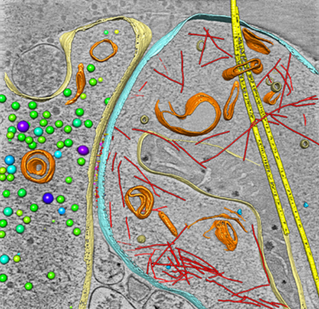
Macropinosomes are key players in early shigella invasion and vacuolar escape in epithelial cells
Intracellular pathogens include all viruses, many bacteria and parasites capable of invading and surviving within host cells. Key to survival is the subversion of host cell pathways by the pathogen for the purpose of propagation and evading the immune system. The intracellular bacterium Shigella flexneri, the causative agent of bacillary dysentery, invades host cells in a vacuole that is subsequently ruptured to allow growth of the pathogen within the host cytoplasm…
Read more
Allon Weiner , Nora Mellouk , Noelia Lopez-Montero , Yuen-Yan Chang, Célia Souque, Christine Schmitt, Jost Enninga
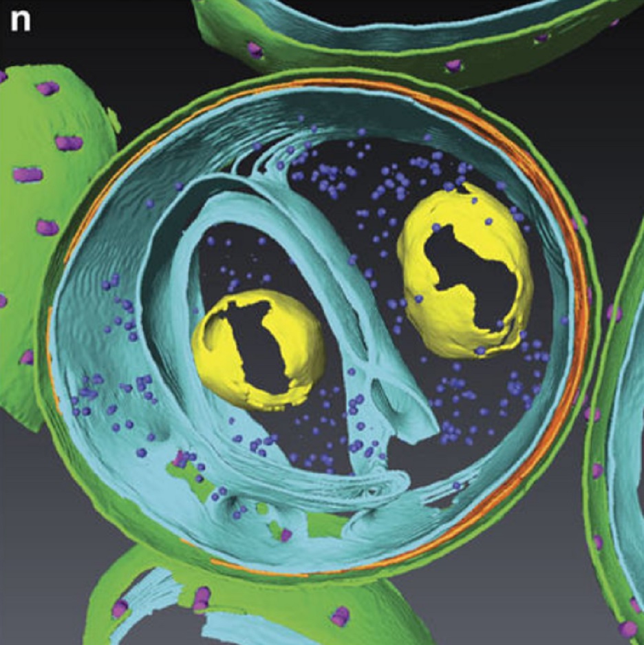
Determining the bacterial cell biology of planctomycetes
Bacteria of the phylum Planctomycetes have been previously reported to possess several features that are typical of eukaryotes, such as cytosolic compartmentalization and endocytosis-like macromolecule uptake. However, recent evidence points towards a Gram-negative cell plan for Planctomycetes, although in-depth experimental analysis has been hampered by insufficient genetic tools…
Read more
Christian Boedeker, Margarete Schüler, Greta Reintjes, Olga Jeske, Muriel C. F. van Teeseling et al.
Fast and precise targeting of single tumor cells in vivo by multimodal correlative microscopy
Intravital microscopy provides dynamic understanding of multiple cell biological processes, but its limited resolution has so far precluded structural analysis. Because it is difficult to capture rare and transient events, only a few attempts have been made to observe specific developmental and pathological processes in animal models using electron microscopy. The multimodal correlative approach that we propose here combines intravital microscopy, microscopic X-ray computed tomography and thr... Read more
Matthia A. Karreman, Luc Mercier, Nicole L. Schieber, Gergely Solecki, Guillaume Allio, Frank Winkler, Bernhard Ruthensteiner, Jacky G. Goetz, Yannick Schwab
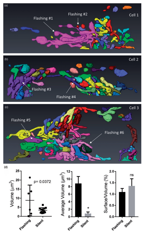
Ultrastructural Characterization of Flashing Mitochondria
Mitochondria undergo spontaneous transient elevations in matrix pH associated with drops in mitochondrial membrane potential. These mitopHlashes require a functional respiratory chain and the profusion protein optic atrophy 1, but their mechanistic basis is unclear. To gain insight on the origin of these dynamic events, we resolved the ultrastructure of flashing mitochondria by correlative light and electron microscopy. HeLa cells expressing the matrix-targeted pH probe mitoSypHer were screen... Read more
Manon Rosselin, Paula Nunes-Hasler, and Nicolas Demaurex
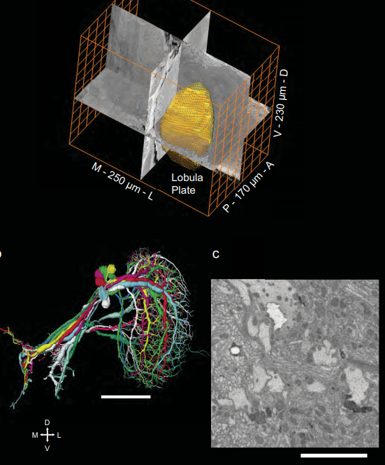
Full reconstruction of large lobula plate tangential cells in Drosophila from a 3D EM dataset
With the advent of neurogenetic methods, the neural basis of behavior is presently being analyzed in more and more detail. This is particularly true for visually driven behavior of Drosophila melanogaster where cell-specific driver lines exist that, depending on the combination with appropriate effector genes, allow for targeted recording, silencing and optogenetic stimulation of individual cell-types. Together with detailed connectomic data of large parts of the fly optic lobe, this has rece... Read more
Kevin M. Boergens , Christoph Kapfer, Moritz Helmstaedter, Winfried Denk, Alexander Borst
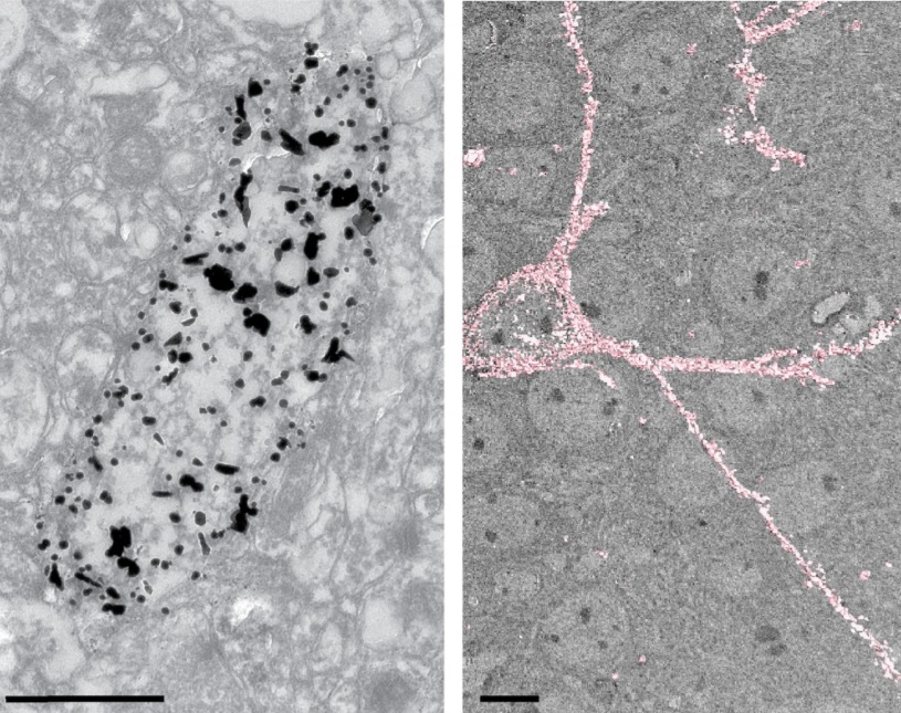
Analysis of neuronal arborization and connections is a powerful tool in fundamental and clinical neuroscience. Changes in neuronal morphology are central to brain development and plasticity and are associated with numerous diseases. Golgi staining is a classical technique based on a deposition of metal precipitate in a random set of neurons. Despite their versatility, Golgi methods have limitations that largely precluded their use in advanced microscopy. We combined Golgi staining with fluore... Read more
Katlijn Vints, Dorien Vandael, Pieter Baatsen, Benjamin Pavie, Frank Vernaillen, Nikky Corthout, Vasily Rybakin, Sebastian Munck & Natalia V. Gounko
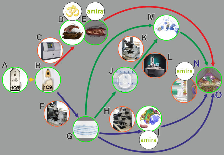
In biomedical research, a huge variety of different techniques is currently available for the structural examination of small specimens, including conventional light microscopy (LM), transmission electron microscopy (TEM), confocal laser scanning microscopy (CLSM), microscopic X-ray computed tomography (microCT), and many others. Since every imaging method is physically limited by certain parameters, a correlative use of complementary methods often yields a significant broader range of inform... Read more
Stephan Handschuh, Natalie Baeumler, Thomas Schwaha and Bernhard Ruthensteiner
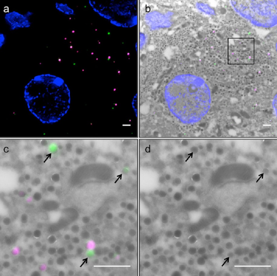
Correlative light and electron microscopy (CLEM) is a powerful approach to investigate the molecular ultrastructure of labeled cell compartments. However, quantitative CLEM studies are rare, mainly due to small sample sizes and the sensitivity of fluorescent proteins to strong fixatives and contrasting reagents for EM. Here, we show that fusion of…
Read more
Andreas Müller, Martin Neukam, Anna Ivanova, Anke Sönmez, Carla Münster, Susanne Kretschmar, Yannis Kalaidzidis, Thomas Kurth, Jean-Marc Verbavatz & Michele Solimena
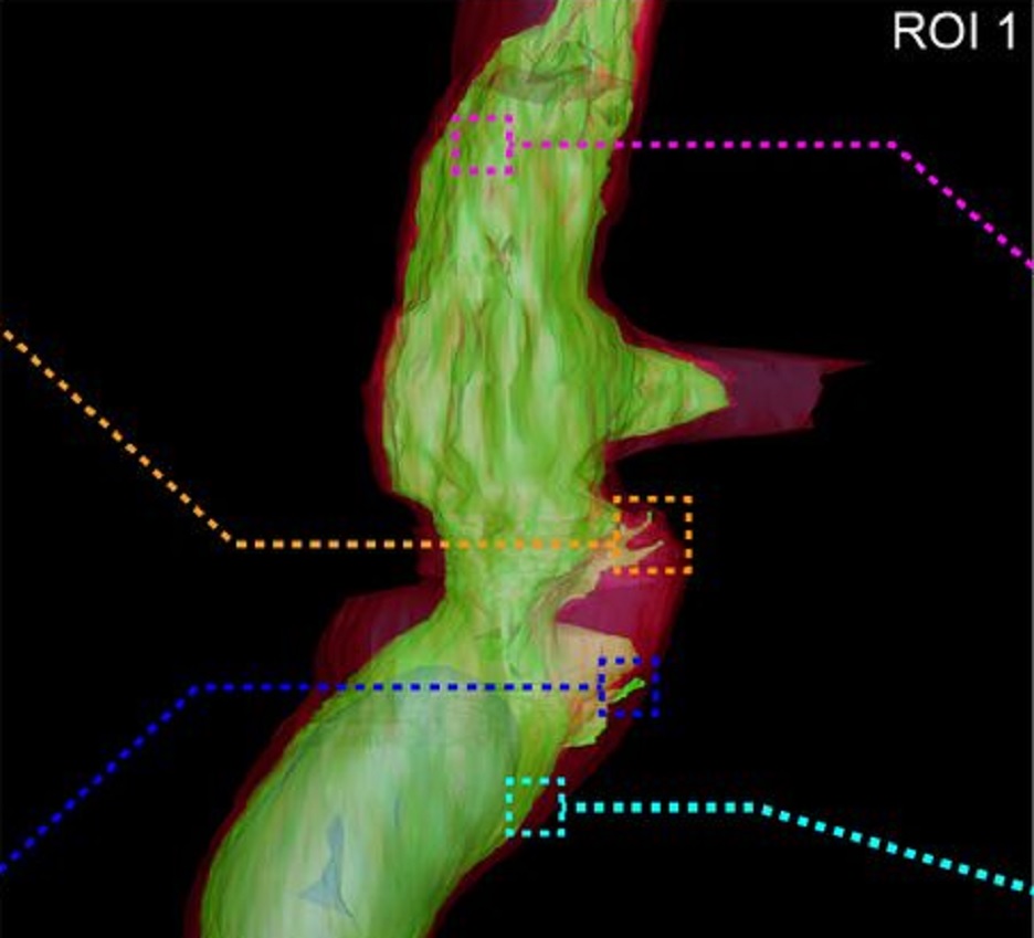
Correlative cryo-electron microscopy reveals the structure of TNTs in neuronal cells
The orchestration of intercellular communication is essential for multicellular organisms. One mechanism by which cells communicate is through long, actin-rich membranous protrusions called tunneling nanotubes (TNTs), which allow the intercellular transport of various cargoes, between the cytoplasm of distant cells in vitro and in vivo. Here, we use correlative FIB-SEM, light- and cryo-electron microscopy approaches to elucidate the structural organization of neuronal TNTs. Our data indicate ... Read more
Anna Sartori-Rupp, Diégo Cordero Cervantes, Anna Pepe, Karine Gousset, Elise Delage, Simon Corroyer-Dulmont, Christine Schmitt, Jacomina Krijnse-Locker & Chiara Zurzolo
Defect detection in 3D printed carbon fibre composites using X-ray Computed Tomography
X-ray Computed Tomography (X-ray CT) has become a vital tool for product quality inspection. The X-ray CT analysis of 3D printed composites, with a layer-by-layer structure of carbon fibre/polyamide and polyamide plies, demonstrates how the void content increases with an increasing number of consecutive carbon fibre layers. Not only the void content, but also the pore
network complexity increases, as more pore types are introduced into the sample. The PolyAmide (PA) matrix has an averag... Read more
Jeroen Soete1, Brice Badoux, Yentl Swolfs, Larissa Gorbatikh, Martine Wevers
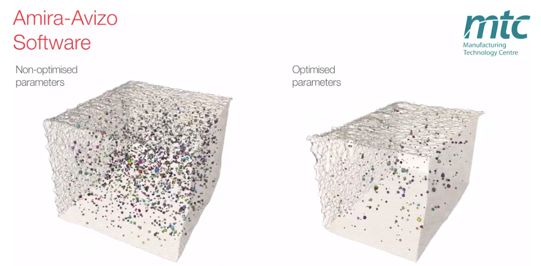
Electron Beam Melting (EBM) is an Additive Manufacturing (AM) process which can be used to fabricate complex 3D metal components. Ti-6Al-4V is one of the high value aerospace materials that has been widely used in the EBM process. However, defects such as gas pores and lack of fusion are often found in the as-built components, which can make a significant difference to the fatigue resistance of the parts. The Manufacturing Technology Centre (MTC) uses X-ray Computed Tomography (XCT) and 3D im... Read more
Dr Shuai Hou, Mélanie Bombardiere, Nathanael Turner, Jonathan Settle, Emmanuel Muzangaza, Dr David Brackett
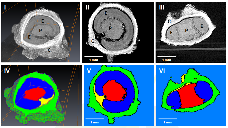
GEVES uses Avizo software to study and control plant seeds
At GEVES, Microtomography (micro-CT), which is a technique using X-rays to investigate internal anatomy and morphology of organisms without destruction, is applied to plant seed quality control and phenotyping. Several applications have been developed using micro-CT coupled with Avizo software development, such as for example in the Measurements of internal and external sugar beet seed structures for seed phenotyping
Read more
Ghassen Trigui, Laurence Le Corre, Dominique Honoré and Karima Boudehri-Giresse - 2D/3D Imaging - R&D team, GEVES
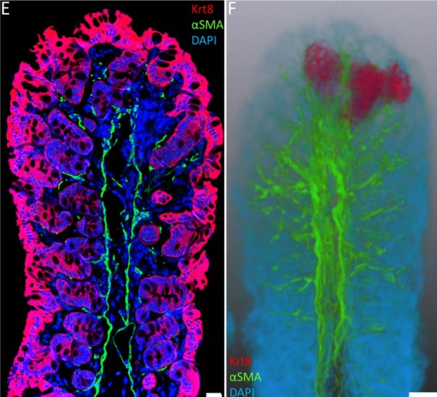
Immunofluorescence tomography is a high-resolution 3-D reconstruction method based on methacrylate embedding and serial-sectioning, where 2-D images of immuno-stained serial-sections are computationally aligned into image stacks, and the 3-D volume rendered. Butyl-Methyl Methacrylate (BMMA) plastic was adopted as it preserves excellent tissue morphology and can be de-plasticized easily using an organic solvent, which enables immuno-staining of serial-sections without antibody penetration issu... Read more
Parfitt, Geraint J
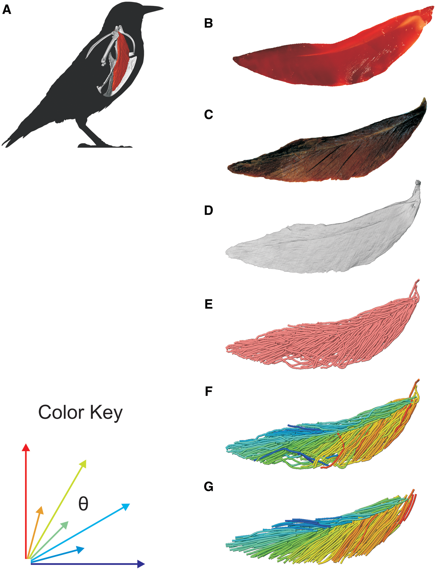
3D Muscle Architecture of the Pectoral Muscles of European Starling (Sturnus vulgaris)
Avian flight is achieved through a number of modifications to the body, including the pectoral girdle (…). Muscle architecture is a critical variable in determining the biomechanical function of the vertebrate musculoskeletal system; however, accurate three-dimensional (3D) understanding of muscle architecture has been historically difficult to acquire. Here, we present a musculoskeletal model of a European starling (Sturnus vulgaris) pectoral girdle generated from iodine contr... Read more
S.P. Sullivan, F.R. McGechie, K.M. Middleton, C.M. Holliday
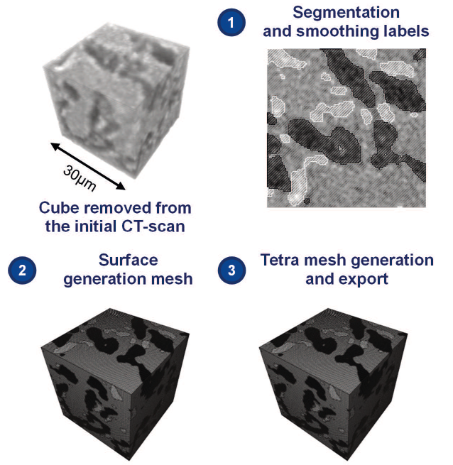
Current materials such nickel based superalloys cannot be used anymore and new materials are thus considered. For the hottest parts of jet engines, eutectic ceramics have potentially interesting features. In order to assess the thermo-mechanical properties of this material, numerical multi-scale analyses may be performed. Thus, a 3D finite element model was generated from a CT scan, representative of the microstructure and with a similar volume fraction. Effective elastic properties were calc... Read more
S. Gourdin, L. Marcin, M. Podgorski, M. Cherif, L. Carroz
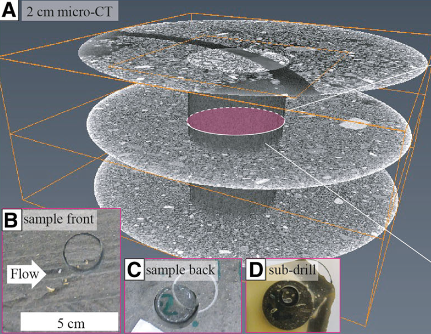
Sediment plates (a type of lacquer peels) represent a sampling method whereby a thin plate of undisturbed sediments is obtained directly from the outcrop. A low-viscosity, hardening epoxy resin is applied to a freshly exposed cross-section of an unconsolidated deposit and impregnates a surface layer of the cross-section via capillary forces before solidifying. Upon hardening, a solid plate (0.5–5 cm thick and up to 2 m in length) of the sedimentary formation can be recovered and transported... Read more
Guilhem Amin Douillet, Ulrich Kueppers, Célia Mato, Quentin Chaffaut, Mélanie Bouysson, Renate Reschetizka, Inga Hoelscher, Patrick Witting, Kai-Uwe Hess, Alexander Cerwenka, Donald B Dingwell and Benjamin Bernard