Welcome to the Amira-Avizo Software Use Case Gallery
Below you will find a collection of use cases of our 3D data visualization and analysis software. These use cases include scientific publications, articles, papers, posters, presentations or even videos that show how Amira-Avizo Software is used to address various scientific and industrial research topics.
Use the Domain selector to filter by main application area, and use the Search box to enter keywords related to specific topics you are interested in.
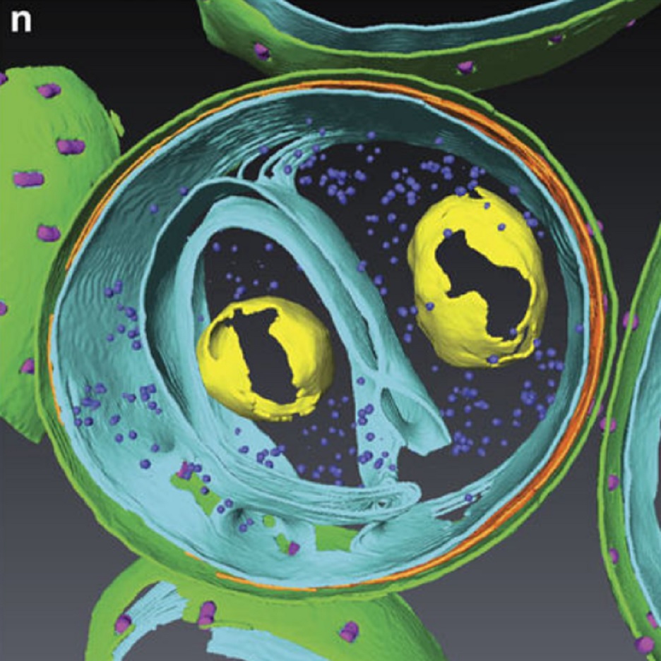
Determining the bacterial cell biology of planctomycetes
Bacteria of the phylum Planctomycetes have been previously reported to possess several features that are typical of eukaryotes, such as cytosolic compartmentalization and endocytosis-like macromolecule uptake. However, recent evidence points towards a Gram-negative cell plan for Planctomycetes, although in-depth experimental analysis has been hampered by insufficient genetic tools…
Read more
Christian Boedeker, Margarete Schüler, Greta Reintjes, Olga Jeske, Muriel C. F. van Teeseling et al.
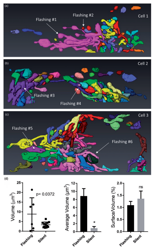
Ultrastructural Characterization of Flashing Mitochondria
Mitochondria undergo spontaneous transient elevations in matrix pH associated with drops in mitochondrial membrane potential. These mitopHlashes require a functional respiratory chain and the profusion protein optic atrophy 1, but their mechanistic basis is unclear. To gain insight on the origin of these dynamic events, we resolved the ultrastructure of flashing mitochondria by correlative light and electron microscopy. HeLa cells expressing the matrix-targeted pH probe mitoSypHer were screen... Read more
Manon Rosselin, Paula Nunes-Hasler, and Nicolas Demaurex
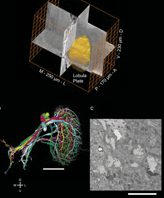
Full reconstruction of large lobula plate tangential cells in Drosophila from a 3D EM dataset
With the advent of neurogenetic methods, the neural basis of behavior is presently being analyzed in more and more detail. This is particularly true for visually driven behavior of Drosophila melanogaster where cell-specific driver lines exist that, depending on the combination with appropriate effector genes, allow for targeted recording, silencing and optogenetic stimulation of individual cell-types. Together with detailed connectomic data of large parts of the fly optic lobe, this has rece... Read more
Kevin M. Boergens , Christoph Kapfer, Moritz Helmstaedter, Winfried Denk, Alexander Borst
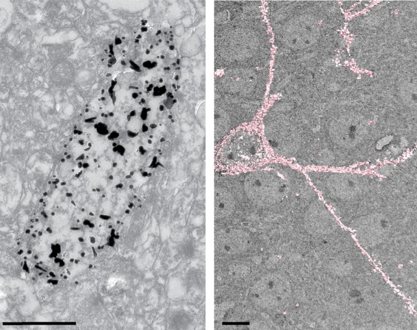
Analysis of neuronal arborization and connections is a powerful tool in fundamental and clinical neuroscience. Changes in neuronal morphology are central to brain development and plasticity and are associated with numerous diseases. Golgi staining is a classical technique based on a deposition of metal precipitate in a random set of neurons. Despite their versatility, Golgi methods have limitations that largely precluded their use in advanced microscopy. We combined Golgi staining with fluore... Read more
Katlijn Vints, Dorien Vandael, Pieter Baatsen, Benjamin Pavie, Frank Vernaillen, Nikky Corthout, Vasily Rybakin, Sebastian Munck & Natalia V. Gounko

Cell-type specific innervation of cortical pyramidal cells at their apical tufts
We investigated the synaptic innervation of apical tufts of cortical pyramidal cells in a region between layers 1 and 2 using 3-D electron microscopy (3D-EM) applied to four cortical regions in mouse. Across all cortices, we found the relative inhibitory input at the apical dendrite’s main bifurcation to be more than 3-fold stronger for layer 2 pyramidal cells than for all other pyramidal cells. Towards the distal tuft dendrites in upper layer 1, however, the relative inhibitory input was a... Read more
Ali Karimi, Jan Odenthal, Florian Drawitsch, Kevin M. Boergens, Moritz Helmstaedter
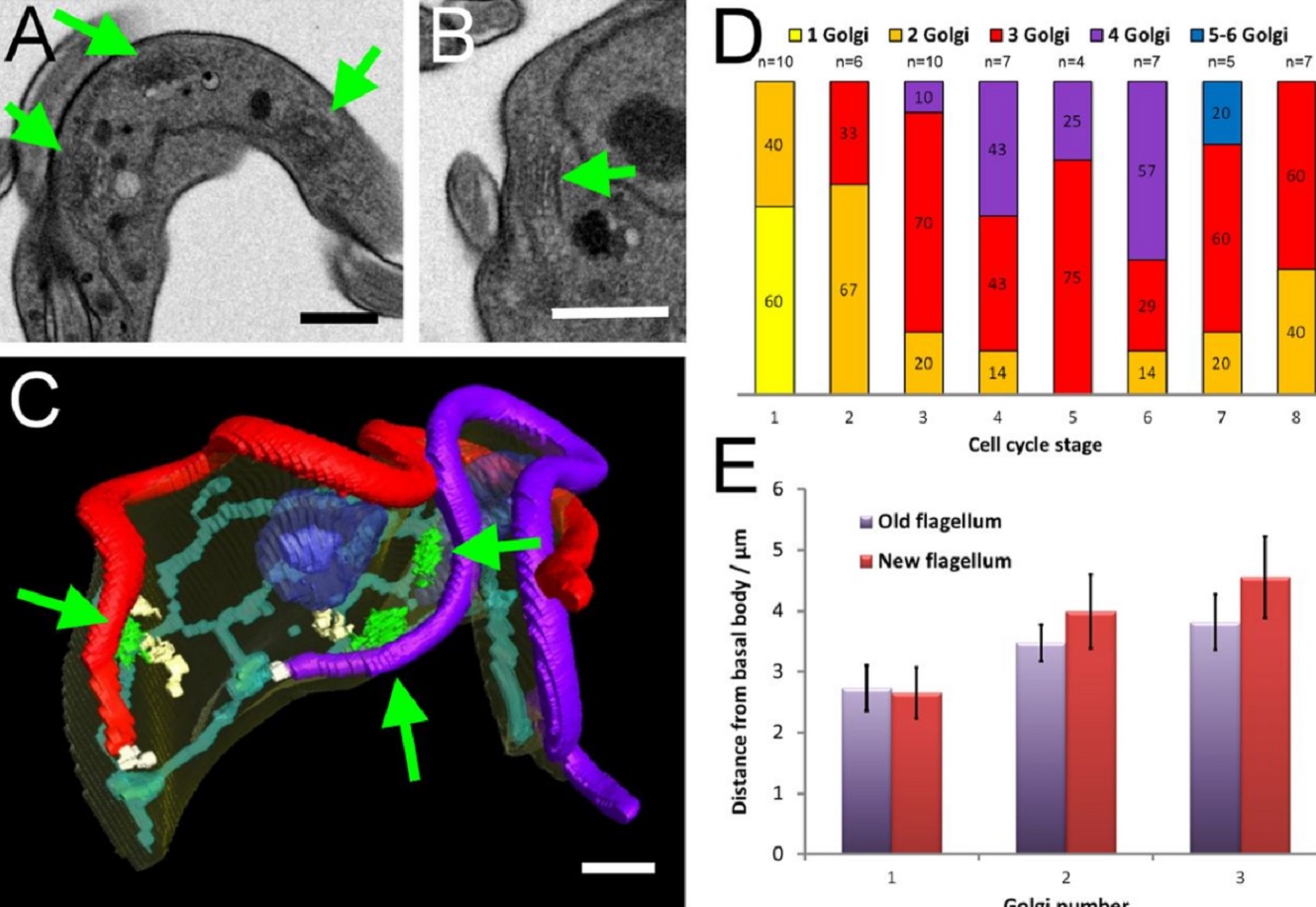
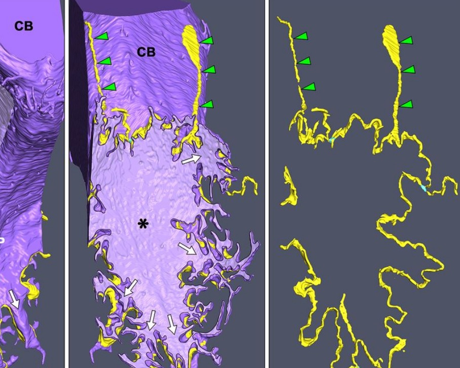
The major mammalian bloodstream form of the African sleeping sickness parasite Trypanosoma bruceimultiplies rapidly, and it is important to understand how these cells divide. Organelle inheritance involves complex spatiotemporal re-arrangements to ensure correct distribution to daughter cells…
Read more
Louise Hughes, Samantha Borrett, Katie Towers, Tobias Starborg, Sue Vaughan
Morphological process of podocyte development revealed by block-face scanning electron microscopy
Podocytes present a unique 3D architecture specialized for glomerular filtration. However, several 3D morphological aspects on podocyte development remain partially understood because they are difficult to reveal using conventional scanning electron microscopy (SEM). Here, we adopted serial block-face SEM imaging…
Read more
Koichiro Ichimura, Soichiro Kakuta, Yuto Kawasaki, Takayuki Miyaki, Takahiro Nonami, Naoyuki Miyazaki, Tomoyo Nakao, Sakiko Enomoto, Shigeo Arai, Masato Koike, Kazuyoshi Murata, Tatsuo Sakai
Juvenile Ovine Ex Vivo Larynges: Phonatory, Histologic, and Micro CT Based Anatomic Analyses
It is well known that the phonatory process changes during the life span. However, detailed investigations on potential factors concerned are rare. To deal with this issue, we performed extended biomechanical, macro anatomical, and histological analyses of the contributing laryngeal structures in ex vivo juvenile sheep models. Altogether twelve juvenile sheep larynges were analyzed within the phonatory experiments. Three different elongation levels and 16 different flow levels were applied to... Read more
Michael Döllinger, Olaf Wendler, Claus Gerstenberger, Tanja Grossmann, Marion Semmler, Hossein Sadeghi, and Markus Gugatschka
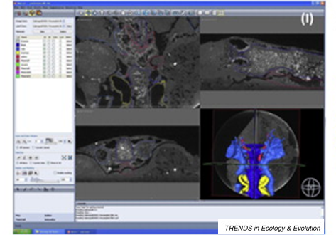
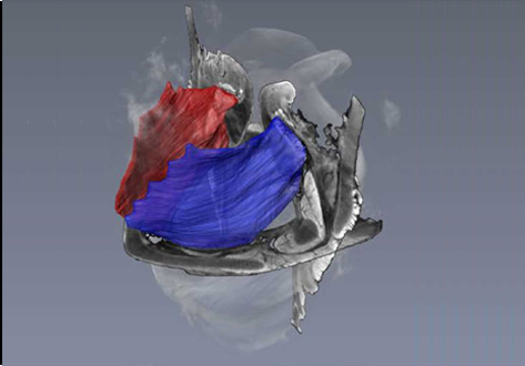
A virtual world of paleontology
Computer-aided visualization and analysis has revolutionized the study of fossils. Fossils can now be characterized in three dimensions and in unprecedented detail. The resulting digital reconstructions can be used in rigorous functional analyses. Hypotheses regarding the function of extinct organisms can therefore be tested.
Read more
Trends in Ecology & Evolution
Aetosauria is a clade of heavily armored, quadrupedal omnivorous to herbivorous archosaurs known from the Late Triassic across what was the supercontinent of Pangea. Their abundance in many deposits relative to the paucity of other Triassic herbivores indicates that they were key components of Late Triassic ecosystems. However, their evolutionary relationships remain contentious due, in large part, to their extensive dermal armor, which often obstructs observation of internal skeletal anatomy... Read more
Devin K. Hoffman, Andrew B. Heckert, Lindsay E. Zanno
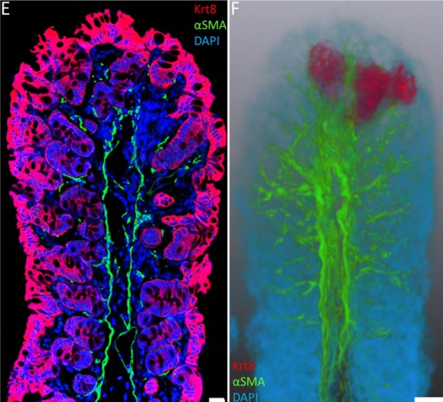
Immunofluorescence tomography is a high-resolution 3-D reconstruction method based on methacrylate embedding and serial-sectioning, where 2-D images of immuno-stained serial-sections are computationally aligned into image stacks, and the 3-D volume rendered. Butyl-Methyl Methacrylate (BMMA) plastic was adopted as it preserves excellent tissue morphology and can be de-plasticized easily using an organic solvent, which enables immuno-staining of serial-sections without antibody penetration issu... Read more
Parfitt, Geraint J
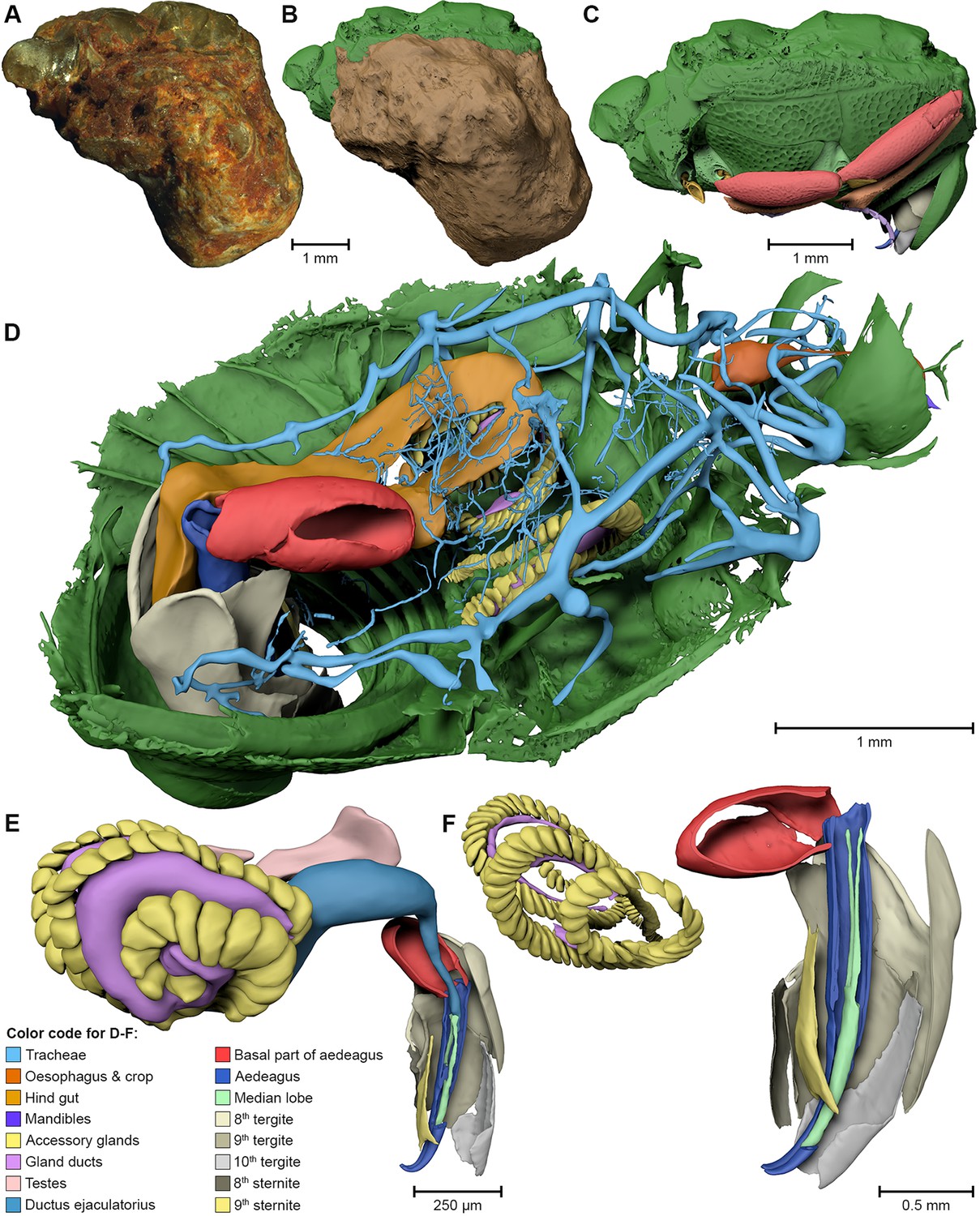
External and internal morphological characters of extant and fossil organisms are crucial to establishing their systematic position, ecological role and evolutionary trends. (…) We found well-preserved three-dimensional anatomy in mineralized arthropods from Paleogene fissure fillings and demonstrate the value of these fossils by utilizing digitally reconstructed anatomical structure of a hister beetle. The new anatomical data facilitate a refinement of the species diagnosis and allowed... Read more
Achim H Schwermann, Tomy dos Santos Rolo, Michael S Caterino, Gunter Bechly, Heiko Schmied, Tilo Baumbach, Thomas van de Kamp
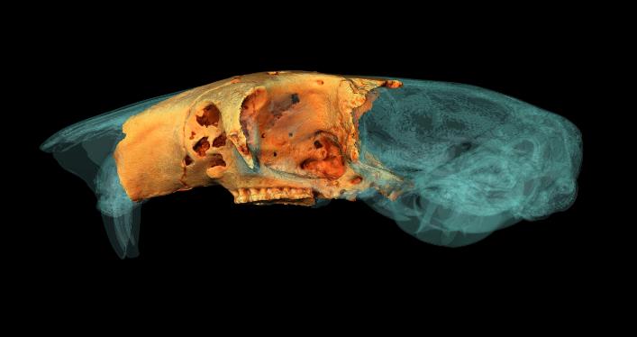
University of York student wins Anatomical Society Best Image Prize using Amira-Avizo Software
PhD student Jesse Hennekam wins for his reconstruction of the skull of a giant dormouse.
A York PhD student has won a prestigious award for his work reconstructing the skull of a giant rodent.
Jesse Hennekam, from the Centre for Anatomical and Human Sciences at the Hull York Medical School, created a digital reconstruction of the skull of a gigantic dormouse (Leithia melitensis), which roamed on the island of Sicily during the Pleistocene, roughly 2 million years ago... Read more
Jesse Hennekam
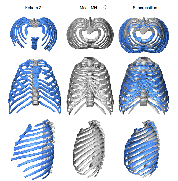
3D virtual reconstruction of the Kebara 2 Neandertal thorax
The size and shape of the Neandertal thorax has been debated since the first discovery of Neandertal ribs more than 150 years ago, with workers proposing different interpretations ranging from a Neandertal thoracic morphology that is indistinguishable from modern humans, to one that was significantly different from them. Here, we provide a virtual 3D reconstruction of the thorax of the adult male Kebara 2 Neandertal. Our analyses reveal that the Kebara 2 thorax is significantly different but ... Read more
Asier Gomez-Olivencia, Alon Barash, Daniel Garcia-Martinez, Mikel Arlegi, Patricia Kramer, Markus Bastir, Ella Been
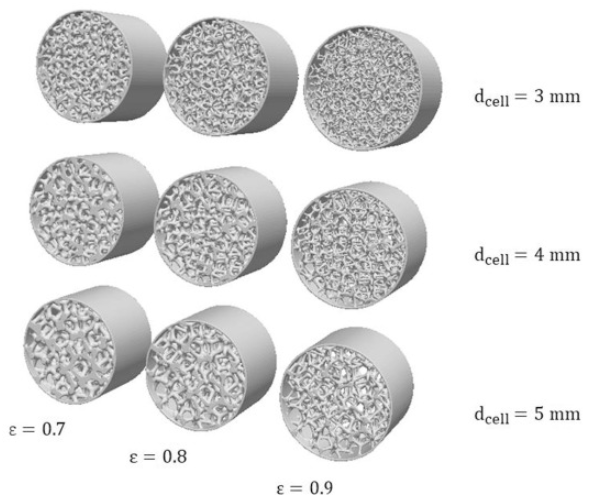
Open-cell foams as structured catalyst supports are promising candidates for the design of high throughput catalytic processes. In this contribution, we employ a coupled numerical and experimental approach to assess the pressure losses in foams. (…) we explore virtually-generated foam models and their 3D printed replicas for a combined CFD and experimental study of fluid dynamics in foams. In particular, we focus our analysis on the low Reynolds number regime, where deviations between t... Read more
Mauro Bracconi, Matteo Ambrosetti, Obinna Okafor, Victor Sans, Xun Zhang, Xiaoxia Ou, Claudio Pereira Da Fonte, Xiaolei Fan, Matteo Maestri, Gianpiero Groppi, Enrico Tronconi
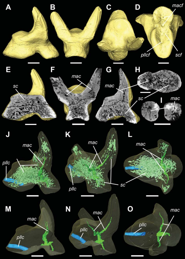
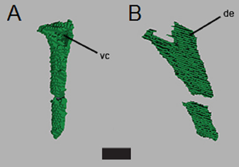
Vascular structure of the earliest shark teeth
Here we use synchrotron tomography to characterise dental vasculature in the oldest known tooth-bearing sharks, Leonodus carlsi Mader, 1986 and Celtiberina maderi Wang, 1993. Three dimensional reconstruction of the vascular system and microstructure of both taxa revealed a complex and dense network of canals, including horizontal, ascending and secondary bifurcated canals, as well as histological features consistent with an osteodont histotype. However, L. carlsi and C. maderi also exhibit si... Read more
Carlos Martinez-Perez, Alba Martin-Lazaro, Humberto G Ferron, Martina Kirstein, Philip C.J. Donoghue, Hector Botella
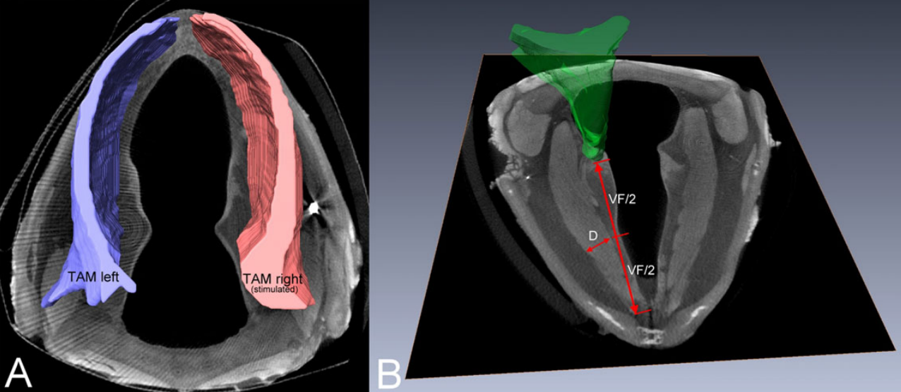
Functional Electrical Stimulation Leads to Increased Volume of the Aged Thyroarytenoid Muscle
A stimulation electrode was placed unilaterally near the terminal adduction branch of the recurrent laryngeal nerve (RLN) adjacent to the right cricothyroid joint. The electrode was connected to an implant located subcutaneously at the neck region. Predesigned training patterns were automatically delivered by a bidirectional radio frequency link using a programming device and were repeated automatically by the implant every other day over 11 weeks in the awake animal. Outcome parameters compr... Read more
Markus Gugatschka, MD, DMSci, Jonathan C. Jarvis, PhD, Justin D. Perkins, MSc, Vladimir Bubalo, PhD, Iris Wiederstein-Grasser, PhD, Hermann Lanmüller, PhD, Claus Gerstenberger, MSc and Michael Karbiener, PhD
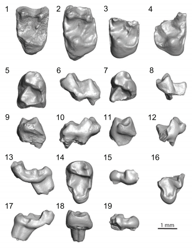
Paromomyidae has been thought to represent the longest-lived group of stem primates (plesiadapiforms), extending from the early Paleocene to late Eocene. We analyzed primate material from the late-middle Eocene of southern California that had initially been ascribed to cf. Phenacolemur shifrae. This material falls at the lowest end of the size range for the family. The Californian specimens also exhibit several dental features that are atypical for paromomyids, such as a strong paraconid on t... Read more
Sergi López-Torres, Mary T. Silcox, and Patricia A. Holroyd
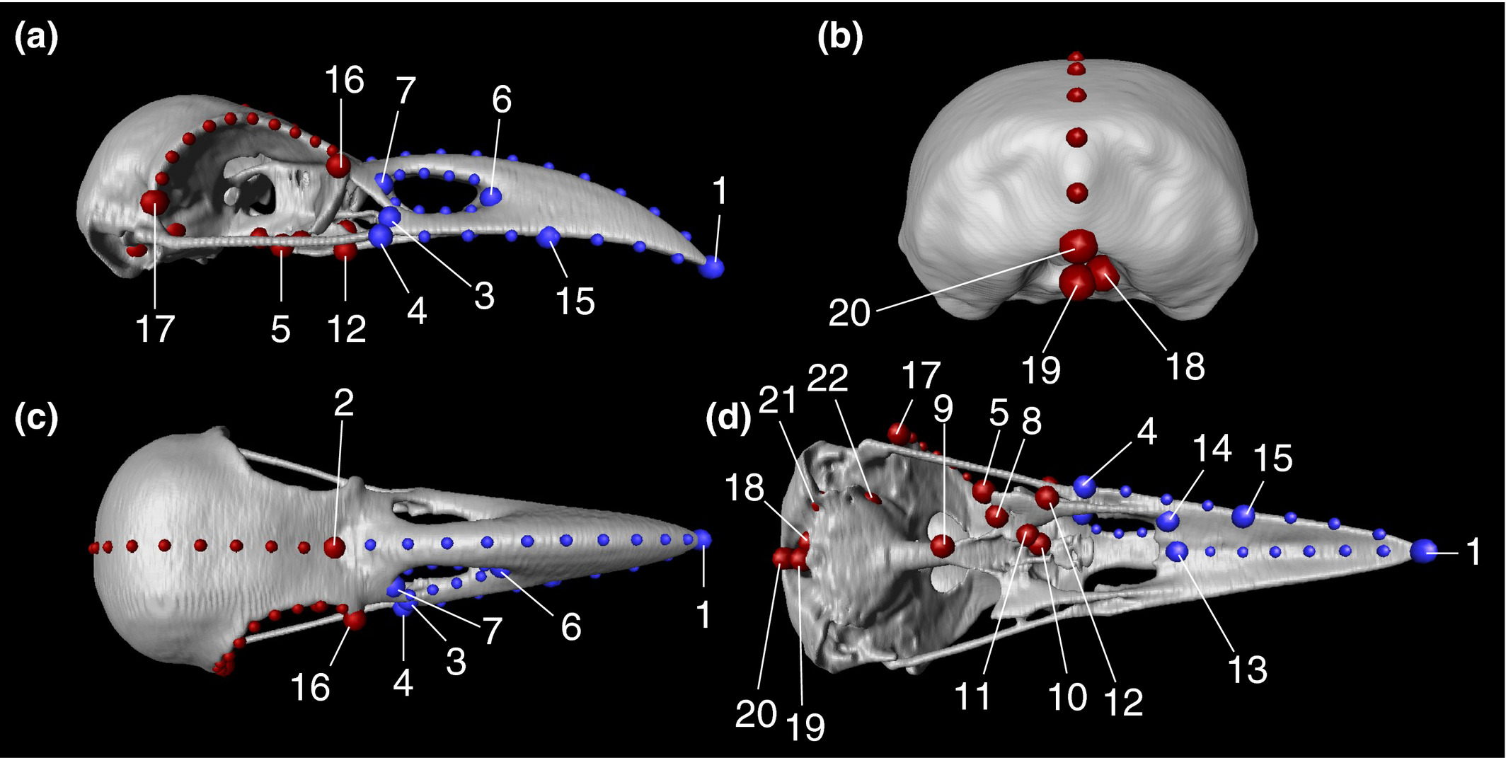
Here, to evaluate the intensity of evolutionary constraints on avian beak shape more appropriately, we selected large billed (Corvus macrorhynchos) and carrion crows (Corvus corone) as study objects. These landbird species seem to experience selection pressures favoring a departure from an allometric trajectory. A landmark based geometric morphometric approach using three dimensional reconstructions of CT scan images revealed that only 45.4% of the total shape variation was explained by allom... Read more
Takeshi Yamasaki, Sou Aoki, Masayoshi Tokita
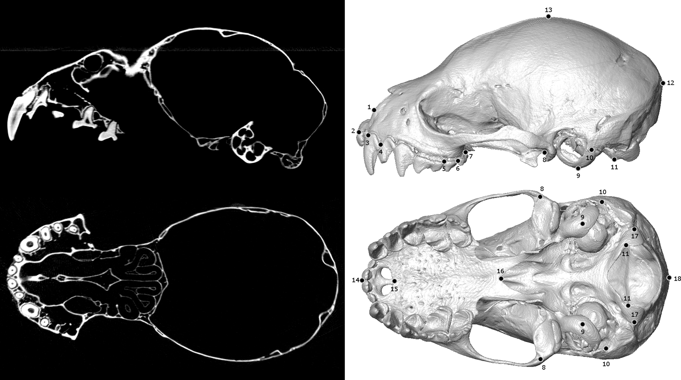
Biological specimens are primary records of organismal ecology and history. As such, museum collections are invaluable repositories for testing ecological and evolutionary hypotheses across the tree of life. Digitizing and broadly sharing the phenotypic data from these collections serves to expand the traditional reach of museums, enabling widespread data sharing, collaboration, and education at an unprecedented scale. In recent years, μCT-scanning has been adopted as one way for efficiently... Read more
Jeff J. Shi, Erin P. Westeen, Daniel L. Rabosky
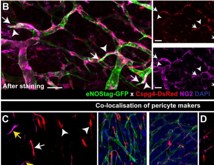
Endothelial cells and pericytes are integral cellular components of the vasculature with distinct interactive functionalities. To study dynamic interactions between these two cells we created two transgenic animal lines. A truncated eNOS (endothelial nitric oxide synthase) construct was used as a GFP tag for endothelial cell evaluation and an inducible Cre-lox recombination, under control of the Pdgfrb (platelet derived growth factor receptor beta) promoter, was created for pericyte assessmen... Read more
Ann L. B. Seynhaeve, Douwe Oostinga, Rien van Haperen, Hanna M. Eilken, Susanne Adams, Ralf H. Adams & Timo L. M. ten Hagen