Welcome to the Amira-Avizo Software Use Case Gallery
Below you will find a collection of use cases of our 3D data visualization and analysis software. These use cases include scientific publications, articles, papers, posters, presentations or even videos that show how Amira-Avizo Software is used to address various scientific and industrial research topics.
Use the Domain selector to filter by main application area, and use the Search box to enter keywords related to specific topics you are interested in.
Lithium-ion (Li-ion) batteries operate via electrochemical reactions between positive and negative electrodes, formed by complex porous microstructures. An improved understanding of these materials can lead to a greater insight into the link between microscopic electrode morphology and macroscopic performance. The practice of calendering electrodes after manufacturing has been widely used to increase the volumetric energy density and improve the electrical contact between electrode... Read more
S. R. Daemi,X. Lu, D. Sykes, J. Behnsen, C. Tan, A. Palacios-Padros, J. Cookson, E. Petrucco, P. J. Withers, D. J. L. Brett and P. R. Shearing
Four-Dimensional Studies of Morphology Evolution in Lithium–Sulfur Batteries
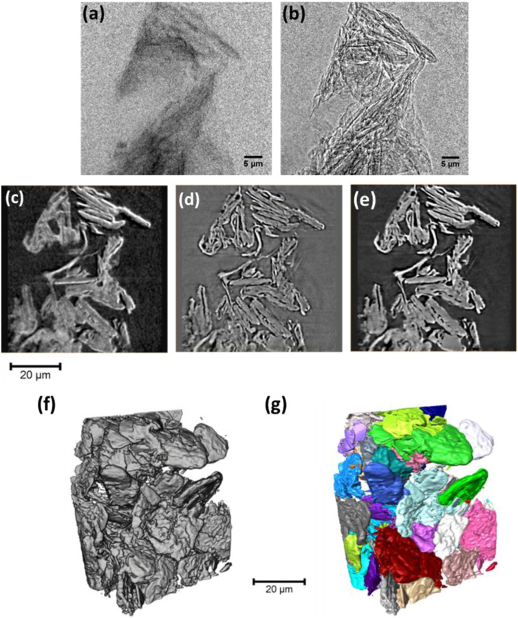
Lithium sulfur (Li–S) batteries have great potential as a successor to Li-ion batteries, but their commercialization has been complicated by a multitude of issues stemming from their complex multiphase chemistry. In situ X-ray tomography investigations enable direct observations to be made about a battery, providing unprecedented insight into the microstructural evolution of the sulfur cathode and shedding light on the reaction kinetics of the sulfur phase. Here, for the first time, the mor... Read more
Chun Tan, Thomas M. M. Heenan, Ralf F. Ziesche, Sohrab R. Daemi, Jennifer Hack, Maximilian Maier, Shashidhara Marathe, Christoph Rau, Daniel J. L. Brett, Paul R. Shearing
The electrode microstructural properties significantly influence the efficiency and durability of many electrochemical devices including solid oxide fuel cells. Despite the possibility of simulating the electrochemical phenomena within real three-dimensional microstructures, the potential of such 3D microstructural information has not yet been fully exploited. We introduce here a completely new methodology for the advanced characterization of inhomogeneous current distribution base... Read more
A.Bertei, V.Yufit, F.Tariq, N.P.Brandon
The use of contrast enhancement techniques in X-ray imaging of lithium–ion battery electrodes
Understanding the microstructural morphology of Li–ion battery electrodes is crucial to improving the electrochemical performance of current Li–ion battery systems and in developing next-generation power systems. The use of 3D X-ray imaging techniques, which are continuously evolving, provides a noninvasive platform to study the relationship between electrode microstructure and performance at various time and length scales. In addition to characterizing a weakly (X-ray) absorbing graphite... Read more
Oluwadamilola O. Taiwo , Donal P. Finegan , Jeff Gelb , Christian Holzner , Daniel J.L. Brett , Paul R. Shearing
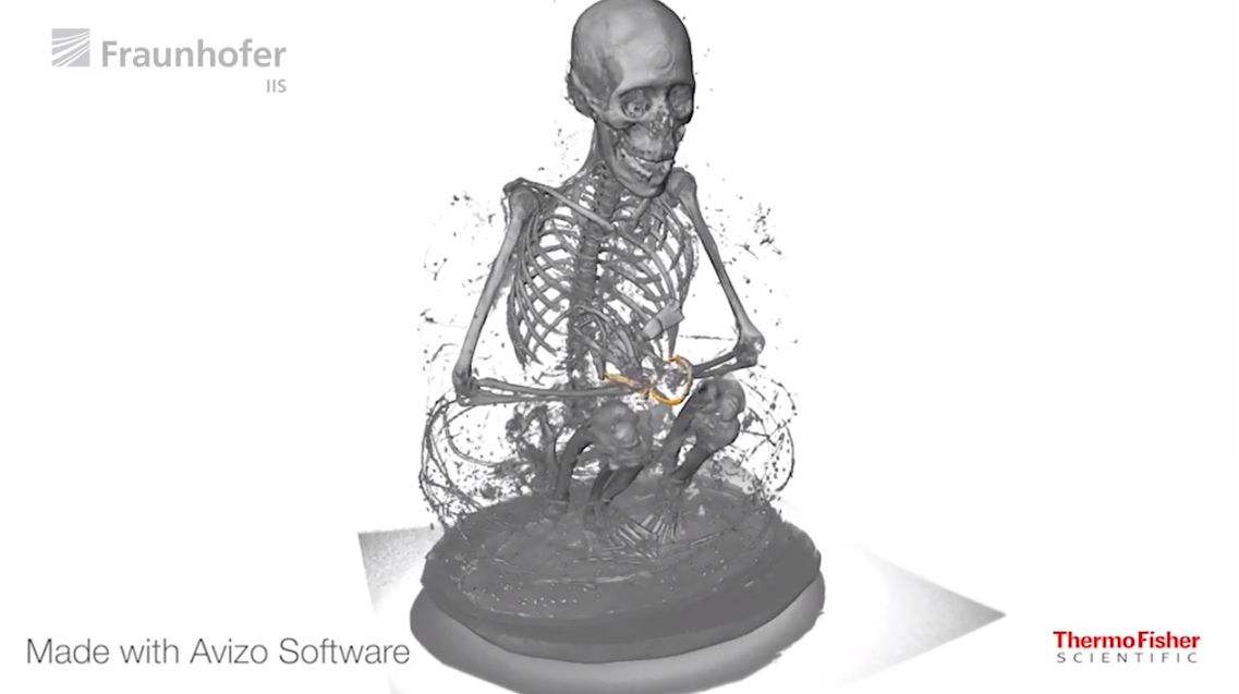
The Fraunhofer Institute uses Avizo to explore a Peruvian mummy CT-scan
As part of the Big Picture project, researchers at Fraunhofer IIS are working on three dimensions images of objects with great precision. In order to demonstrate the progress of the project, which has begun in February 2018, they made the 3d Xray-CT acquisition of a mummy from the Linden-Museum Stuttgart collection, probably from the 11th-15th century.
Modern measurement systems are producing increasingly large volumes of extremely complex data that must be stored, processed and convert... Read more
Prof. Dr. Tomas Sauer
Biodegradable materials, such as collagen scaffolds, are used extensively in clinical medicine for tissue regeneration and/or as an implantable drug delivery vehicle. However, available methods to study biomaterial degradation are typically invasive, destructive, and/or non-volumetric. Therefore, the objective of this study was to investigate a new method for nondestructive, longitudinal, and volumetric measurement of collagen scaffold degradation. Gold nanoparticles (Au NPs) were covalently ... Read more
Tyler A. Finamore, Tyler E. Curtis, James V. Tedesco, Kathryn Grandfield, Ryan K. Roeder
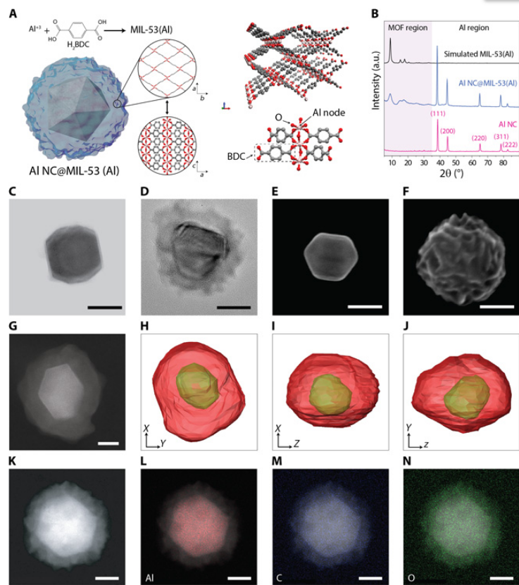
Metal-organic frameworks tailor the properties of aluminum nanocrystals
Metal-organic frameworks (MOFs) and metal nanoparticles are two classes of materials that have received considerable recent attention, each for controlling chemical reactivities, albeit in very different ways. Here, we report the growth of MOF shell layers surrounding aluminum nanocrystals (Al NCs), an Earth-abundant metal with energetic, plasmonic, and photocatalytic properties. The MOF shell growth proceeds by means of dissolution-and-growth chemistry that uses the intrinsic surface oxide o... Read more
Hossein Robatjazi, Daniel Weinberg, Dayne F. Swearer, Christian Jacobson, Ming Zhang, Shu Tian, Linan Zhou, Peter Nordlander, Naomi J. Halas
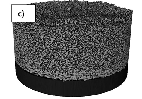
Aging of a Pt/Al2O3 exhaust gas catalyst monitored by quasi in situ X-ray micro computed tomography
Catalyst aging effects were analyzed using X-ray absorption micro-computed tomography in combination with conventional characterization methods on various length scales ranging from nm to μm to gain insight into deactivation mechanisms.
For this purpose, a 4 wt% Pt/Al2O3 model exhaust gas catalyst was coated on a cordierite honeycomb and subjected to sequential thermal aging in static air at 950 °C for 4, 8, 12 and 24 hours. The ag... Read more
Georg Hofmann, Amélie Rochet, Elen Ogel, Maria Casapu, Stephan Ritter, Malte Ogurreck and Jan-Dierk Grunwaldt
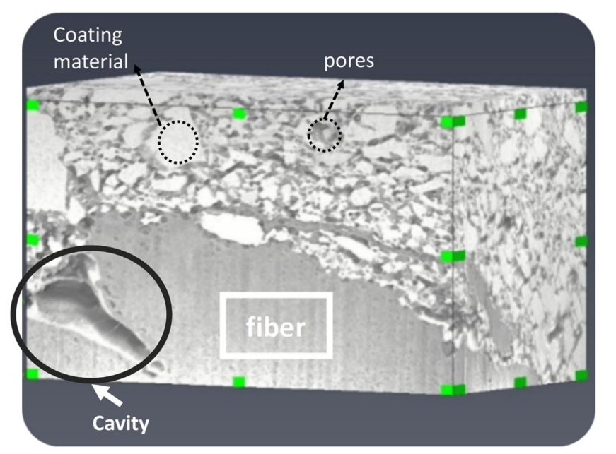
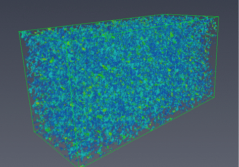
Characterization of the Interface Between Coating and Fibrous Layers of Paper
Coated paper is an example of a multi-layer porous medium, involving a coating layer along the two surfaces of the paper and a fibrous layer in the interior of the paper. The interface between these two media needs to be characterized in order to develop relevant modeling tools. After careful cutting of the paper, a cross section was imaged using focused ion beam scanning electron microscopy. The resulting image was analyzed to characterize the coating layer and its transition to the fibrous ... Read more
H. Aslannejad, S. M. Hassanizadeh, M. A. Celia
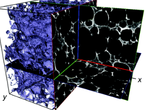
The development of focused ion beam-scanning electron microscopy (FIB-SEM) techniques has allowed high-resolution 3D imaging of nanometre-scale porous materials. These systems are of important interest to the oil and gas sector, as well as for the safe long-term storage of carbon and nuclear waste. This work focuses on validating the accurate representation of sample pore space in FIB-SEM-reconstructed volumes and the predicted permeability of these systems from subsequent single-phase flow s... Read more
Department of Chemical Engineering, Qatar Carbonates and Carbon Storage Research Centre, Imperial College London | Department of Applied Mathematics and Theoretical Physics, Cambridge University
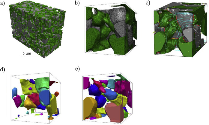
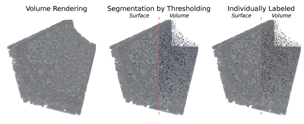
Visualizing the Carbon Binder Phase of Battery Electrodes in Three Dimensions
This study presents a technique to directly characterize the carbon and binder domain (CBD) in lithium-ion (Li-ion) battery electrodes in three dimensions and use it to determine the effective transport properties of a LiNi0.33Mn0.33Co0.33O2 (NMC) electrode. X-ray nanocomputed tomography (nano-CT) is used to image an electrode composed solely of carbon and binder, whereas focused ion beam–scanning electron microscopy is used to analyze cross-sect... Read more
Sohrab R. Daemi, Chun Tan, Tobias Volkenandt, Samuel J. Cooper, Anna Palacios-Padros, James Cookson, Dan J. L. Brett, and Paul R. Shearing
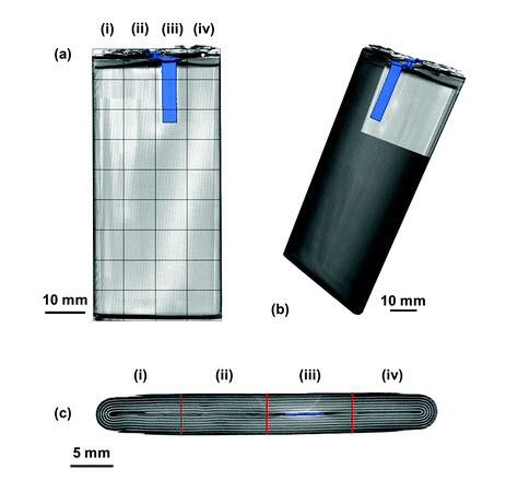
Spatially resolved ultrasound diagnostics of Li-ion battery electrodes
The importance of reliable battery diagnostic systems has grown substantially in recent years as a result of the use of high power Li-ion battery packs in an increasingly diverse range of applications. Here, spatially resolved ultrasound acoustic measurements are used to analyse the condition of Li-ion electrodes. Ultrasonic measurements are performed on a commercial mobile phone battery over the full operating voltage window with the lithiation and delithiation of electrodes o... Read more
James B. Robinson, Maximilian Maier , George Alster , Tomos Compton , Dan J. L. Brett and Paul R. Shearing
As key functional units in neural circuits, different types of neuronal synapses play distinct roles in brain information processing, learning, and memory. Synaptic abnormalities are believed to underlie various neurological and psychiatric disorders. Here, by combining cryo-electron tomography and cryo-correlative light and electron microscopy, we distinguished intact excitatory and inhibitory synapses of cultured hippocampal neurons, and visualized the in situ 3D organization of ... Read more
Chang-Lu Tao, Yun-Tao Liu, Rong Sun, Bin Zhang, Lei Qi, Sakar Shivakoti, Chong-Li Tian, Peijun Zhang, Pak-Ming Lau, Z. Hong Zhou and Guo-Qiang Bi
Corpora amylacea are cell-derived structures that appear physiologically in the aged human brain. While their histological identification is straightforward, their ultrastructural composition and microenvironment at the nanoscale have remained unclear so far, as has their relevance to aging and certain disease states that involve the sequestration of toxic cellular metabolites. Here, we apply correlative serial block-face scanning electron microscopy and transmission electron tomograp... Read more
Paula P. Navarro, Christel Genoud, Daniel Castaño-Díez, Alexandra Graff-Meyer, Amanda J. Lewis, Yvonne de Gier, Matthias E. Lauer, Markus Britschgi, Bernd Bohrmann, Stephan Frank, Jürgen Hench, Gabriel Schweighauser, Annemieke J. M. Rozemuller, Wilma D. J. van de Berg, Henning Stahlberg & Sarah H. Shahmoradian
Novel Sulfolobus Virus with an Exceptional Capsid Architecture
A novel archaeal virus, denoted Sulfolobus ellipsoid virus 1 (SEV1), was isolated from an acidic hot spring in Costa Rica. The morphologically unique virion of SEV1 contains a protein capsid with 16 regularly spaced striations and an 11-nm-thick envelope. The capsid exhibits an unusual architecture in which the viral DNA, probably in the form of a nucleoprotein filament, wraps around the longitudinal axis of the virion in... Read more
Haina Wang, Zhenqian Guo, Hongli Feng, Yufei Chen, Xiuqiang Chen, Zhimeng Li, Walter Hernández-Ascencio, Xin Dai, Zhenfeng Zhang, Xiaowei Zheng, Marielos Mora-López, Yu Fu, Chuanlun Zhang, Ping Zhu, Li Huang
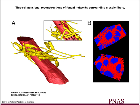
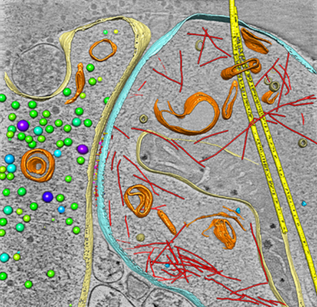
3D visualization and deep-learning reveal complex parasite networks in behaviorally manipulated ants
Microbial parasites may behave collectively to manipulate their host’s behavior. We examine adaptations of a microbial parasite in its natural environment: the body of its coevolved and manipulated host.
Electron microscopy and 3D reconstructions of host and parasite tissues reveal that this fungus invades muscle fibers throughout the ant’s body but leaves the brain intact, and that the fungal cells connect to form extensive networks.
Read more
Maridel A. Fredericksena, Yizhe Zhangb, Missy L. Hazenc, Raquel G. Loretoa,d, Colleen A. Mangoldd,e, Danny Z. Chenb, and David P. Hughes, Department of Entomology, Pennsylvania State University
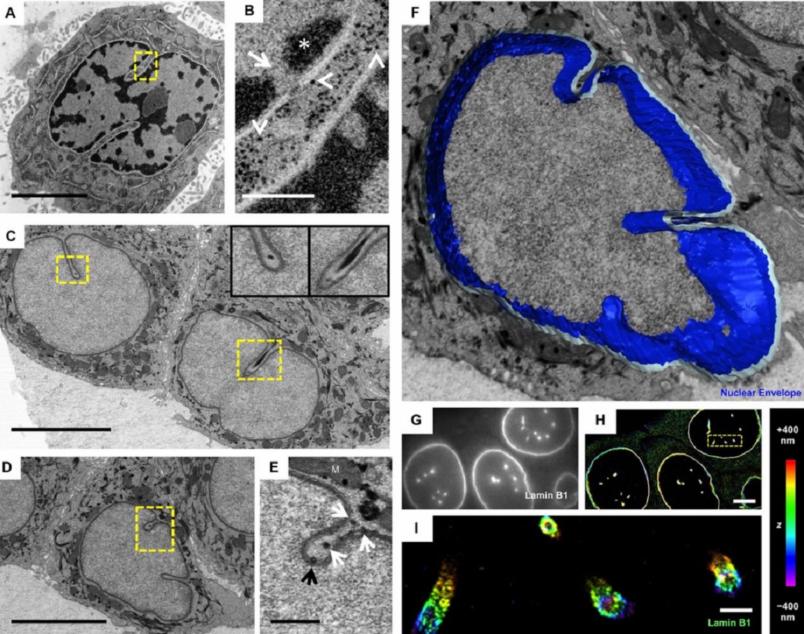
The importance of context in regulation of gene expression is now an accepted principle; yet the mechanism by which the microenvironment communicates with the nucleus and chromatin in healthy tissues is poorly understood. A functional role for nuclear and cytoskeletal architecture is suggested by the phenotypic differences observed between epithelial and mesenchymal cells…
Read more
Danielle M. Jorgens, Jamie L. Inman, Michal Wojcik, Claire Robertson, Hildur Palsdottir, Wen-Ting Tsai, Haina Huang, Alexandre Bruni-Cardoso, Claudia S. López, Mina J. Bissell, Ke Xu, Manfred Auer

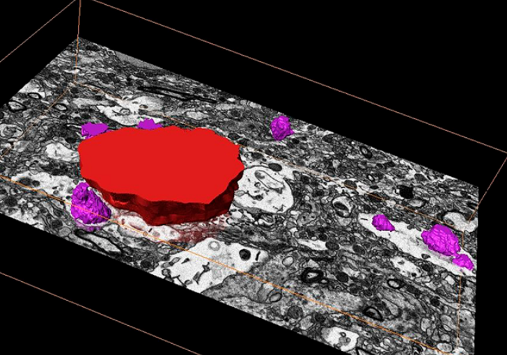
Macropinosomes are key players in early shigella invasion and vacuolar escape in epithelial cells
Intracellular pathogens include all viruses, many bacteria and parasites capable of invading and surviving within host cells. Key to survival is the subversion of host cell pathways by the pathogen for the purpose of propagation and evading the immune system. The intracellular bacterium Shigella flexneri, the causative agent of bacillary dysentery, invades host cells in a vacuole that is subsequently ruptured to allow growth of the pathogen within the host cytoplasm…
Read more
Allon Weiner , Nora Mellouk , Noelia Lopez-Montero , Yuen-Yan Chang, Célia Souque, Christine Schmitt, Jost Enninga
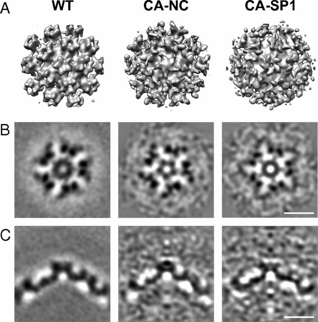
HIV-1 maturation occurs via multiple proteolytic cleavages of the Gag polyprotein, causing rearrangement of the virus particle required for infectivity. (…) How individual cleavages contribute to changes in protein structure and interactions, and how the mature, conical capsid forms, are poorly understood. Here, we employed cryoelectron tomography to determine morphology and high-resolution CA lattice structures for HIV1 derivatives in which Gag cleavage sites are mutated. These analyse... Read more
Simone Mattei, Aaron Tan, Barbel Glass, Barbara Muller, Hans-Georg Krausslich, and John A. G. Briggs
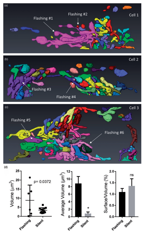
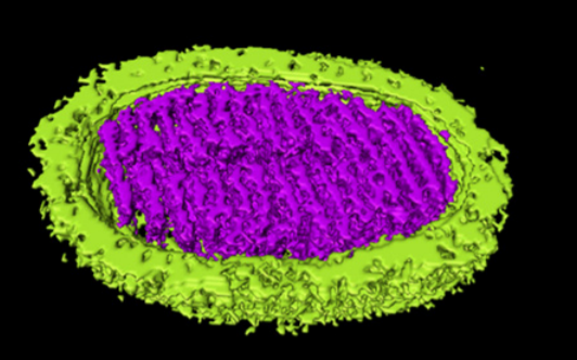
Ultrastructural Characterization of Flashing Mitochondria
Mitochondria undergo spontaneous transient elevations in matrix pH associated with drops in mitochondrial membrane potential. These mitopHlashes require a functional respiratory chain and the profusion protein optic atrophy 1, but their mechanistic basis is unclear. To gain insight on the origin of these dynamic events, we resolved the ultrastructure of flashing mitochondria by correlative light and electron microscopy. HeLa cells expressing the matrix-targeted pH probe mitoSypHer were screen... Read more
Manon Rosselin, Paula Nunes-Hasler, and Nicolas Demaurex
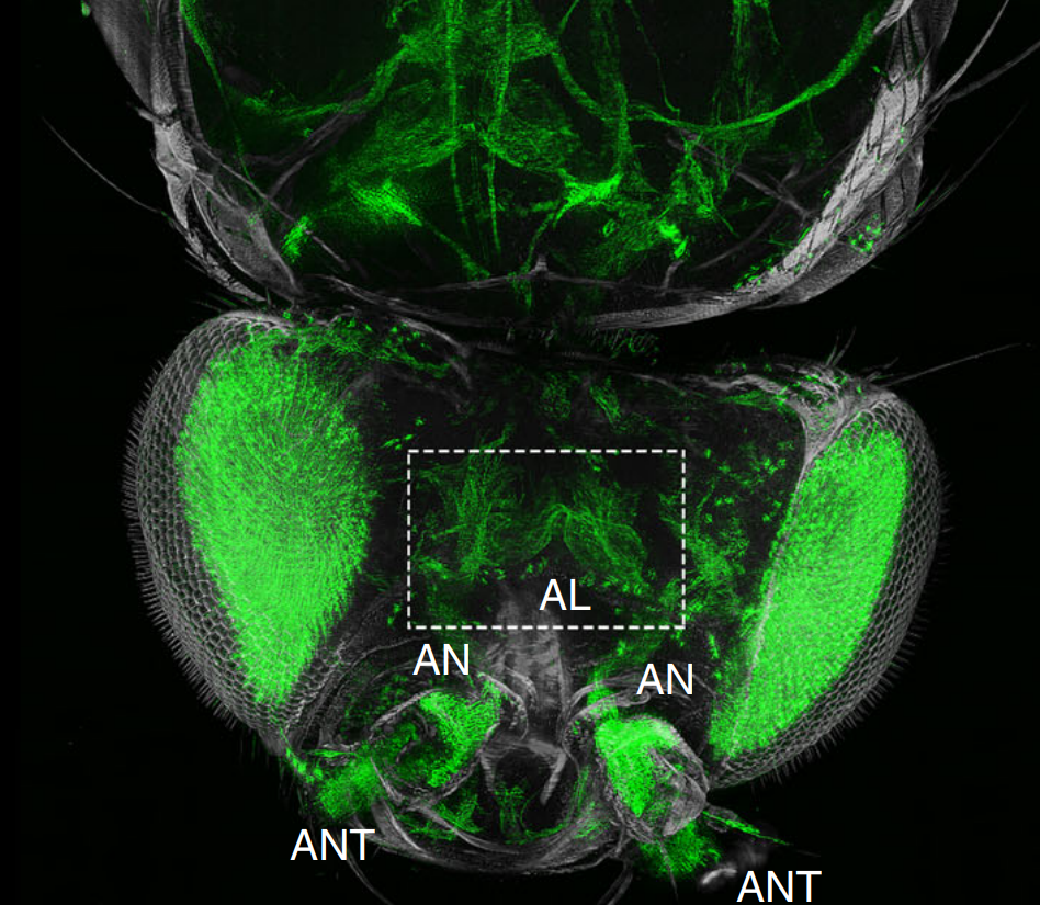
The fruit fly, Drosophila melanogaster, is an important experimental model to address central questions in neuroscience at an organismic level. However, imaging of neural circuits in intact fruit flies is limited due to structural properties of the cuticle. Here we present a novel approach combining tissue clearing, ultramicroscopy, and data analysis that enables the visualisation of neuronal networks with single-cell resolution from the larval stage up to the adult Drosophila. (…) This... Read more
Marko Pende, Klaus Becker, Martina Wanis, Saiedeh Saghafi, Rashmit Kaur, Christian Hahn, Nika Pende, Massih Foroughipour, Thomas Hummel & Hans-Ulrich Dodt