Welcome to the Amira-Avizo Software Use Case Gallery
Below you will find a collection of use cases of our 3D data visualization and analysis software. These use cases include scientific publications, articles, papers, posters, presentations or even videos that show how Amira-Avizo Software is used to address various scientific and industrial research topics.
Use the Domain selector to filter by main application area, and use the Search box to enter keywords related to specific topics you are interested in.
Objectives
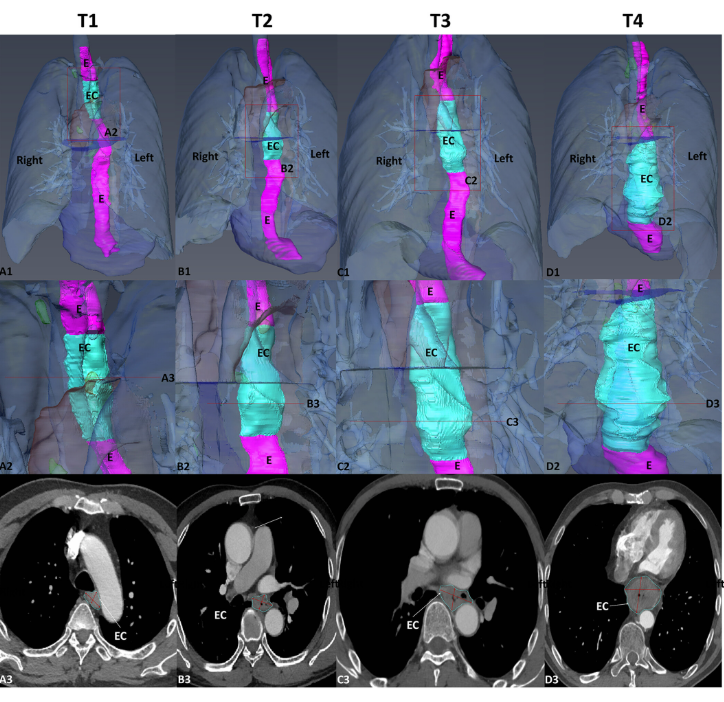
To statistically study the 3D shape of oesophageal cancer (EC) and its spatial relationships based on computed tomography angiography (CTA) 3D reconstruction, to determine its relationship with T-stages, and to create an optimal T-stage diagnosis protocol based on CTA calculation.
Runyuan Wang, Xiaoqin Zhang, Wei Wu, Jinfeng Ma, Jincheng Chen, Zhu Zhang, Liqun Liu, Shanshan Xu, Ximei Cao, Yi Wu, Huilin Cui
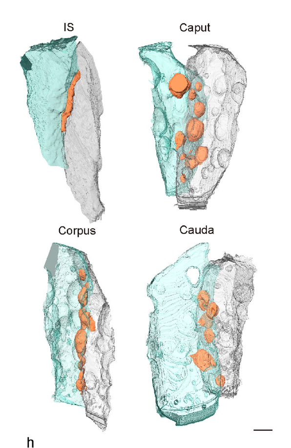
Mammalian epididymal epithelial cells are crucial for sperm maturation. Historically,
vacuole-like ultrastructures in epididymal epithelial cells were
observed via transmission electron microscopy but were undefined. Here, we
utilize volume electron microscopy (vEM) to generate 3D reconstructions of
epididymal epithelial cells and identify these vacuoles as intercellular organelle
reservoirs (IORs) in the lateral intercellular space (LIS), which contains
pr... Read more
Xia Li, Feng Qiao, Jiansheng Guo, Ting Jiang, Huifang Lou, Huixia Li, Gangcai Xie, Hangjun Wu, Weizhen Wang, Ruoyu Pei, Sha Liu, Mei Ye, Jin Li, Shiqin Huang, Mengya Zhang, Chaoye Ma, Yiwen Huang, Shushu Xu, Xiaofeng Li, Xiao Sun, Jun Yu, Kin Lam Fok, Shumin Duan & Hao Chen
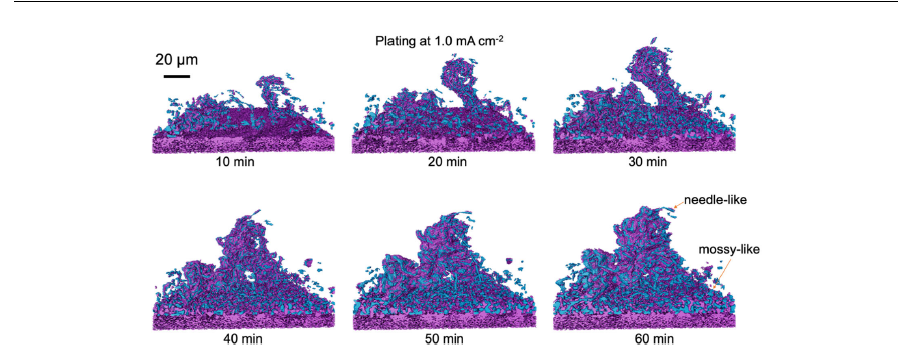
Li metal is considered as the most important negative electrode active material for Li-based batteries because of its high theoretical specific capacity of 3860mAh g-1, which is an order of magnitude higher than the currently used graphite, and by being the most electropositive metal. When coupled with high-capacity cathodes, either Li insertion materials or conversion chemistries, or applied in a solid-sate configuration, a leap in energy density can be obtained. The main challenge in the di... Read more
Matthew Sadd, Shizhao Xiong, Jacob R. Bowen, Federica Marone, Aleksandar Matic
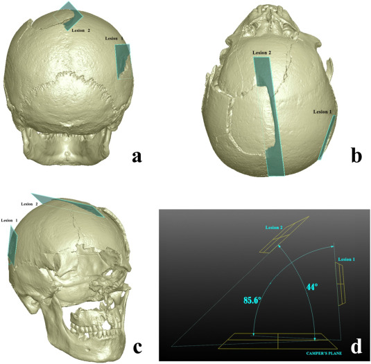
Human skeletal remains from archaeological contexts occasionally present signs of traumatic injuries from weapons, revealing, for example, the degree of interpersonal violence, the type of weapon and the sequence of events of a specific historical context.
Traumatic lesions are generally analyzed using macroscopic and microscopic methods, which are not necessarily integrated in the same study. In this study, we employed a multi-analytical approach to determine i... Read more
Antonino Vazzana, Lucia Martina Scalise, Mirko Traversari, Carla Figus, Salvatore Andrea Apicella, Laura Buti, Gregorio Oxilia, Rita Sorrentino, Silvia Pellegrini, Chiara Matteucci, Lucio Calcagnile, Raffaele Savigni, Robin N.M.Feeney, Giorgio Gruppioni, Stefano Benazziah
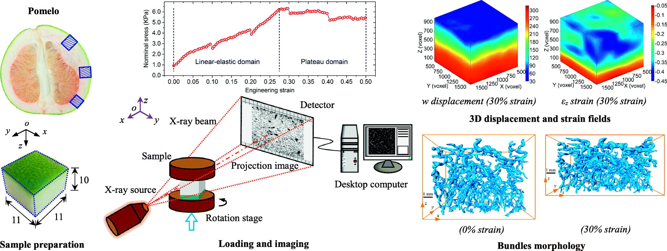
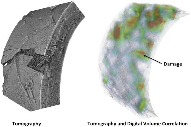
Among natural cellular materials, pomelo peels, having a foam-like hierarchical microstructure, represent an ideal model for developing materials with high energy absorption efficiency. In this work, by combining X-ray tomographic imaging technique and digital volume correlation (DVC), in-situ stepwise uniaxial compression tests were performed to quantify the internal morphological evolution and kinematic responses of pomelo peel samples during compression.
Read more
B.Wang, B.Pan, G.Lubineau
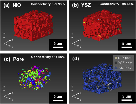
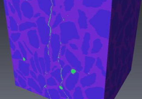
Improving microstructural quantification in FIB/SEM nanotomography
Advanced nanotomographic analysis is still far from routine, and a number of challenges remain in data acquisition and post-processing. In this work, we present a number of techniques to improve the quality of the acquired data, together with easy-to-implement methods to obtain “advanced” microstructural quantifications. The techniques are applied to a solid oxide fuel cell cathode of interest to the electrochemistry community, but the methodologies are easily adaptable to a wide range of... Read more
Joshua A.Taillon, Christopher Pellegrinelli, Yi-Lin Huang, Eric D.Wachsman, Lourdes G. Salamanca-Riba
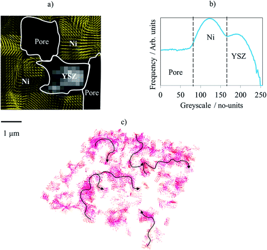
Nickel-yttria-stabilized zirconia (Ni-YSZ) cermet is widely used as an anode material in solid oxide fuel cells (SOFCs); however, Ni re-oxidation causes critical problems due to volume expansion, which causes high thermal stress. We fabricated a Ni-YSZ anode functional layer (AFL), which is an essential component in high-performance SOFCs, and re-oxidized it to investigate the related three-dimensional (3D) microstructural and thermo-mechanical effects. A 3D model of the re-oxidized AFL ... Read more
Jun Woo Kim, Kiho Bae, Hyun Joong Kim, Ji-won Son, Namkeun Kim, Stefan Stenfelt, Fritz B. Prinz, Joon Hyung Shim
The three-dimensional (3D) characterization of nuclear fuel with X-ray microscopy has historically proven difficult, due to uranium’s high attenuation of easily accessible X-rays, both in a laboratory setting and at a synchrotron user facility. However, this imaging modality provides nondestructive information that can be used to investigate morphological changes arising from external stimuli (e.g., neutron irradiation, high-temperature testing).
Using an appropriate X-ray energy spe... Read more
Nikolaus L. Cordes, William C. Chuirazzi & Joshua J. Kane - John D. Stempien
In situ observation of mechanical damage within a SiC-SiC ceramic matrix composite
SiC-SiC ceramic matrix composites are candidate materials for fuel cladding in Generation IV nuclear fission reactors and as accident tolerant fuel clad in current generation plant.
Experimental methods are needed that can detect and quantify the development of mechanical damage, to support modelling and qualification tests for these critical components. In situ observations of damage development have been obtained of tensile and C-ring mechanical test specimens of a braided nuclear gr... Read more
L. Saucedo-Mora, T. Lowe, S. Zhao, P.D. Lee, P.M. Mummery, T.J. Marrow

Protocols for Generating Surfaces and Measuring 3D Organelle Morphology Using Amira
High-resolution 3D images of organelles are of paramount importance in cellular biology. Although light microscopy and transmission electron microscopy (TEM) have provided the standard for imaging cellular structures, they cannot provide 3D images.
However, recent technological advances such as serial block-face scanning electron microscopy (SBF-SEM) and focused ion beam scanning electron microscopy (FIB-SEM) provide the tools to create 3D images for the ultrastructural analysis of org... Read more
Edgar Garza-Lopez, Zer Vue, Prasanna Katti, Kit Neikirk, Michelle Biete, Jacob Lam, Heather K. Beasley, Andrea G. Marshall, Taylor A. Rodman, Trace A. Christensen, Jeffrey L. Salisbury, Larry Vang, Margaret Mungai, Salma Ash Shareef, Sandra A. Murray, Jianqiang Shao, Jennifer Streeter, Brian Glancy, Renata O. Pereira1, E. Dale Abel, and Antentor Hinton, Jr.
Precise methods for quantifying drug accumulation in brain tissue are currently very limited, challenging the development of new therapeutics for brain disorders. Transcardial perfusion is instrumental for removing the intravascular fraction of an injected compound, thereby allowing for ex vivo assessment of extravasation into the brain. However, pathological remodeling of tissue microenvironment can affect the efficiency of transcardial perfusion, which has been largely overlooked.
We... Read more
Serhii Kostrikov, Kasper B. Johnsen, Thomas H. Braunstein, Johann M. Gudbergsson, Frederikke P. Fliedner, Elisabeth A. A. Obara, Petra Hamerlik, Anders E. Hansen, Andreas Kjaer, Casper Hempel & Thomas L. Andresen
Influenza A matrix protein M1 is sufficient to induce lipid membrane deformation
The matrix protein M1 of the Influenza A virus is considered to mediate viral assembly and budding at the plasma membrane (PM) of infected cells. In order for a new viral particle to form, the PM lipid bilayer has to bend into a vesicle towards the extracellular side. Studies in cellular models have proposed that different viral proteins might be responsible for inducing membrane curvature in this context (including M1), but a clear consensus has not been reached. In this study, we use a comb... Read more
Ismail Dahmani, Kai Ludwig, Salvatore Chiantia
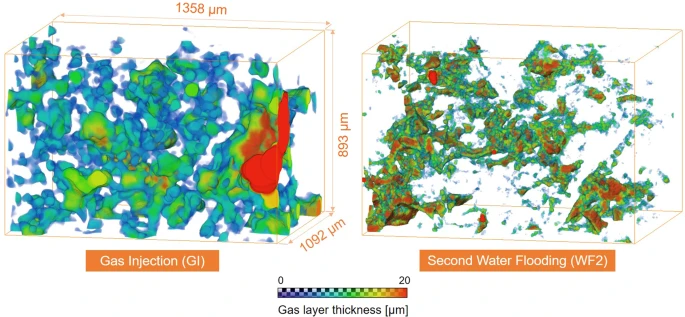
Pore-scale mechanisms of CO2 storage in oilfields
Rapid implementation of global scale carbon capture and storage is required to limit temperature rises to 1.5 °C this century. Depleted oilfields provide an immediate option for storage, since injection infrastructure is in place and there is an economic benefit from enhanced oil recovery. To design secure storage, we need to understand how the fluids are configured in the microscopic pore spaces of the reservoir rock. We use high-resolution X-ray imaging to study the flow of oil, water and ... Read more
Abdulla Alhosani, Alessio Scanziani, Qingyang Lin, Ali Q. Raeini, Branko Bijeljic & Martin J. Blunt
Decommissioning of the damaged Chernobyl nuclear reactor Unit 4 is a top priority for the global community. Before such operations begin, it is crucial to understand the behaviour of the hazardous materials formed during the accident. Since those materials formed under extreme and mostly unquantified conditions, modelling alone is insufficient to accurately predict their physical, chemical and, predominantly, mechanical behaviour. Meanwhile, knowledge of the mechanical characteristics of thos... Read more
C.Paraskevoulakos, J.P.Forna-Kreutzer, K.R.Hallam, C.P.Jones, T.B.Scott, C.Gausse, D.J.Bailey, C.A.Simpson, D.Liu, C.Reinhard, C.L.Corkhill, M.Mostafavi
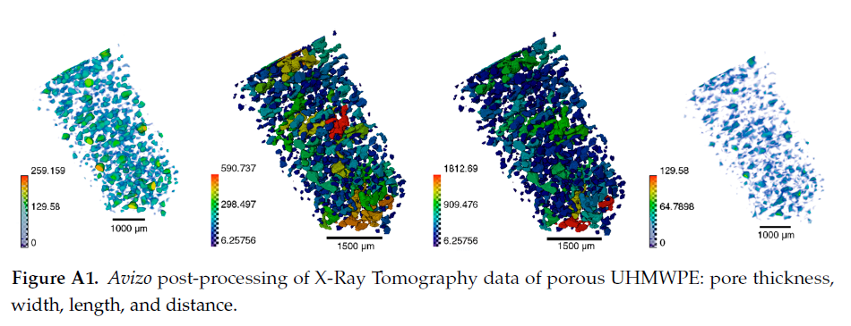
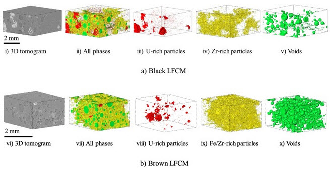
Since its invention and commercialization in the 1950s, ultra-high molecular weight polyethylene (UHMWPE) has been known as a high-performance polymer successfully applied in diverse engineering systems ranging from strong ropes for naval demands and wear-resistant liners in bearings, transportation belts and heavy trucks in mines and quarries, through the lining of chemical vessels and disposable bags in bioreactors, to sophisticated products such as orthopaedic implants and replacements of ... Read more
Eugene S. Statnik, Codrutza Dragu, Cyril Besnard, Alexander J.G. Lunt, Alexey I. Salimon, Aleksey Maksimkin and Alexander M. Korsunsky
Quantifying Microstructural Evolution in Moving Magma
Many of the grand challenges in volcanic and magmatic research are focused on understanding the dynamics of highly heterogeneous systems and the critical conditions that enable magmas to move or eruptions to initiate. However, we are usually unable to observe the processes directly. Here we give a short synopsis of the new capabilities and highlight the potential insights that in situ observation can provide. We present the first 3D data showing the evolving textural heterogeneity within a sh... Read more
Katherine J. Dobson1, Anja Allabar, Eloise Bretagne, Jason Coumans, Mike Cassidy, Corrado Cimarelli, Rebecca Coats, Thomas Connolley, Loic Courtois, Donald B. Dingwell, Danilo Di Genova, Benjamin Fernando, Julie L. Fife, Frey Fyfe, Stephan Gehne, Thomas Jones, Jackie E. Kendrick, Helen Kinvig, Stephan Kolzenburg, Yan Lavallée, Emma Liu, Edward W. Llewellin, Amber Madden-Nadeau, Kamel Madi, Federica Marone, Cerith Morgan, Julie Oppenheimer, Anna Ploszajski, Gavin Reid, Jenny Schauroth, Christian M. Schlepütz, Catriona Sellick, Jérémie Vasseur, Felix W. von Aulock, Fabian B. Wadsworth, Sebastian Wiesmaier and Kaz Wanelik
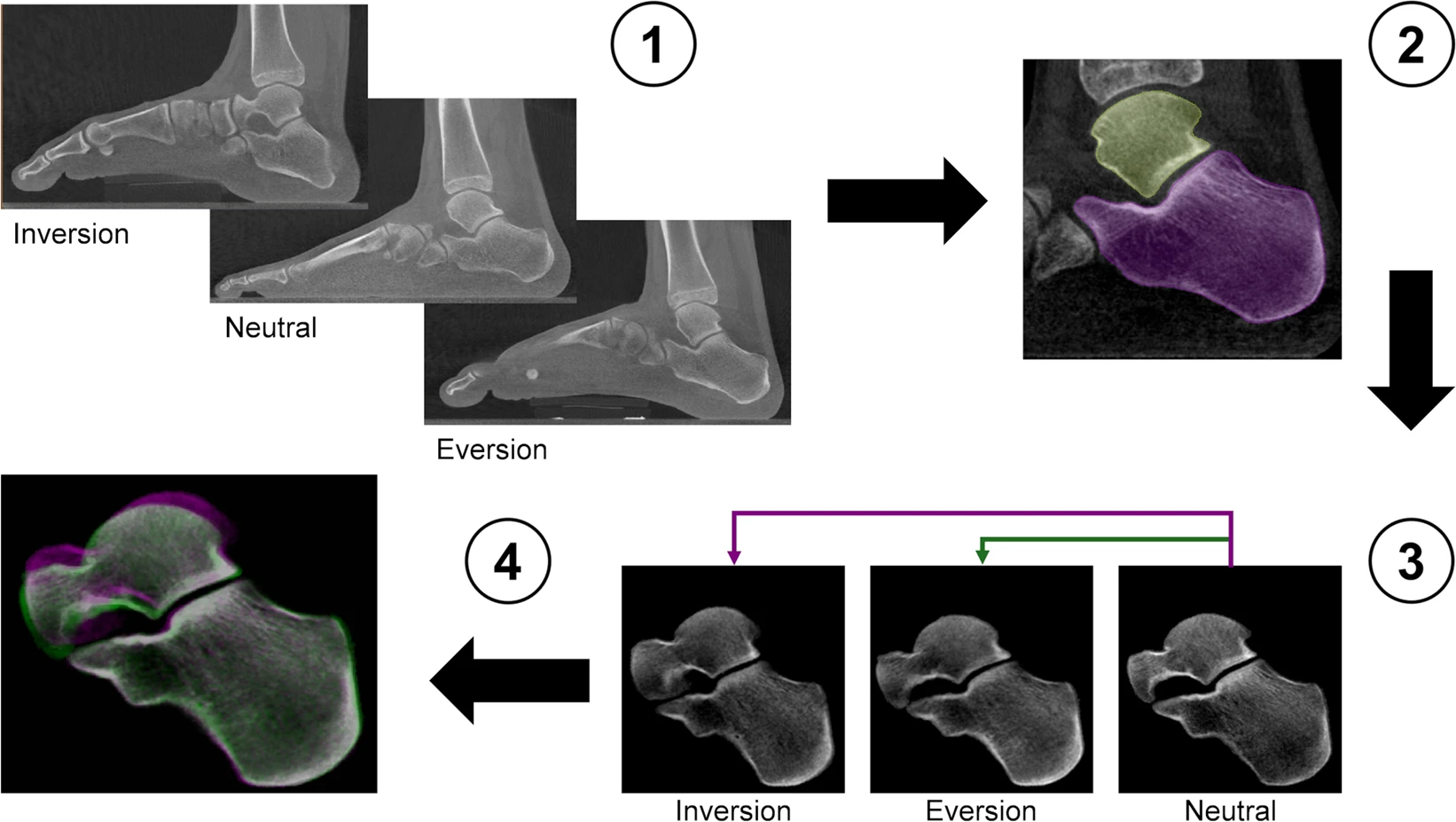
Centre of Rotation of the Human Subtalar Joint Using Weight-Bearing Clinical Computed Tomography
The subtalar joint describes an articulation between talus and calcaneus, forming one of two joints of the hindfoot with the tibiotalar or ankle joint above the talus and the subtalar joint below. The talus comprises of three facets (anterior, middle and posterior) that articulate with the mating facets on the calcaneus at the subtalar joint. The bones are connected by a complex of ligamentous structures that connect the talus to the calcaneus and both structures to the adjacent navicular bon... Read more
Marta Peña Fernández, Dorela Hoxha, Oliver Chan, Simon Mordecai, Gordon W. Blunn, Gianluca Tozzi & Andy Goldberg
X-ray Computed Tomography (XCT) is a powerful technology that can accurately image the internal structures of composite and heterogeneous materials in three-dimensions (3D). In this study, in-situ micro XCT tests of concrete specimens under progressive compressive loading are carried out. The aim of the observations is to gain a better understanding of 3D fracture and failure mechanisms at the meso-scale. To characterise the fracture evolution as the deformation increases, two methods are use... Read more
College of Civil Engineering and Architecture, Zhejiang University | School of Mechanical, Aerospace and Civil Engineering, the University of Manchester | Manchester X-ray Imaging Facility | Oxford Martin School and Department of Materials
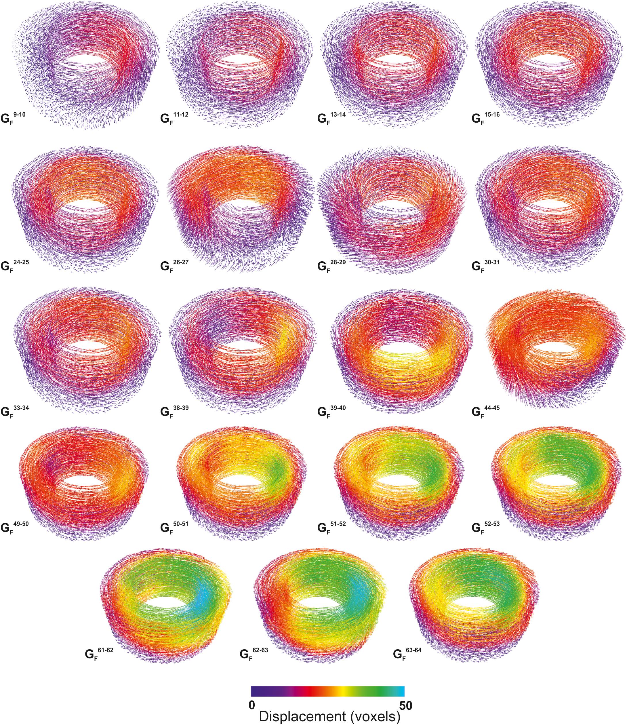
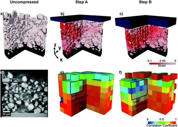
Evaluating microstructure evolution in an SOFC electrode using digital volume correlation
Degradation mechanisms within solid oxide fuel cells (SOFC) during thermal cycling limit operational start-up times and cell lifetime, and must therefore be better understood and mitigated. This work explores such mechanisms using digital volume correlation (DVC) techniques applied to lab-based X-ray tomograms where the microstructural evolution is evaluated during the operational cycling of a Ni–YSZ/YSZ cell. To emulate reduced start-up times, five tomograms were collected over four operat... Read more
T. M. M. Heenan, X. Lu,, D. P. Finegan,, J. Robinson, F. Iacoviello, J. J. Bailey, D. J. L. Brett and P. R. Shearing
Lithium-ion (Li-ion) batteries operate via electrochemical reactions between positive and negative electrodes, formed by complex porous microstructures. An improved understanding of these materials can lead to a greater insight into the link between microscopic electrode morphology and macroscopic performance. The practice of calendering electrodes after manufacturing has been widely used to increase the volumetric energy density and improve the electrical contact between electrode... Read more
S. R. Daemi,X. Lu, D. Sykes, J. Behnsen, C. Tan, A. Palacios-Padros, J. Cookson, E. Petrucco, P. J. Withers, D. J. L. Brett and P. R. Shearing

Macropinosomes are key players in early shigella invasion and vacuolar escape in epithelial cells
Intracellular pathogens include all viruses, many bacteria and parasites capable of invading and surviving within host cells. Key to survival is the subversion of host cell pathways by the pathogen for the purpose of propagation and evading the immune system. The intracellular bacterium Shigella flexneri, the causative agent of bacillary dysentery, invades host cells in a vacuole that is subsequently ruptured to allow growth of the pathogen within the host cytoplasm…
Read more
Allon Weiner , Nora Mellouk , Noelia Lopez-Montero , Yuen-Yan Chang, Célia Souque, Christine Schmitt, Jost Enninga