Welcome to the Amira-Avizo Software Use Case Gallery
Below you will find a collection of use cases of our 3D data visualization and analysis software. These use cases include scientific publications, articles, papers, posters, presentations or even videos that show how Amira-Avizo Software is used to address various scientific and industrial research topics.
Use the Domain selector to filter by main application area, and use the Search box to enter keywords related to specific topics you are interested in.
Nickel-yttria-stabilized zirconia (Ni-YSZ) cermet is widely used as an anode material in solid oxide fuel cells (SOFCs); however, Ni re-oxidation causes critical problems due to volume expansion, which causes high thermal stress. We fabricated a Ni-YSZ anode functional layer (AFL), which is an essential component in high-performance SOFCs, and re-oxidized it to investigate the related three-dimensional (3D) microstructural and thermo-mechanical effects. A 3D model of the re-oxidized AFL ... Read more
Jun Woo Kim, Kiho Bae, Hyun Joong Kim, Ji-won Son, Namkeun Kim, Stefan Stenfelt, Fritz B. Prinz, Joon Hyung Shim
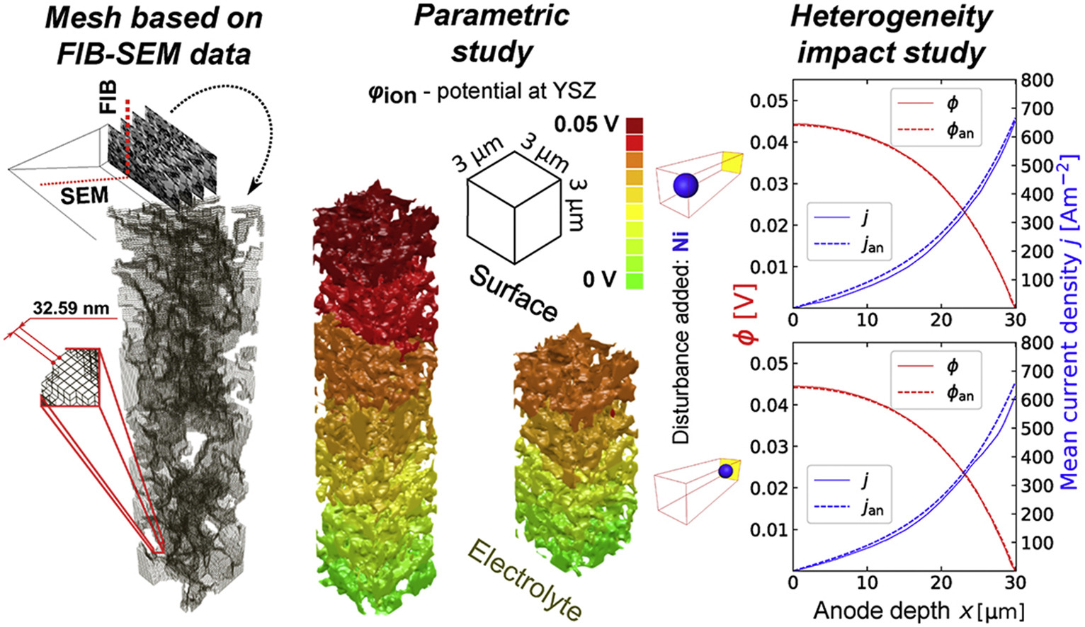
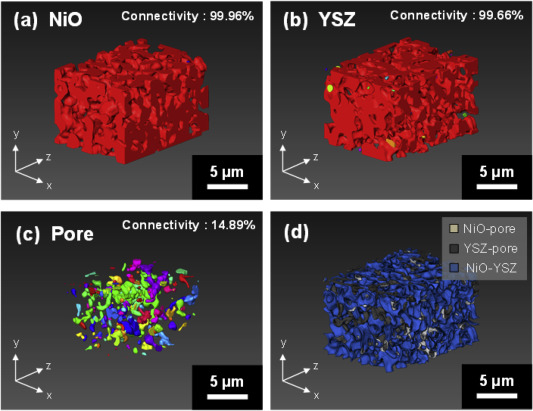
In this paper a fully three dimensional, multiphase, micro-scale solid oxide fuel cellanode transport phenomena numerical model is proposed and verified. The Butler-Volmer model was combined with empirical relations for conductivity and diffusivity – notably the Fuller-Shetler-Giddings equation, and the Fickian modelfor transport of gas reagents. FIB-SEM tomography of a commercial SOFC stack anode was performed and the resulting images were processed to acquire input data. ... Read more
Tomasz A. Prokop, Katarzyna Berent, Hiroshi Iwai, Janusz S.Szmyd, Grzegorz Brus
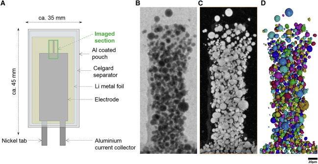
Cryo-STEM mapping of solid–liquid interfaces and dendrites in lithium-metal batteries
Solid–liquid interfaces are important in a range of chemical, physical and biological processes but are often not fully understood owing to the lack of high-resolution characterization methods that are compatible with both solid and liquid components. For example, the related processes of dendritic deposition of lithium metal and the formation of solid–electrolyte interphase layers are known to be key determinants of battery safety and performance in high-energy-density lithium-metal bat... Read more
Michael J. Zachman, Zhengyuan Tu, Snehashis Choudhury, Lynden A. Archer & Lena F. Kourkoutis
Growing popularity and rapid development of Solid Oxide Fuel Cells (SOFCs) stem for their potential to become a gamechanger in the field of clean power generation technologies.
In this paper, a transient microstructure-oriented numerical simulation of a planar Direct Internal Reforming Solid Oxide Fuel Cell (DIR-SOFC) is delivered. The performance criteria in a direct steam reforming for a fuel starvation scenario are analyzed in order to optimize the underlying process. The proposed t... Read more
Maciej Chalusiak, Michal Wrobel, Marcin Mozdzierz, Katarzyna Berent, Janusz S. Szmyd, Shinji Kimijima, Grzegorz Brus
A unique approach to correlating an evolving 3D microstructure in an Al-Cu alloyand its micro-scale mechanical properties has been introduced. Using these nanoscale three-dimensional microstructures derived from Transmission X-rayMicroscopy (TXM), individual contributions from different strengthening mechanisms were quantified. The spatial distribution and morphology of the individual θ′ and θ phases were seen to play an important role in influencing dislocation storage. Uniaxi... Read more
C. Shashank Kaira, Christopher Kantzos, Jason J. Williams, Vincent De Andrade, Francesco De Carlo, Nikhilesh Chawlaa
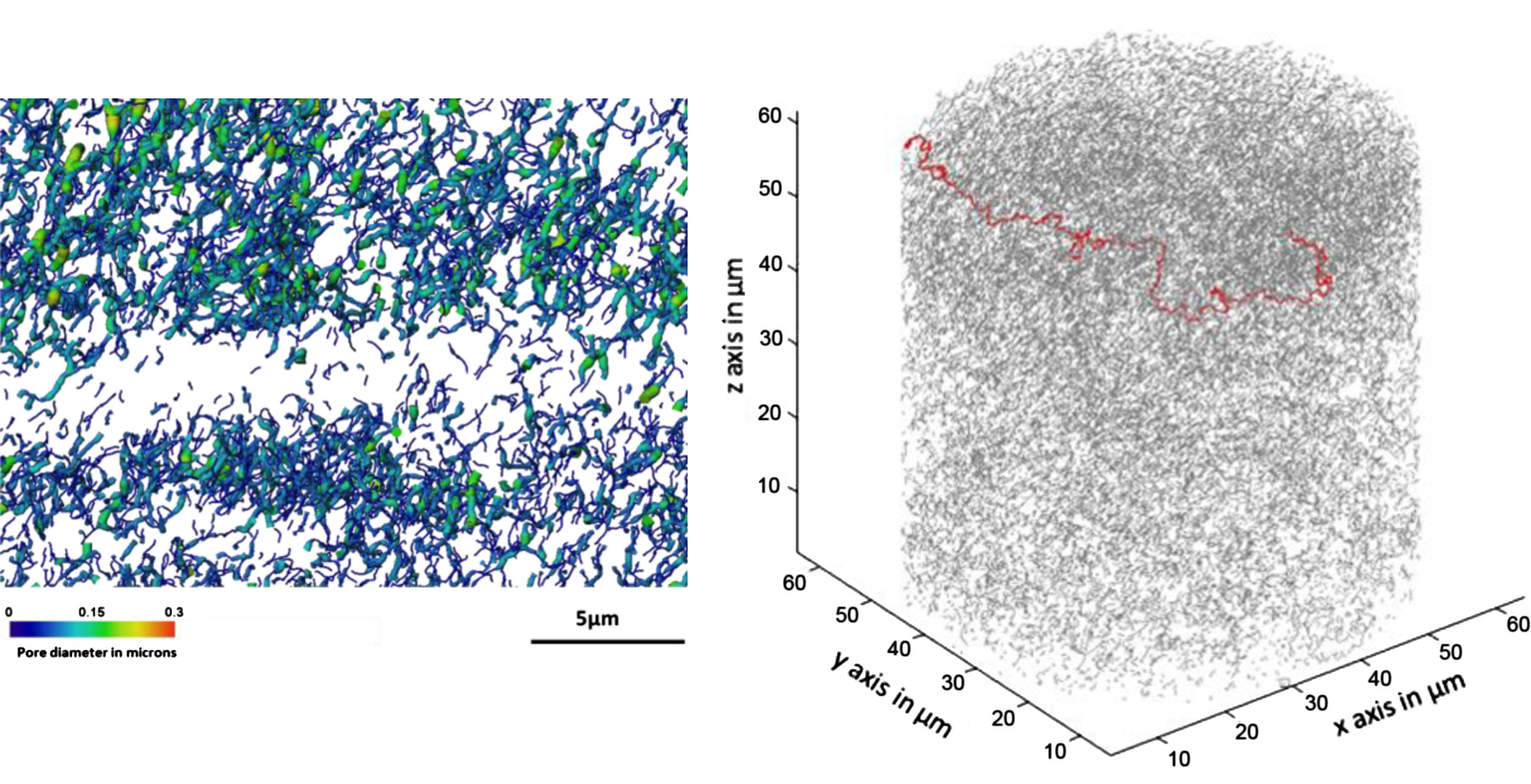
Mesoscale characterization of local property distributions in heterogeneous electrodes
The performance of electrochemical devices depends on the three-dimensional (3D) distributions of microstructural features in their electrodes. Several mature methods exist to characterize 3D microstructures over the microscale (tens of microns), which are useful in understanding homogeneous electrodes. However, methods that capture mesoscale (hundreds of microns) volumes at appropriate resolution (tens of nm) are lacking, though they are needed to understand more common, less ideal electrode... Read more
Tim Hsu, William K. Epting, Rubayyat Mahbub, Noel T. Nuhfer, Sudip Bhattachary, Yinkai Lei, Herbert M. Miller, Paul R. Ohodnicki, Kirk R. Gerdes, Harry W. Abernathy, Gregory A. Hackett, Anthony D. Rollett, Marc De Graef, Shawn Litster, Paul A. Salvador
Laser processing of metal surfaces by ultrafast Read more
Edwin Peng, Alexander Roth, Craig A. Zuhlke, Soodabeh Azadehranjbar, Dennis R. Alexander, George Gogos, Jeffrey E. Shield
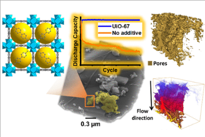
Porous Metal–Organic Frameworks for Enhanced Performance Silicon Anodes in Lithium-Ion Batteries
Maintaining the physical integrity of electrode microstructures in Li-ion batteries is critical to significantly extend their cycle life. This is especially important for high-capacity anode materials such as silicon, whose operational volume expansion exerts huge internal stress within the anode, resulting in electrode destruction and capacity fade. In this study, we demonstrate that by incorporating metal–organic frameworks (MOFs) with carboxylate organic linkers into Si-based anodes, a s... Read more
Romeo Malik, Melanie. J. Loveridge, Luke J. Williams, Qianye Huang, Geoff West, Paul R. Shearing, Rohit Bhagat, Richard I. Walton
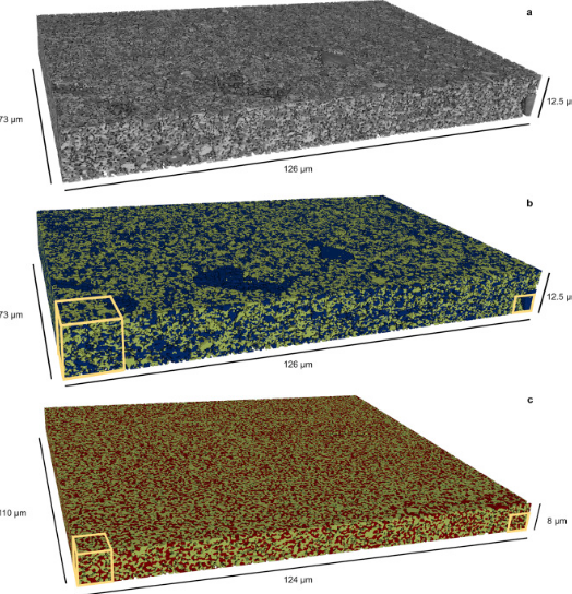
Microstructural analysis of TRISO particles using multi-scale X-ray computed tomography
TRISO particles, a composite nuclear fuel built up by ceramic and graphitic layers, have outstanding high temperature resistance. TRISO fuel is the key technology for High Temperature Reactors (HTRs) and the Generation IV Very High Temperature Reactor (VHTR) variant.
TRISO offers unparalleled containment of fission products and is extremely robust during accident conditions. An understanding of the thermal performance and mechanical properties of TRISO fuel requires a detailed knowledg... Read more
T. Lowe, R.S. Bradley, S. Yue, K. Barii, J. Gelb, N. Rohbeck, J. Turner, P.J. Withers
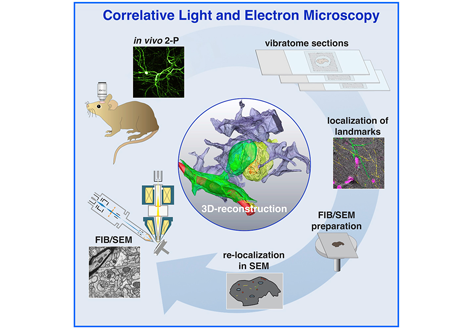
Label-free 3D-CLEM using endogenous tissue landmarks
We demonstrate feasibility of the workflow by combining in vivo 2-photon microscopy and focused ion beam scanning electron microscopy (FIB/SEM) to dissect the role of astrocytic coverage in the persistence of dendritic spines.
Emerging 3D correlative light and electron microscopy (CLEM) approaches enable studying neuronal structure-function relations at unprecedented depth and precision. However, established protocols for the correlation of light and electron micrographs rely ... Read more
Manja Luckner,Steffen Burgold, Severin Filser, Maximilian Scheungrab, Yilmaz Niyaz, Eric Hummel, Gerhard Wanner, Jochen Herms
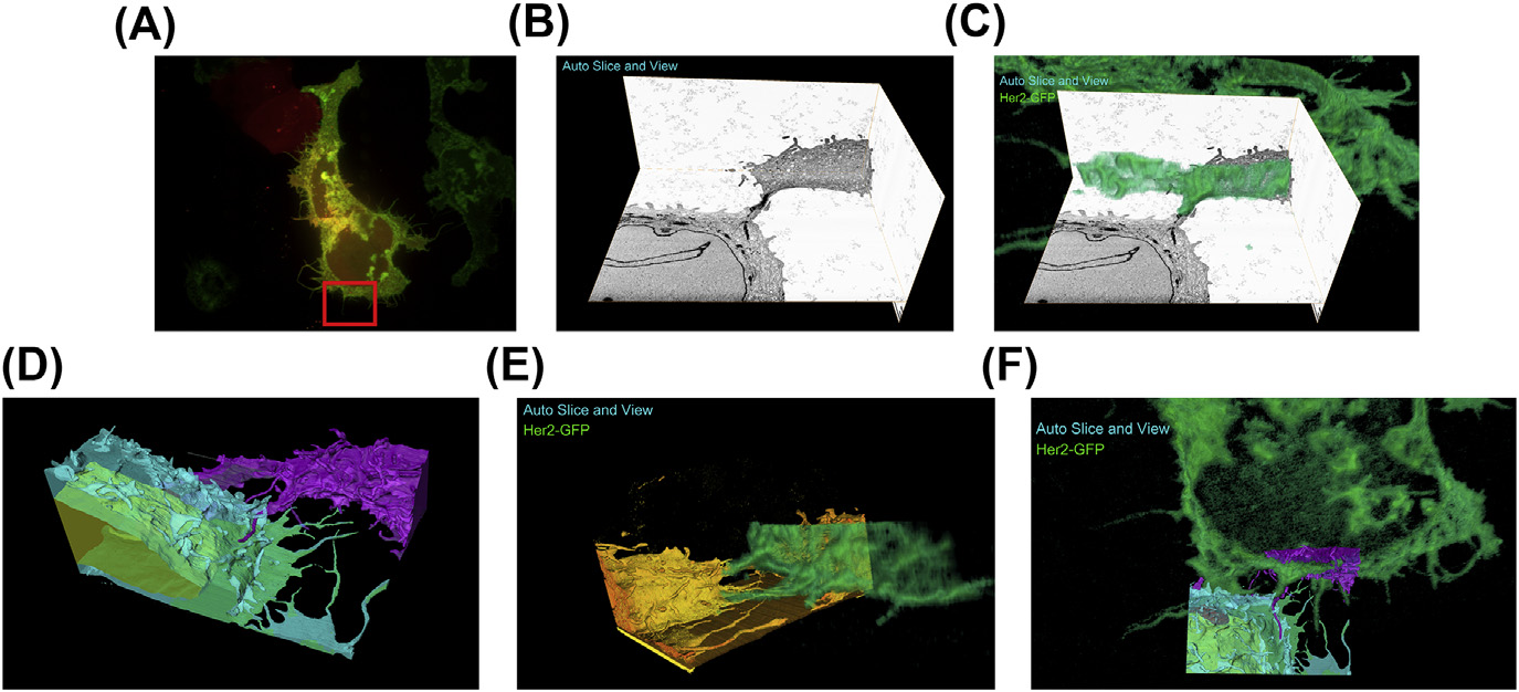
A fully integrated, three-dimensional fluorescence to electron microscopy correlative workflow
While fluorescence microscopy provides tools for highly specific labeling and sensitive detection, its resolution limit and lack of general contrast has hindered studies of cellular structure and protein localization. Recent advances in correlative light and electron microscopy (CLEM), including the fully integrated CLEM workflow instrument, the Thermo Scientific CorrSight with MAPS, have allowed for a more reliable, reproducible, and quicker approach to correlate three-dimensional time-lapse... Read more
Claudia S. Lopez, Cedric Bouchet-Marquis, Christopher P. Arthur, Jessica L. Riesterer, Gregor Heiss, Guillaume Thibault, Lee Pullan, Sunjong Kwon, Joe W. Gray
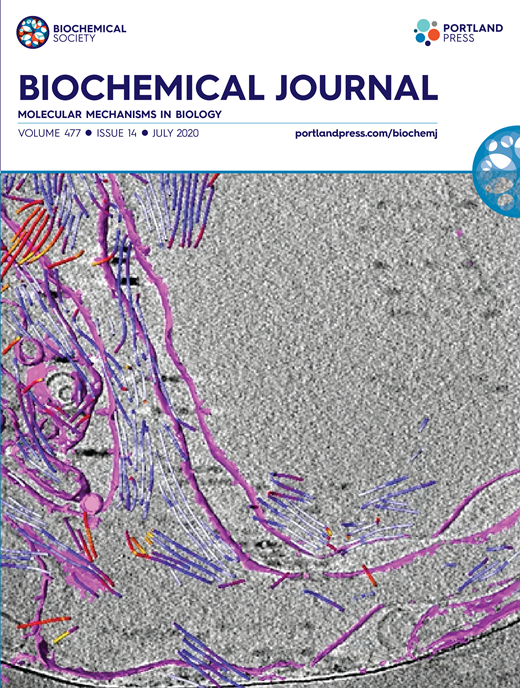
Synergistic role of nucleotides and lipids for the self-assembly of Shs1 septin oligomers
Amira capacities for membranes and filaments segmentation in cryo-TEM images are featured on the front cover of Biochemical Journal, July 2020.
Budding yeast septins are essential for cell division and polarity. (…) [The authors] have dissected, here, for the first time, the behavior of the Shs1 protomer bound to membranes at nanometer resolution, in complex with the other septins. Using electron microscopy, [the authors] have shown that on membranes, Shs1 protomers self-assembl... Read more
Cyntia Taveneau, Rémi Blanc, Gerard Pehau-Arnaudet, Aurélie Cicco, Aurélie Bertin
Microscopic organisms that penetrate calcareous structures by actively dissolving the carbonate matrix, namely microendoliths, have an important influence on the breakdown of marine carbonates.
Microscopic organisms that penetrate calcareous structures by actively dissolving the carbonate matrix, namely microendoliths, have an important influence on the breakdown of marine carbonates. The study of these microorganisms and the bioerosion traces they produce is crucial for understanding ... Read more
Philipp-Konrad Schätzle, Max Wisshak, Andreas Bick, André Freiwald, Alexander Kieneke
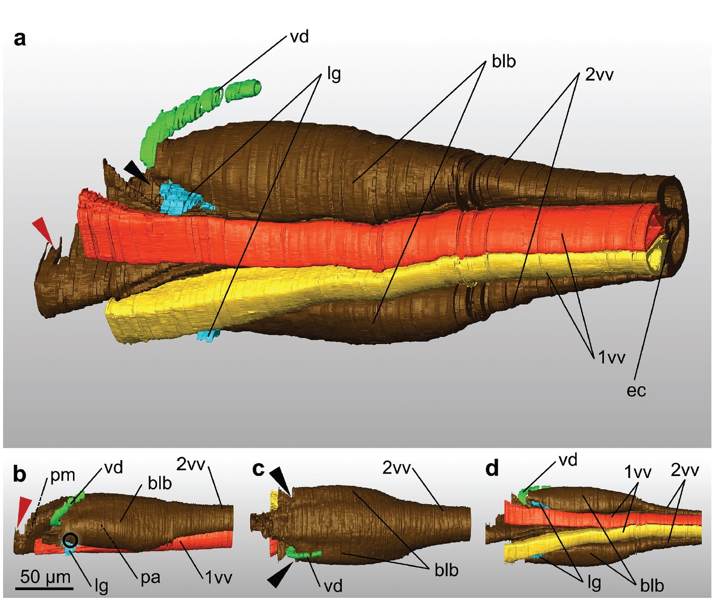
Ovipositor of the braconid wasp Habrobracon hebetor: structural and functional aspects
The Braconidae are a megadiverse and ecologically highly important group of insects. The vast majority of braconid wasps are parasitoids of other insects, usually attacking the egg or larval stages of their hosts. The ovipositor plays a crucial role in the assessment of the potential host and precise egg laying. We used lightand electron-microscopic techniques to investigate all inherent cuticular elements of the ovipositor (the female 9th abdominal tergum, two pairs of valvifers, and three p... Read more
Michael Csader, Karin Mayer, Oliver Betz, Stefan Fischer, Benjamin Eggs

Protocols for Generating Surfaces and Measuring 3D Organelle Morphology Using Amira
High-resolution 3D images of organelles are of paramount importance in cellular biology. Although light microscopy and transmission electron microscopy (TEM) have provided the standard for imaging cellular structures, they cannot provide 3D images.
However, recent technological advances such as serial block-face scanning electron microscopy (SBF-SEM) and focused ion beam scanning electron microscopy (FIB-SEM) provide the tools to create 3D images for the ultrastructural analysis of org... Read more
Edgar Garza-Lopez, Zer Vue, Prasanna Katti, Kit Neikirk, Michelle Biete, Jacob Lam, Heather K. Beasley, Andrea G. Marshall, Taylor A. Rodman, Trace A. Christensen, Jeffrey L. Salisbury, Larry Vang, Margaret Mungai, Salma Ash Shareef, Sandra A. Murray, Jianqiang Shao, Jennifer Streeter, Brian Glancy, Renata O. Pereira1, E. Dale Abel, and Antentor Hinton, Jr.
The molecular basis for sarcomere organization in vertebrate skeletal muscle
Sarcomeres are force-generating and load-bearing devices of muscles. A precise molecular picture of how sarcomeres are built underpins understanding their role in health and disease. Here, we determine the molecular architecture of native vertebrate skeletal sarcomeres by electron cryo-tomography.
Our reconstruction reveals molecular details of the three-dimensional organization and interaction of actin and myosin in the A-band, I-band, and Z-disc and demonstrates that α-actinin cros... Read more
Zhexin Wang, Michael Grange, Thorsten Wagner, Ay Lin Kho, Mathias Gautel, Stefan Raunser
Automatic whole cell organelle segmentation in volumetric electron microscopy
Cells contain hundreds of different organelle and macromolecular assemblies intricately organized relative to each other to meet any cellular demands. Obtaining a complete understanding of their organization is challenging and requires nanometer-level, three-dimensional reconstruction of whole cells. Even then, the immense size of datasets and large number of structures to be characterized requires generalizable, automatic methods.
To meet this challenge, we developed an analy... Read more
Larissa Heinrich, Davis Bennett, David Ackerman, Woohyun Park, John Bogovic, View ORCID ProfileNils Eckstein, Alyson Petruncio, Jody Clements, C. Shan Xu, Jan Funke, Wyatt Korff, Harald F. Hess, Jennifer Lippincott-Schwartz, Stephan Saalfeld, Aubrey V. Weigel, COSEM Project Team
Nickel-rich transition metal oxide materials […] are of great interest for achieving immediate improvements in the energy density of Li-ion batteries and for risk reduction within the Li-ion battery supply chain.
[…] An increase in Ni content in NMC materials leads to accelerated degradation […]. This potentially complicates their adoption in applications requiring extended cycle life such as in electric vehicles.
Recent developments in X-ray characterization to... Read more
ChunTan, Andrew S.Leach, Thomas M.M. Heenan, Huw Parks, Rhodri Jervis, Johanna Nelson Weker, Daniel J.L. Brett, Paul R.Shearing
Given the urgent need to move to low-carbon technologies, batteries are being increasingly used in a range of applications. Lithium-ion batteries are the most widely used chemistry, but to meet the growing demand, there is a need to move beyond lithium towards alternative battery chemistries. Metal–air batteries are a group of such battery alternatives that hold promise, especially for stationary power and flexible electronics applications.
However, barriers to their widespread adopt... Read more
Jennifer Hack, Drasti Patel, Josh J Bailey, Francesco Iacoviello, Paul R Shearing, and Dan J L Brett
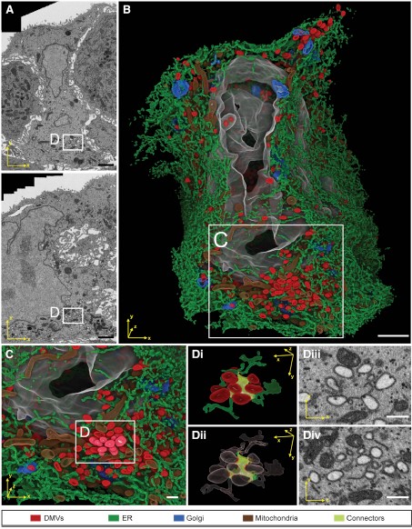
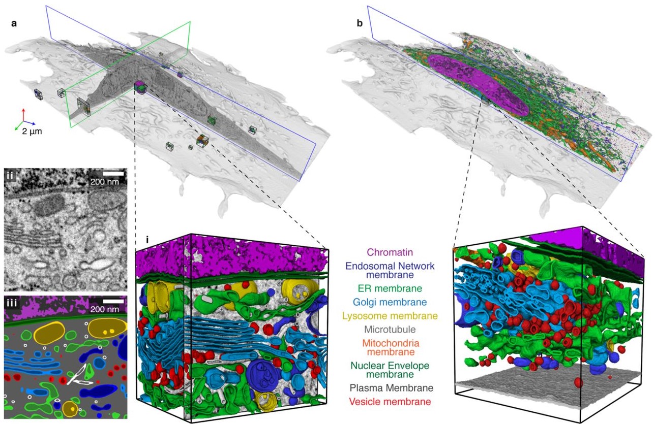
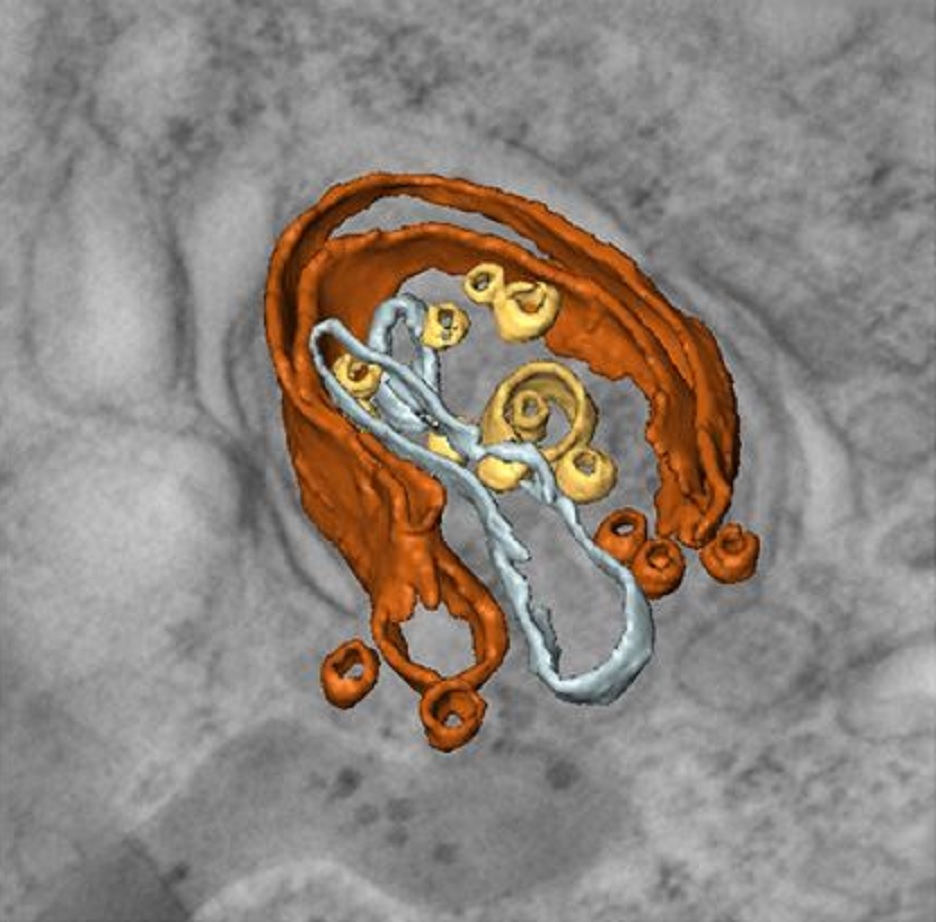
Integrative Imaging Reveals SARS-CoV-2-Induced Reshaping of Subcellular Morphologies
Cortese et al. use integrative imaging techniques to generate a publicly available repository of morphological alterations induced by SARS-CoV-2 in lung cells. Accumulation of ER-derived double-membrane vesicles, the viral replication organelle, occurs concomitantly with cytoskeleton remodeling and Golgi fragmentation. Pharmacological alteration of cytoskeleton dynamics restricts viral replication and spread.
Pathogenesis induced by SARS-CoV-2 is thought to result from both an inflamma... Read more
Mirko Cortese, Ji-Young Lee, Berati Cerikan, ..., Laurent Chatel-Chaix, Yannick Schwab, Ralf Bartenschlager
Macroautophagy is morphologically characterized by autophagosome formation. Autophagosomes are double-membraned vesicles that sequester cytoplasmic components for further degradation in the lysosome. Basal autophagy is paramount for intracellular quality control in post-mitotic cells but, surprisingly, the number of autophagosomes in post-mitotic neurons is very low, suggesting that alternative degradative structures could exist in neurons…
Read more
Maria Rosario Fernandez-Fernandez, Desire Ruiz-Garcia, Eva Martin-Solana, Francisco Javier Chichon, Jose L. Carrascosa, Jose-Jesus Fernandez