Welcome to the Amira-Avizo Software Use Case Gallery
Below you will find a collection of use cases of our 3D data visualization and analysis software. These use cases include scientific publications, articles, papers, posters, presentations or even videos that show how Amira-Avizo Software is used to address various scientific and industrial research topics.
Use the Domain selector to filter by main application area, and use the Search box to enter keywords related to specific topics you are interested in.
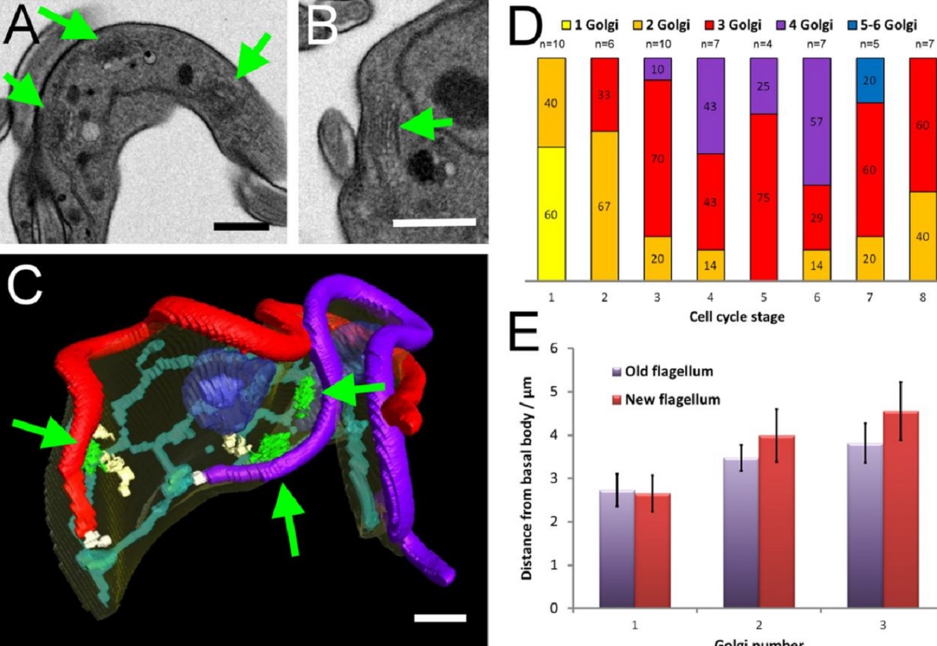
The major mammalian bloodstream form of the African sleeping sickness parasite Trypanosoma bruceimultiplies rapidly, and it is important to understand how these cells divide. Organelle inheritance involves complex spatiotemporal re-arrangements to ensure correct distribution to daughter cells…
Read more
Louise Hughes, Samantha Borrett, Katie Towers, Tobias Starborg, Sue Vaughan
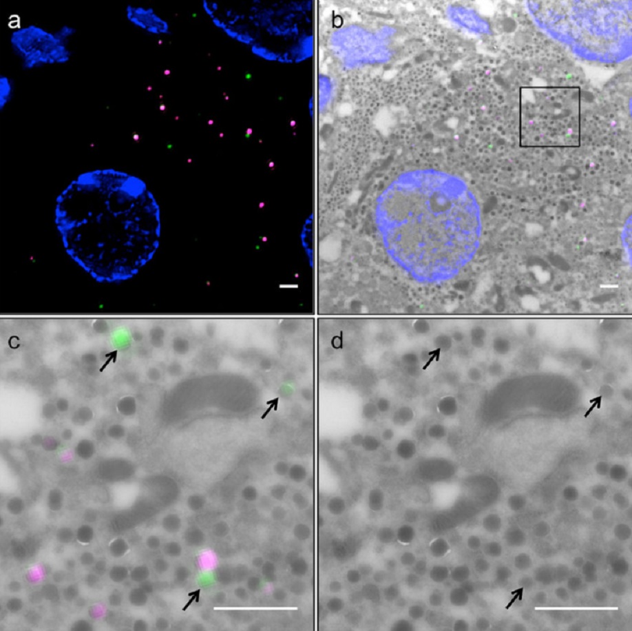
Correlative light and electron microscopy (CLEM) is a powerful approach to investigate the molecular ultrastructure of labeled cell compartments. However, quantitative CLEM studies are rare, mainly due to small sample sizes and the sensitivity of fluorescent proteins to strong fixatives and contrasting reagents for EM. Here, we show that fusion of…
Read more
Andreas Müller, Martin Neukam, Anna Ivanova, Anke Sönmez, Carla Münster, Susanne Kretschmar, Yannis Kalaidzidis, Thomas Kurth, Jean-Marc Verbavatz & Michele Solimena
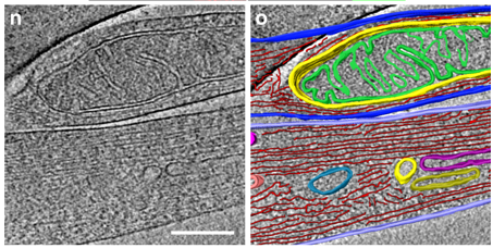
Correlative cryo-electron microscopy reveals the structure of TNTs in neuronal cells
The orchestration of intercellular communication is essential for multicellular organisms. One mechanism by which cells communicate is through long, actin-rich membranous protrusions called tunneling nanotubes (TNTs), which allow the intercellular transport of various cargoes, between the cytoplasm of distant cells in vitro and in vivo. Here, we use correlative FIB-SEM, light- and cryo-electron microscopy approaches to elucidate the structural organization of neuronal TNTs. Our data indicate ... Read more
Anna Sartori-Rupp, Diégo Cordero Cervantes, Anna Pepe, Karine Gousset, Elise Delage, Simon Corroyer-Dulmont, Christine Schmitt, Jacomina Krijnse-Locker & Chiara Zurzolo
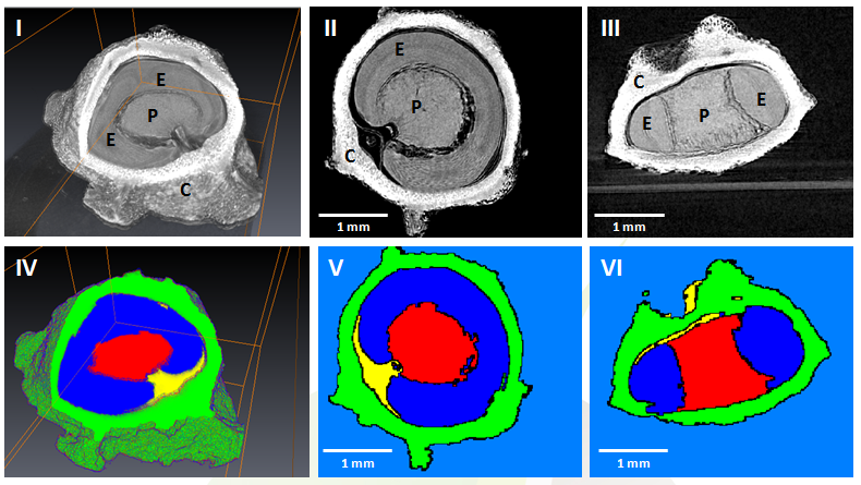
GEVES uses Avizo software to study and control plant seeds
At GEVES, Microtomography (micro-CT), which is a technique using X-rays to investigate internal anatomy and morphology of organisms without destruction, is applied to plant seed quality control and phenotyping. Several applications have been developed using micro-CT coupled with Avizo software development, such as for example in the Measurements of internal and external sugar beet seed structures for seed phenotyping
Read more
Ghassen Trigui, Laurence Le Corre, Dominique Honoré and Karima Boudehri-Giresse - 2D/3D Imaging - R&D team, GEVES
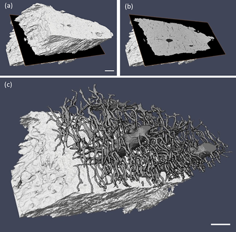
Computed tomography is an increasingly popular technique for the non-destructive study of fossils. Whilst the science of X-ray computed tomography (CT) has greatly matured since its first fossil applications in the early 1980s, the applications and limitations of neutron tomography (NT) remain relatively unexplored in palaeontology. These highest resolution neutron tomographic scans in palaeontology to date were conducted on a specimen of Austrosequoia novae-zeelandiae (Ettingshausen) Mays an... Read more
Chris Mays, Joseph J. Bevitt, and Jeffrey D. Stilwell
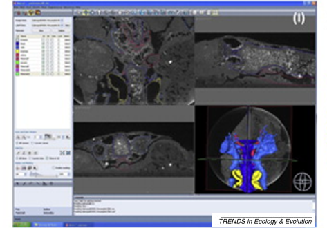
A virtual world of paleontology
Computer-aided visualization and analysis has revolutionized the study of fossils. Fossils can now be characterized in three dimensions and in unprecedented detail. The resulting digital reconstructions can be used in rigorous functional analyses. Hypotheses regarding the function of extinct organisms can therefore be tested.
Read more
Trends in Ecology & Evolution
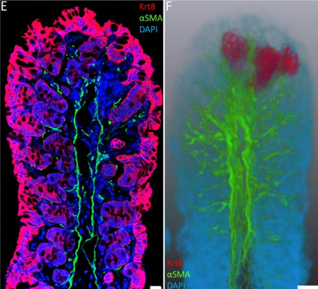
Immunofluorescence tomography is a high-resolution 3-D reconstruction method based on methacrylate embedding and serial-sectioning, where 2-D images of immuno-stained serial-sections are computationally aligned into image stacks, and the 3-D volume rendered. Butyl-Methyl Methacrylate (BMMA) plastic was adopted as it preserves excellent tissue morphology and can be de-plasticized easily using an organic solvent, which enables immuno-staining of serial-sections without antibody penetration issu... Read more
Parfitt, Geraint J
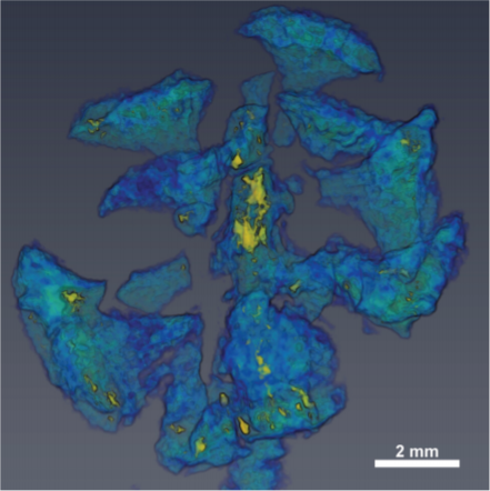
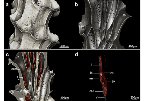
Microbial-tubeworm associations in a 440 million year old hydrothermal vent community
Microorganisms are the chief primary producers within present-day deep-sea hydrothermal vent ecosystems, and play a fundamental role in shaping the ecology of these environments. (…) The oldest known hydrothermal vent community that includes metazoans is preserved within the Ordovician to early Silurian Yaman Kasy massive sulfide deposit, Ural Mountains, Russia. (…) A re-examination of these fossils using a range of microscopy, chemical analysis and nano-tomography techniques re... Read more
Magdalena N. Georgieva , Crispin T. S. Little , Russell J. Bailey , Alexander D. Ball and Adrian G. Glover
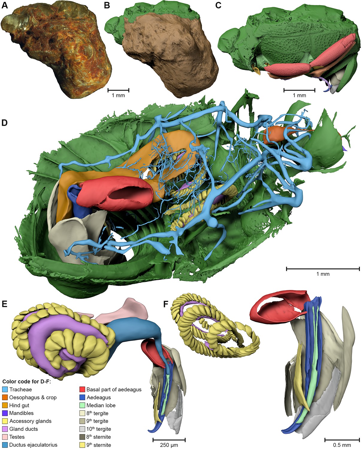
External and internal morphological characters of extant and fossil organisms are crucial to establishing their systematic position, ecological role and evolutionary trends. (…) We found well-preserved three-dimensional anatomy in mineralized arthropods from Paleogene fissure fillings and demonstrate the value of these fossils by utilizing digitally reconstructed anatomical structure of a hister beetle. The new anatomical data facilitate a refinement of the species diagnosis and allowed... Read more
Achim H Schwermann, Tomy dos Santos Rolo, Michael S Caterino, Gunter Bechly, Heiko Schmied, Tilo Baumbach, Thomas van de Kamp
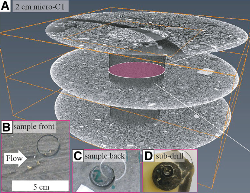
Sediment plates (a type of lacquer peels) represent a sampling method whereby a thin plate of undisturbed sediments is obtained directly from the outcrop. A low-viscosity, hardening epoxy resin is applied to a freshly exposed cross-section of an unconsolidated deposit and impregnates a surface layer of the cross-section via capillary forces before solidifying. Upon hardening, a solid plate (0.5–5 cm thick and up to 2 m in length) of the sedimentary formation can be recovered and transported... Read more
Guilhem Amin Douillet, Ulrich Kueppers, Célia Mato, Quentin Chaffaut, Mélanie Bouysson, Renate Reschetizka, Inga Hoelscher, Patrick Witting, Kai-Uwe Hess, Alexander Cerwenka, Donald B Dingwell and Benjamin Bernard
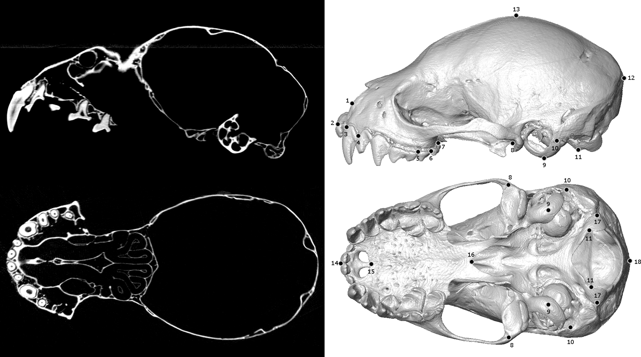
Biological specimens are primary records of organismal ecology and history. As such, museum collections are invaluable repositories for testing ecological and evolutionary hypotheses across the tree of life. Digitizing and broadly sharing the phenotypic data from these collections serves to expand the traditional reach of museums, enabling widespread data sharing, collaboration, and education at an unprecedented scale. In recent years, μCT-scanning has been adopted as one way for efficiently... Read more
Jeff J. Shi, Erin P. Westeen, Daniel L. Rabosky
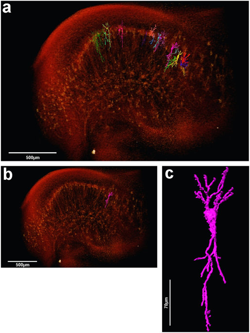
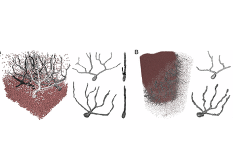
The assessment of neuronal number, spatial organization and connectivity is fundamental for a complete understanding of brain function. However, the evaluation of the three-dimensional (3D) brain cytoarchitecture at cellular resolution persists as a great challenge in the field of neuroscience. In this context, X-ray microtomography has shown to be a valuable non-destructive tool for imaging a broad range of samples, from dense materials to soft biological specimens, arisen as a new method fo... Read more
Matheus de Castro Fonseca, Bruno Henrique Silva Araujo, Carlos Sato Baraldi Dias, Nathaly Lopes Archilha, Dionísio Pedro Amorim Neto, Esper Cavalheiro, Harry Westfahl Jr, Antônio José Roque da Silva, Kleber Gomes Franchini
Ptychographic X-ray computed tomography (PXCT) is a quantitative imaging modality that non-destructively maps the 3D electron density inside an object with tens of nanometers spatial resolution. This method provides unique access to the morphology and structure of the osteocyte lacuno-canalicular network (LCN) and nanoscale density of the tissue in the vicinity of an osteocyte lacuna. Our findings indicate that PXCT can non-destructively provide detailed, nanoscale information on the 3D org... Read more
Antonia Ciani, Hechmi Toumi, Stéphane Pallu, Esther H.R.Tsai, Ana Diaz, Manuel Guizar-Sicairos, Mirko Holler, Eric Lespessailles, Cameron M.Kewish | Synchrotron Soleil, France
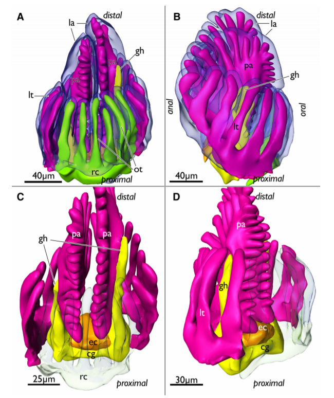
Cyclostome bryozoans are an ancient group of marine colonial suspension-feeders comprising approximately 700 extant species. Previous morphological studies are mainly restricted to skeletal characters whereas data on soft tissues obtained by state-of-the-art methods are still lacking. In order to contribute to issues related to cyclostome ground pattern reconstruction, we analyzed the morphology of the neuromuscular system Cinctipora elegans by means of immunocytochemical staining,... Read more
Thomas F. Schwaha, Stephan Handschuh, Andrew N. Ostrovsky, Andreas Wanninger
Three-dimensional virtual histology of human cerebellum by X-ray phase-contrast tomography
To quantitatively evaluate brain tissue and its corresponding function, knowledge of the 3D cellular distribution is essential. The gold standard to obtain this information is histology, a destructive and labor-intensive technique where the specimen is sliced and examined under a light microscope, providing 3D information at nonisotropic resolution. To overcome the limitations of conventional histology, we use phase-contrast X-ray tomography with optimized optics, reconstruction, and image an... Read more
Mareike Töpperwien, Franziska van der Meer, Christine Stadelmann, and Tim Salditt
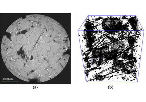
In this study, chemically bonded phosphate ceramic (CBPC) fiber reinforced composites were made at indoor temperatures. The mechanical properties and microstructure of the CBPC composites were studied. The CBPC matrix of aluminum phosphate binder, metakaolin, and magnesia with different Si/P ratios was prepared. The results show that when the Si/P ratio was 1.2, and magnesia content in the CBPC was 15%, CBPC reached its maximum flexural strength. The fiber reinforced CBPC composites were prep... Read more
Zhu Ding; Yu-Yu Li; Can Lu; Jian Liu
Phylactolaemate bryozoans are the sister-group to all remaining bryozoan taxa. Consequently, their study is essential to reveal and analyze ancestral traits of Phylactolaemata and Bryozoa in general.
They are the only bryozoans to possess an epistome which traditionally has been regarded as shared with phoronids and brachiopods. Contrary to older observations, an epistome was recently reported to be missing in the early branching phylactolaemate Lophopus crystallinus. In this... Read more
Thomas Schwaha
Composite membranes with defective metal–organic frameworks (MOFs) connect the emerging fields of MOF topological modification, MOF-polymer interfacial engineering and composite material functionalization.
Although defective MOFs can be fabricated via thermal or chemical treatment, the relationship between hierarchical MOF structure and their performance in a polymeric membrane matrix has so far not been investigated. Here we show how a modula... Read more
Weibin Liang, Lin Li, Jingwei Hou, Nicholas D. Shepherd, Thomas D. Bennett, Deanna M. D'Alessandro and Vicki Chen
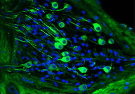
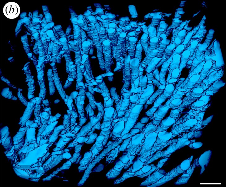
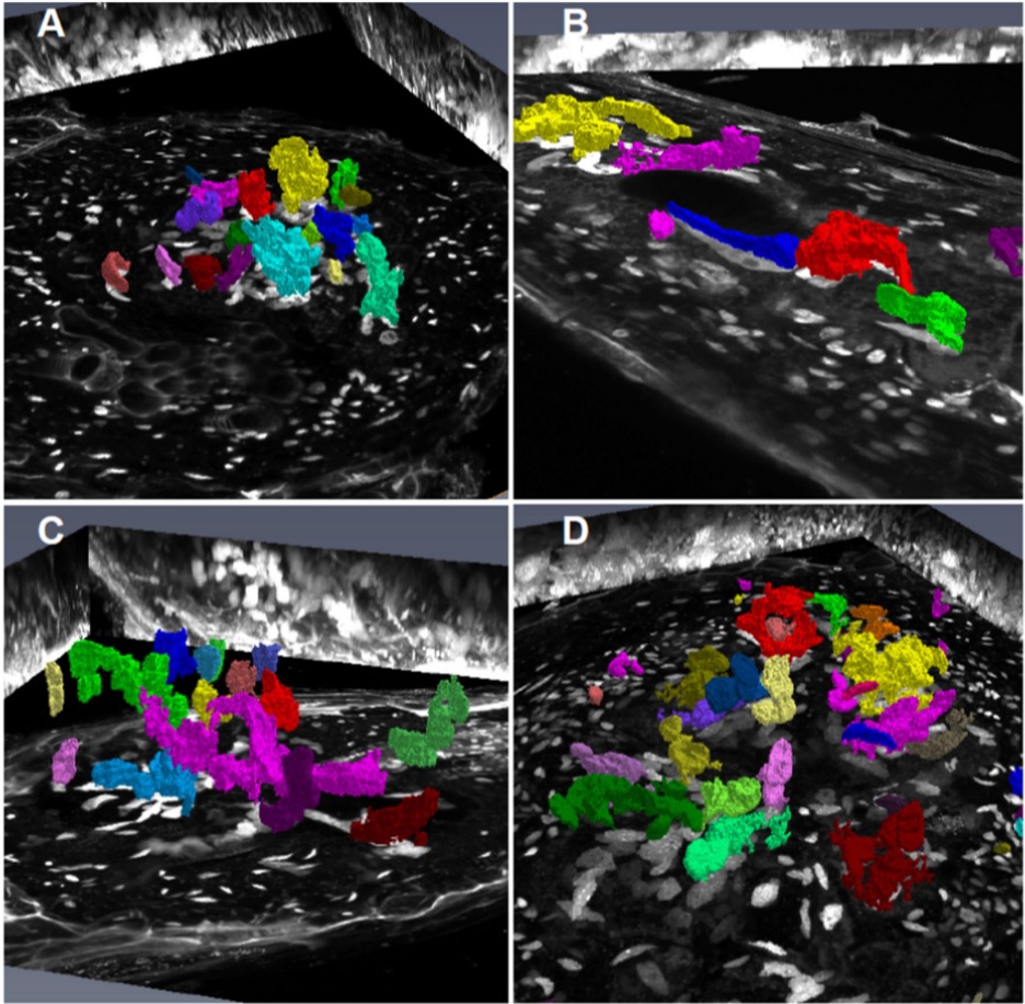
Chronic cigarette smoke exposure drives spiral ganglion neuron loss in mice
Tobacco use is associated with an increased risk of hearing loss in older individuals, suggesting cigarette smoke (CS) exposure may target the peripheral auditory organs. However, the effects of CS exposure on general cochlear anatomy have not previously been explored.
Here we compare control and chronic CS exposed cochleae from adult mice to assess changes in structure and cell survival. Two-photon imaging techniques, including the imaging of second harmonic generation (SHG) and two-p... Read more
Stephen T. Paquette, Ryan P. Dawes, Isaac K. Sundar, Irfan Rahman, Edward B. Brown & Patricia M. White
Root-knot nematodes induce galls that contain giant-feeding cells harboring multiple enlarged nuclei within the roots of host plants. It is recognized that the cell cycle plays an essential role in the set-up of a peculiar nuclear organization that seemingly steers nematode feeding site induction and development. Functional studies of a large set of cell cycle genes in transgenic lines of the model host Arabidopsis thaliana have contributed to better understand the role of the cell cycle comp... Read more
Antonino de Souza Junior José Dijair, Pierre Olivier, Coelho Roberta R., Grossi-de-Sa Maria F., Engler Gilbert, de Almeida Engler Janice / Institut National de la Recherche Agronomique, Université Côte d’Azur, Centre National de la Recherche Scientifique, Institut Sophia Agrobiotech, Sophia-Antipolis, France
Volcanogenic Pseudo-Fossils from the ∼3.48 Ga
The ∼3.48 billion-year-old Dresser Formation, Pilbara Craton, Western Australia, is a key geological unit for the study of Earth’s earliest life and the habitats it occupied. Here, we describe a new suite of spheroidal to lenticular microstructures that morphologically resemble some previously reported Archean microfossils. Correlative microscopy shows that these objects have a size distribution, wall ultrastructure, and chemistry that are incompatible with a microfossil origin and in... Read more
Wacey David , Noffke Nora , Saunders Martin , Guagliardo Paul , and Pyle David M