Welcome to the Amira-Avizo Software Use Case Gallery
Below you will find a collection of use cases of our 3D data visualization and analysis software. These use cases include scientific publications, articles, papers, posters, presentations or even videos that show how Amira-Avizo Software is used to address various scientific and industrial research topics.
Use the Domain selector to filter by main application area, and use the Search box to enter keywords related to specific topics you are interested in.
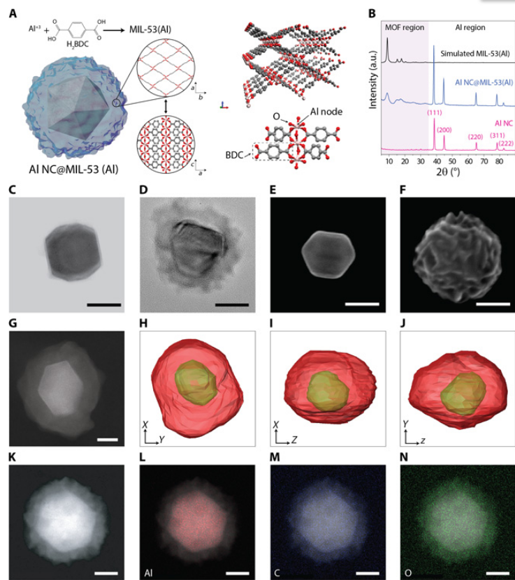
Metal-organic frameworks tailor the properties of aluminum nanocrystals
Metal-organic frameworks (MOFs) and metal nanoparticles are two classes of materials that have received considerable recent attention, each for controlling chemical reactivities, albeit in very different ways. Here, we report the growth of MOF shell layers surrounding aluminum nanocrystals (Al NCs), an Earth-abundant metal with energetic, plasmonic, and photocatalytic properties. The MOF shell growth proceeds by means of dissolution-and-growth chemistry that uses the intrinsic surface oxide o... Read more
Hossein Robatjazi, Daniel Weinberg, Dayne F. Swearer, Christian Jacobson, Ming Zhang, Shu Tian, Linan Zhou, Peter Nordlander, Naomi J. Halas
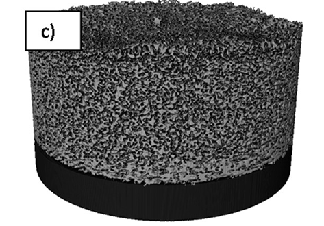
Aging of a Pt/Al2O3 exhaust gas catalyst monitored by quasi in situ X-ray micro computed tomography
Catalyst aging effects were analyzed using X-ray absorption micro-computed tomography in combination with conventional characterization methods on various length scales ranging from nm to μm to gain insight into deactivation mechanisms.
For this purpose, a 4 wt% Pt/Al2O3 model exhaust gas catalyst was coated on a cordierite honeycomb and subjected to sequential thermal aging in static air at 950 °C for 4, 8, 12 and 24 hours. The ag... Read more
Georg Hofmann, Amélie Rochet, Elen Ogel, Maria Casapu, Stephan Ritter, Malte Ogurreck and Jan-Dierk Grunwaldt
The development of focused ion beam-scanning electron microscopy (FIB-SEM) techniques has allowed high-resolution 3D imaging of nanometre-scale porous materials. These systems are of important interest to the oil and gas sector, as well as for the safe long-term storage of carbon and nuclear waste. This work focuses on validating the accurate representation of sample pore space in FIB-SEM-reconstructed volumes and the predicted permeability of these systems from subsequent single-phase flow s... Read more
Department of Chemical Engineering, Qatar Carbonates and Carbon Storage Research Centre, Imperial College London | Department of Applied Mathematics and Theoretical Physics, Cambridge University
Despite the importance of studying the instability of delithiated cathode materials, it remains difficult to underpin the degradation mechanism of lithium-rich cathode materials due to the complication of combined chemical and structural evolutions. Herein, we use state-of-the-art electron microscopy tools, in conjunction with synchrotron X-ray techniques and first-principle calculations to study a 4d-element-containing compound, Li2Ru0.5Mn0.5O3. We find surprisingly, after cycling, ruthenium... Read more
Lin, Ruoqian AU - Hu, Enyuan AU - Liu, Mingjie AU - Wang, Yi AU - Cheng, Hao AU - Wu, Jinpeng AU - Zheng, Jin-Cheng AU - Wu, Qin AU - Bak, Seongmin AU - Tong, Xiao AU - Zhang, Rui AU - Yang, Wanli AU - Persson, Kristin A. AU - Yu, Xiqian AU - Yang, Xiao-Qing AU - Xin, Huolin L. PY
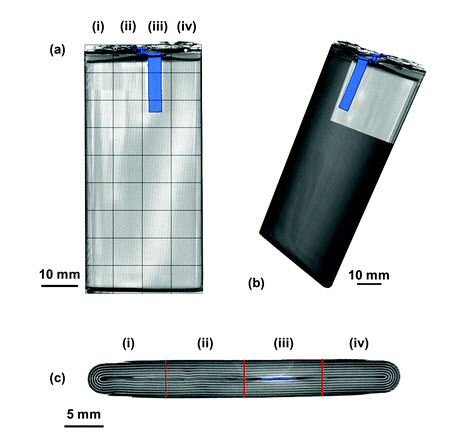
Visualizing the Carbon Binder Phase of Battery Electrodes in Three Dimensions
This study presents a technique to directly characterize the carbon and binder domain (CBD) in lithium-ion (Li-ion) battery electrodes in three dimensions and use it to determine the effective transport properties of a LiNi0.33Mn0.33Co0.33O2 (NMC) electrode. X-ray nanocomputed tomography (nano-CT) is used to image an electrode composed solely of carbon and binder, whereas focused ion beam–scanning electron microscopy is used to analyze cross-sect... Read more
Sohrab R. Daemi, Chun Tan, Tobias Volkenandt, Samuel J. Cooper, Anna Palacios-Padros, James Cookson, Dan J. L. Brett, and Paul R. Shearing
Spatially resolved ultrasound diagnostics of Li-ion battery electrodes
The importance of reliable battery diagnostic systems has grown substantially in recent years as a result of the use of high power Li-ion battery packs in an increasingly diverse range of applications. Here, spatially resolved ultrasound acoustic measurements are used to analyse the condition of Li-ion electrodes. Ultrasonic measurements are performed on a commercial mobile phone battery over the full operating voltage window with the lithiation and delithiation of electrodes o... Read more
James B. Robinson, Maximilian Maier , George Alster , Tomos Compton , Dan J. L. Brett and Paul R. Shearing
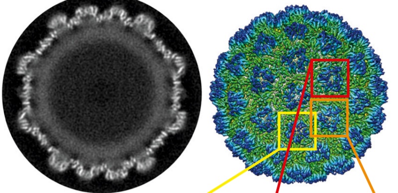
Structure of the Ty3/Gypsy retrotransposon capsid and the evolution of retroviruses
Retroviruses evolved from long terminal repeat (LTR) retrotransposons by acquisition of envelope functions, and subsequently reinvaded host genomes. Together, endogenous retroviruses and LTR retrotransposons represent major components of animal, plant, and fungal genomes. Sequences from these elements have been exapted to perform essential host functions, including placental development, synaptic communication, and transcriptional regulation. They encode a Gag polypeptide, the capsid domains ... Read more
Svetlana O. Dodonova, Simone Prinz, Virginia Bilanchone, Suzanne Sandmeyer, and John A. G. Briggs
As key functional units in neural circuits, different types of neuronal synapses play distinct roles in brain information processing, learning, and memory. Synaptic abnormalities are believed to underlie various neurological and psychiatric disorders. Here, by combining cryo-electron tomography and cryo-correlative light and electron microscopy, we distinguished intact excitatory and inhibitory synapses of cultured hippocampal neurons, and visualized the in situ 3D organization of ... Read more
Chang-Lu Tao, Yun-Tao Liu, Rong Sun, Bin Zhang, Lei Qi, Sakar Shivakoti, Chong-Li Tian, Peijun Zhang, Pak-Ming Lau, Z. Hong Zhou and Guo-Qiang Bi
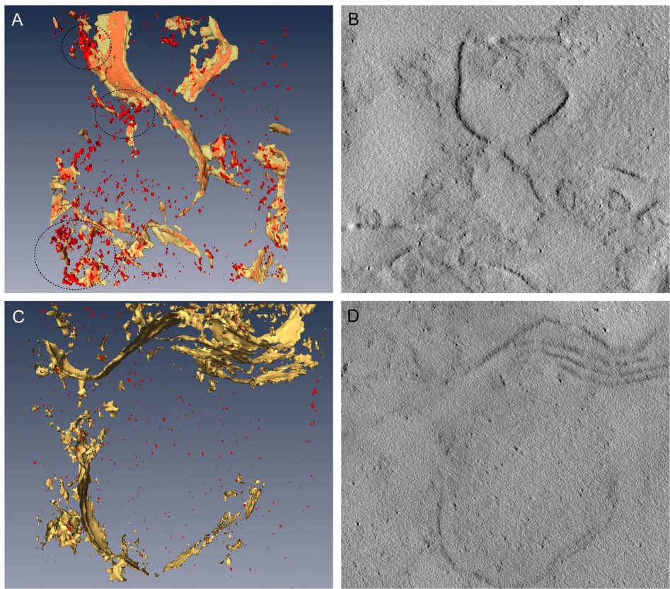
Corpora amylacea are cell-derived structures that appear physiologically in the aged human brain. While their histological identification is straightforward, their ultrastructural composition and microenvironment at the nanoscale have remained unclear so far, as has their relevance to aging and certain disease states that involve the sequestration of toxic cellular metabolites. Here, we apply correlative serial block-face scanning electron microscopy and transmission electron tomograp... Read more
Paula P. Navarro, Christel Genoud, Daniel Castaño-Díez, Alexandra Graff-Meyer, Amanda J. Lewis, Yvonne de Gier, Matthias E. Lauer, Markus Britschgi, Bernd Bohrmann, Stephan Frank, Jürgen Hench, Gabriel Schweighauser, Annemieke J. M. Rozemuller, Wilma D. J. van de Berg, Henning Stahlberg & Sarah H. Shahmoradian
Novel Sulfolobus Virus with an Exceptional Capsid Architecture
A novel archaeal virus, denoted Sulfolobus ellipsoid virus 1 (SEV1), was isolated from an acidic hot spring in Costa Rica. The morphologically unique virion of SEV1 contains a protein capsid with 16 regularly spaced striations and an 11-nm-thick envelope. The capsid exhibits an unusual architecture in which the viral DNA, probably in the form of a nucleoprotein filament, wraps around the longitudinal axis of the virion in... Read more
Haina Wang, Zhenqian Guo, Hongli Feng, Yufei Chen, Xiuqiang Chen, Zhimeng Li, Walter Hernández-Ascencio, Xin Dai, Zhenfeng Zhang, Xiaowei Zheng, Marielos Mora-López, Yu Fu, Chuanlun Zhang, Ping Zhu, Li Huang
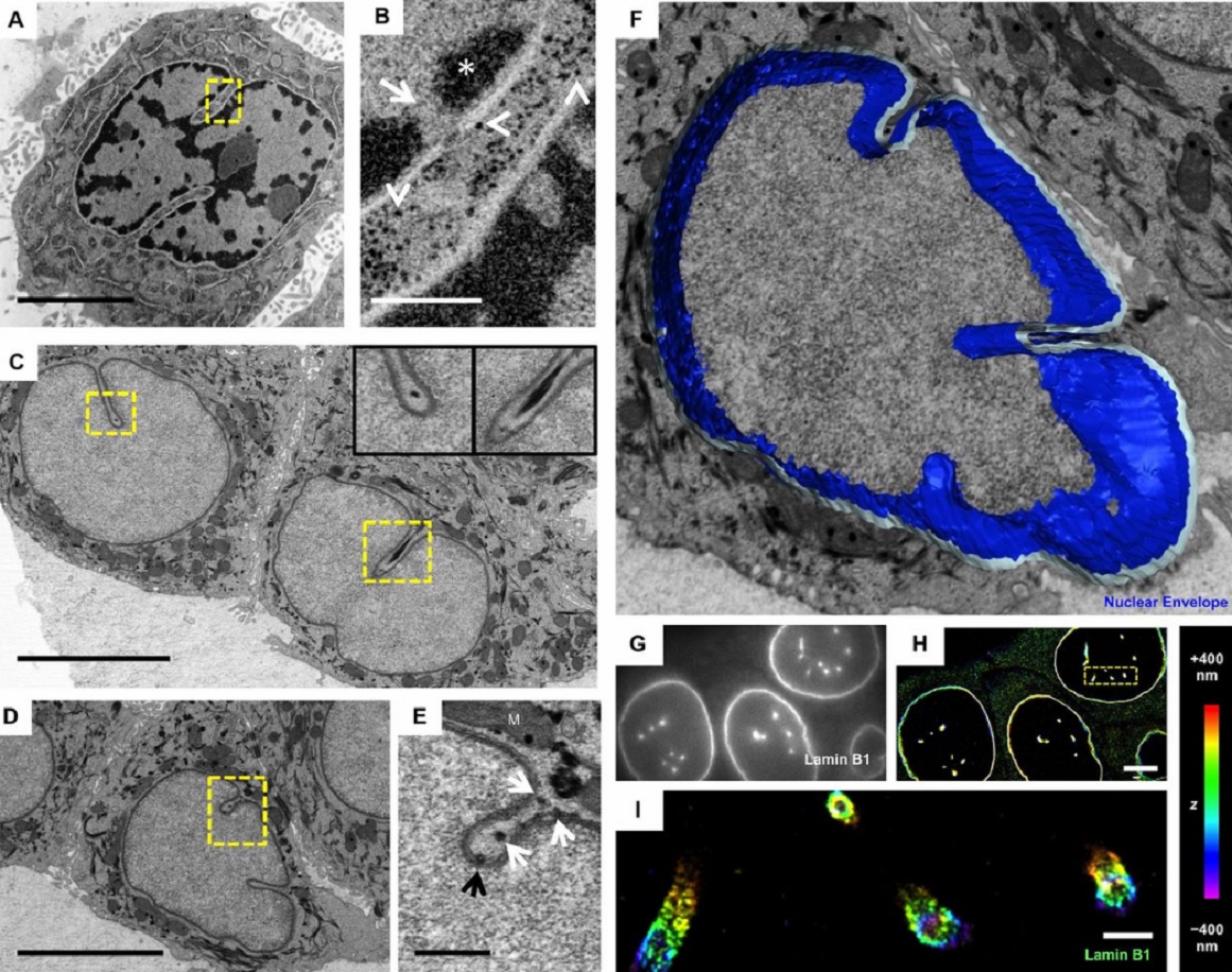
The importance of context in regulation of gene expression is now an accepted principle; yet the mechanism by which the microenvironment communicates with the nucleus and chromatin in healthy tissues is poorly understood. A functional role for nuclear and cytoskeletal architecture is suggested by the phenotypic differences observed between epithelial and mesenchymal cells…
Read more
Danielle M. Jorgens, Jamie L. Inman, Michal Wojcik, Claire Robertson, Hildur Palsdottir, Wen-Ting Tsai, Haina Huang, Alexandre Bruni-Cardoso, Claudia S. López, Mina J. Bissell, Ke Xu, Manfred Auer

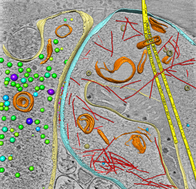
Macropinosomes are key players in early shigella invasion and vacuolar escape in epithelial cells
Intracellular pathogens include all viruses, many bacteria and parasites capable of invading and surviving within host cells. Key to survival is the subversion of host cell pathways by the pathogen for the purpose of propagation and evading the immune system. The intracellular bacterium Shigella flexneri, the causative agent of bacillary dysentery, invades host cells in a vacuole that is subsequently ruptured to allow growth of the pathogen within the host cytoplasm…
Read more
Allon Weiner , Nora Mellouk , Noelia Lopez-Montero , Yuen-Yan Chang, Célia Souque, Christine Schmitt, Jost Enninga
Determining the bacterial cell biology of planctomycetes
Bacteria of the phylum Planctomycetes have been previously reported to possess several features that are typical of eukaryotes, such as cytosolic compartmentalization and endocytosis-like macromolecule uptake. However, recent evidence points towards a Gram-negative cell plan for Planctomycetes, although in-depth experimental analysis has been hampered by insufficient genetic tools…
Read more
Christian Boedeker, Margarete Schüler, Greta Reintjes, Olga Jeske, Muriel C. F. van Teeseling et al.
Three-dimensional imaging of the intracellular assembly of a functional viral RNA replicase complex
Positive-strand RNA viruses, which can be devastating pathogens in humans, animals and plants, replicate their genomes on intracellular membranes. Here, we describe the three-dimensional ultrastructural organization of a tombusvirus replicase in yeast, a valuable model for exploring virus–host interactions…
Read more
Isabel Fernández de Castro, José J. Fernández, Daniel Barajas, Peter D. Nagy, Cristina Risco
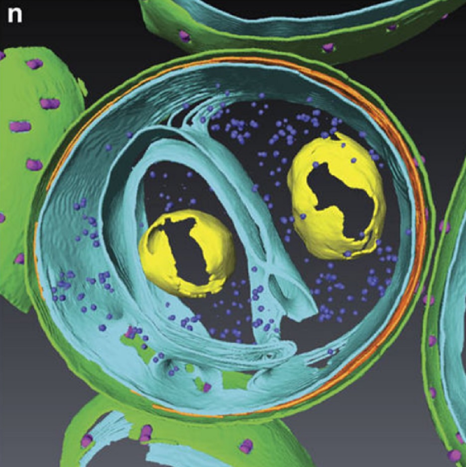
Fast and precise targeting of single tumor cells in vivo by multimodal correlative microscopy
Intravital microscopy provides dynamic understanding of multiple cell biological processes, but its limited resolution has so far precluded structural analysis. Because it is difficult to capture rare and transient events, only a few attempts have been made to observe specific developmental and pathological processes in animal models using electron microscopy. The multimodal correlative approach that we propose here combines intravital microscopy, microscopic X-ray computed tomography and thr... Read more
Matthia A. Karreman, Luc Mercier, Nicole L. Schieber, Gergely Solecki, Guillaume Allio, Frank Winkler, Bernhard Ruthensteiner, Jacky G. Goetz, Yannick Schwab
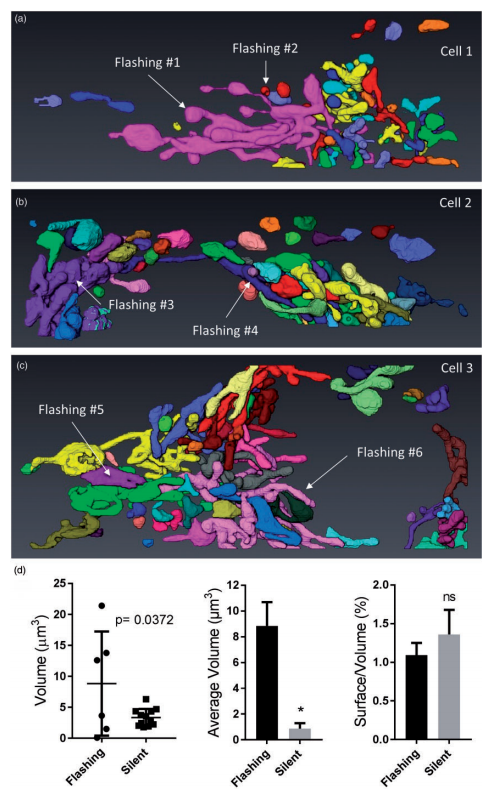
Ultrastructural Characterization of Flashing Mitochondria
Mitochondria undergo spontaneous transient elevations in matrix pH associated with drops in mitochondrial membrane potential. These mitopHlashes require a functional respiratory chain and the profusion protein optic atrophy 1, but their mechanistic basis is unclear. To gain insight on the origin of these dynamic events, we resolved the ultrastructure of flashing mitochondria by correlative light and electron microscopy. HeLa cells expressing the matrix-targeted pH probe mitoSypHer were screen... Read more
Manon Rosselin, Paula Nunes-Hasler, and Nicolas Demaurex
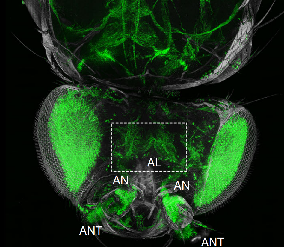
The fruit fly, Drosophila melanogaster, is an important experimental model to address central questions in neuroscience at an organismic level. However, imaging of neural circuits in intact fruit flies is limited due to structural properties of the cuticle. Here we present a novel approach combining tissue clearing, ultramicroscopy, and data analysis that enables the visualisation of neuronal networks with single-cell resolution from the larval stage up to the adult Drosophila. (…) This... Read more
Marko Pende, Klaus Becker, Martina Wanis, Saiedeh Saghafi, Rashmit Kaur, Christian Hahn, Nika Pende, Massih Foroughipour, Thomas Hummel & Hans-Ulrich Dodt
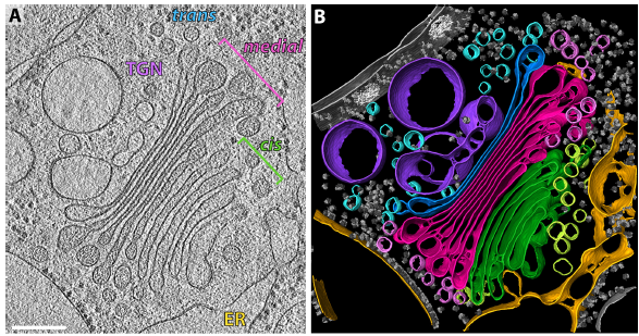
The structure of the COPI coat determined within the cell
COPI-coated vesicles mediate trafficking within the Golgi apparatus and from the Golgi to the endoplasmic reticulum. Here, we applied cryo-focused ion beam milling, cryo-electron tomography and subtomogram averaging to determine the native structure of the COPI coat within vitrified Chlamydomonas reinhardtii cells. The native algal structure resembles the in vitro mammalian structure, but additionally reveals cargo bound beneath beta’–COP. We find that all coat components disassemble... Read more
Yury S Bykov, Miroslava Schaffer, Svetlana O Dodonova, Sahradha Albert, Jurgen M Plitzko, Wolfgang Baumeister, Benjamin D Engel, John AG Briggs
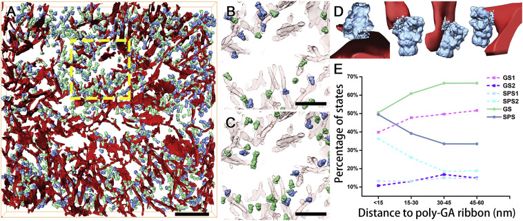
In Situ Structure of Neuronal C9orf72 Poly-GA Aggregates Reveals Proteasome Recruitment
Protein aggregation and dysfunction of the ubiquitin-proteasome system are hallmarks of many neurodegenerative diseases. Here, we address the elusive link between these phenomena by employing cryo-electron tomography to dissect the molecular architecture of protein aggregates within intact neurons at high resolution. We focus on the poly-Gly-Ala (poly-GA) aggregates resulting from aberrant translation of an expanded GGGGCC repeat in C9orf72, the most common genetic cause of amyotrophic latera... Read more
Qiang Guo, Carina Lehmer, Antonio Martinez-Sanchez, Till Rudack, Florian Beck, Hannelore Hartmann, Manuela Perez-Berlanga, Frederic Frottin, Mark S.Hipp, F. Ulrich Hartl, Dieter Edbauer, Wolfgang Baumeister, Ruben Fernandez-Busnadiego
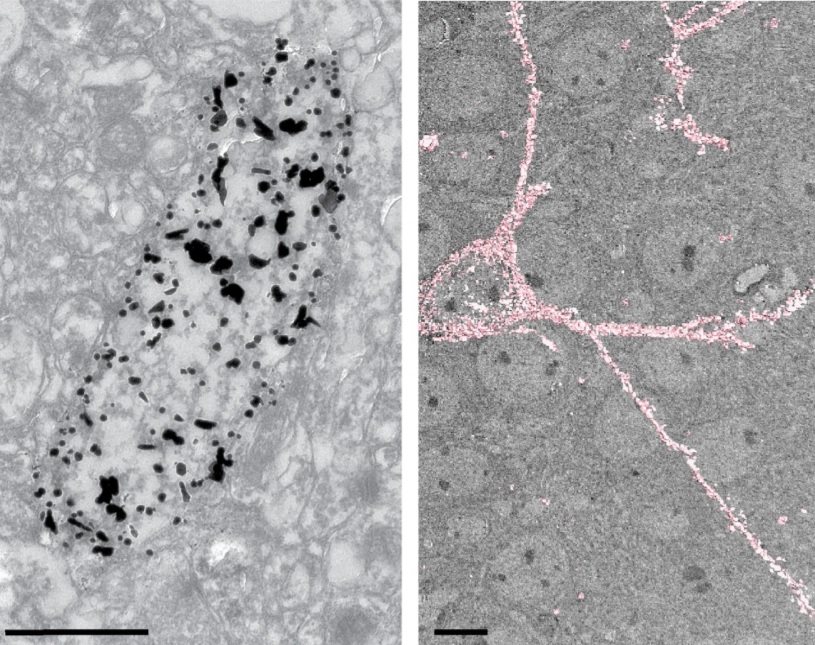
Analysis of neuronal arborization and connections is a powerful tool in fundamental and clinical neuroscience. Changes in neuronal morphology are central to brain development and plasticity and are associated with numerous diseases. Golgi staining is a classical technique based on a deposition of metal precipitate in a random set of neurons. Despite their versatility, Golgi methods have limitations that largely precluded their use in advanced microscopy. We combined Golgi staining with fluore... Read more
Katlijn Vints, Dorien Vandael, Pieter Baatsen, Benjamin Pavie, Frank Vernaillen, Nikky Corthout, Vasily Rybakin, Sebastian Munck & Natalia V. Gounko

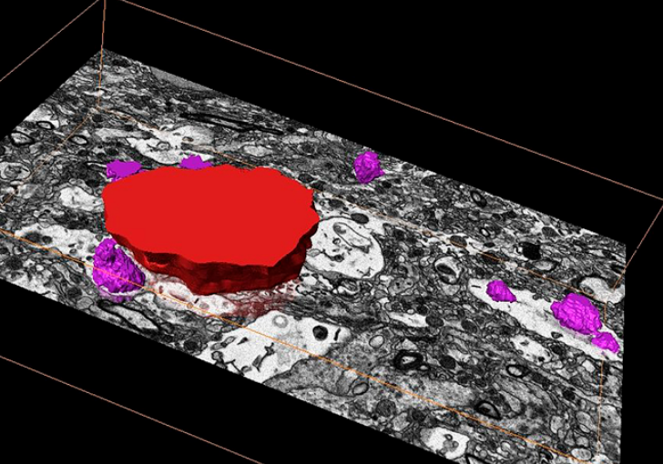
Cell-type specific innervation of cortical pyramidal cells at their apical tufts
We investigated the synaptic innervation of apical tufts of cortical pyramidal cells in a region between layers 1 and 2 using 3-D electron microscopy (3D-EM) applied to four cortical regions in mouse. Across all cortices, we found the relative inhibitory input at the apical dendrite’s main bifurcation to be more than 3-fold stronger for layer 2 pyramidal cells than for all other pyramidal cells. Towards the distal tuft dendrites in upper layer 1, however, the relative inhibitory input was a... Read more
Ali Karimi, Jan Odenthal, Florian Drawitsch, Kevin M. Boergens, Moritz Helmstaedter