Welcome to the Amira-Avizo Software Use Case Gallery
Below you will find a collection of use cases of our 3D data visualization and analysis software. These use cases include scientific publications, articles, papers, posters, presentations or even videos that show how Amira-Avizo Software is used to address various scientific and industrial research topics.
Use the Domain selector to filter by main application area, and use the Search box to enter keywords related to specific topics you are interested in.
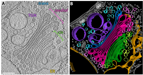
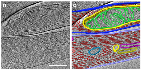
The structure of the COPI coat determined within the cell
COPI-coated vesicles mediate trafficking within the Golgi apparatus and from the Golgi to the endoplasmic reticulum. Here, we applied cryo-focused ion beam milling, cryo-electron tomography and subtomogram averaging to determine the native structure of the COPI coat within vitrified Chlamydomonas reinhardtii cells. The native algal structure resembles the in vitro mammalian structure, but additionally reveals cargo bound beneath beta’–COP. We find that all coat components disassemble... Read more
Yury S Bykov, Miroslava Schaffer, Svetlana O Dodonova, Sahradha Albert, Jurgen M Plitzko, Wolfgang Baumeister, Benjamin D Engel, John AG Briggs
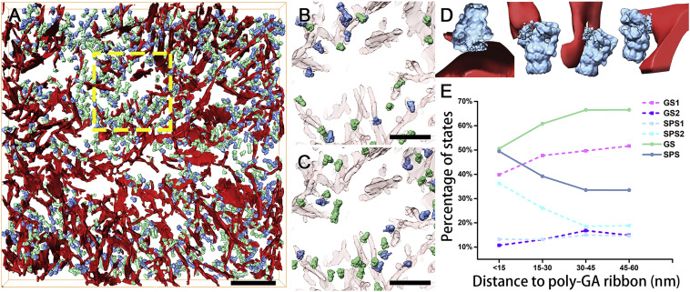
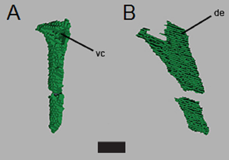
In Situ Structure of Neuronal C9orf72 Poly-GA Aggregates Reveals Proteasome Recruitment
Protein aggregation and dysfunction of the ubiquitin-proteasome system are hallmarks of many neurodegenerative diseases. Here, we address the elusive link between these phenomena by employing cryo-electron tomography to dissect the molecular architecture of protein aggregates within intact neurons at high resolution. We focus on the poly-Gly-Ala (poly-GA) aggregates resulting from aberrant translation of an expanded GGGGCC repeat in C9orf72, the most common genetic cause of amyotrophic latera... Read more
Qiang Guo, Carina Lehmer, Antonio Martinez-Sanchez, Till Rudack, Florian Beck, Hannelore Hartmann, Manuela Perez-Berlanga, Frederic Frottin, Mark S.Hipp, F. Ulrich Hartl, Dieter Edbauer, Wolfgang Baumeister, Ruben Fernandez-Busnadiego
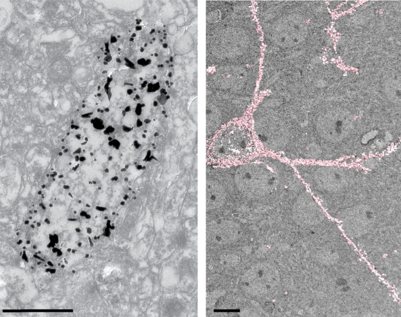
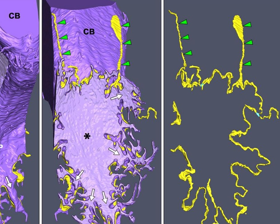
Analysis of neuronal arborization and connections is a powerful tool in fundamental and clinical neuroscience. Changes in neuronal morphology are central to brain development and plasticity and are associated with numerous diseases. Golgi staining is a classical technique based on a deposition of metal precipitate in a random set of neurons. Despite their versatility, Golgi methods have limitations that largely precluded their use in advanced microscopy. We combined Golgi staining with fluore... Read more
Katlijn Vints, Dorien Vandael, Pieter Baatsen, Benjamin Pavie, Frank Vernaillen, Nikky Corthout, Vasily Rybakin, Sebastian Munck & Natalia V. Gounko

Cell-type specific innervation of cortical pyramidal cells at their apical tufts
We investigated the synaptic innervation of apical tufts of cortical pyramidal cells in a region between layers 1 and 2 using 3-D electron microscopy (3D-EM) applied to four cortical regions in mouse. Across all cortices, we found the relative inhibitory input at the apical dendrite’s main bifurcation to be more than 3-fold stronger for layer 2 pyramidal cells than for all other pyramidal cells. Towards the distal tuft dendrites in upper layer 1, however, the relative inhibitory input was a... Read more
Ali Karimi, Jan Odenthal, Florian Drawitsch, Kevin M. Boergens, Moritz Helmstaedter
The major mammalian bloodstream form of the African sleeping sickness parasite Trypanosoma bruceimultiplies rapidly, and it is important to understand how these cells divide. Organelle inheritance involves complex spatiotemporal re-arrangements to ensure correct distribution to daughter cells…
Read more
Louise Hughes, Samantha Borrett, Katie Towers, Tobias Starborg, Sue Vaughan
Morphological process of podocyte development revealed by block-face scanning electron microscopy
Podocytes present a unique 3D architecture specialized for glomerular filtration. However, several 3D morphological aspects on podocyte development remain partially understood because they are difficult to reveal using conventional scanning electron microscopy (SEM). Here, we adopted serial block-face SEM imaging…
Read more
Koichiro Ichimura, Soichiro Kakuta, Yuto Kawasaki, Takayuki Miyaki, Takahiro Nonami, Naoyuki Miyazaki, Tomoyo Nakao, Sakiko Enomoto, Shigeo Arai, Masato Koike, Kazuyoshi Murata, Tatsuo Sakai
In biomedical research, a huge variety of different techniques is currently available for the structural examination of small specimens, including conventional light microscopy (LM), transmission electron microscopy (TEM), confocal laser scanning microscopy (CLSM), microscopic X-ray computed tomography (microCT), and many others. Since every imaging method is physically limited by certain parameters, a correlative use of complementary methods often yields a significant broader range of inform... Read more
Stephan Handschuh, Natalie Baeumler, Thomas Schwaha and Bernhard Ruthensteiner
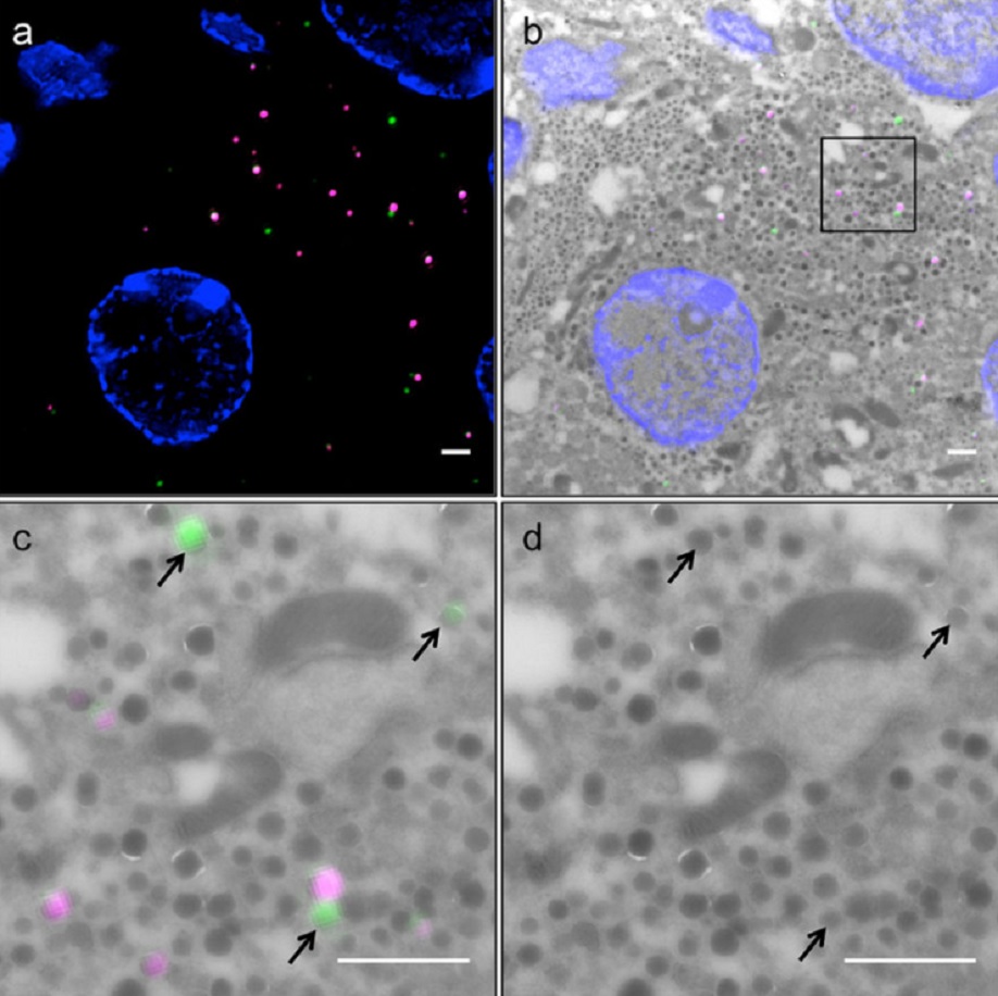
Correlative light and electron microscopy (CLEM) is a powerful approach to investigate the molecular ultrastructure of labeled cell compartments. However, quantitative CLEM studies are rare, mainly due to small sample sizes and the sensitivity of fluorescent proteins to strong fixatives and contrasting reagents for EM. Here, we show that fusion of…
Read more
Andreas Müller, Martin Neukam, Anna Ivanova, Anke Sönmez, Carla Münster, Susanne Kretschmar, Yannis Kalaidzidis, Thomas Kurth, Jean-Marc Verbavatz & Michele Solimena
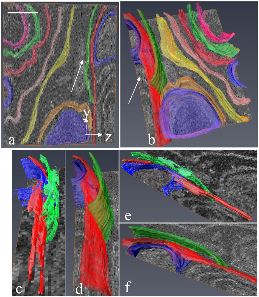
Serial block-face electron microscopy (SBEM) provides nanoscale 3D ultrastructure of embedded and stained cells and tissues in volumes of up to 107 µm3. In SBEM, electrons with 1–3 keV energies are incident on a specimen block, from which backscattered electron (BSE) images are collected with x, y resolution of 5–10 nm in the block-face plane, and successive layers are removed by an in situ ultramicrotome. Sp... Read more
Q. He, M. Hsueh, G. Zhang, D. C. Joy & R. D. Leapman
Correlative cryo-electron microscopy reveals the structure of TNTs in neuronal cells
The orchestration of intercellular communication is essential for multicellular organisms. One mechanism by which cells communicate is through long, actin-rich membranous protrusions called tunneling nanotubes (TNTs), which allow the intercellular transport of various cargoes, between the cytoplasm of distant cells in vitro and in vivo. Here, we use correlative FIB-SEM, light- and cryo-electron microscopy approaches to elucidate the structural organization of neuronal TNTs. Our data indicate ... Read more
Anna Sartori-Rupp, Diégo Cordero Cervantes, Anna Pepe, Karine Gousset, Elise Delage, Simon Corroyer-Dulmont, Christine Schmitt, Jacomina Krijnse-Locker & Chiara Zurzolo
Defect detection in 3D printed carbon fibre composites using X-ray Computed Tomography
X-ray Computed Tomography (X-ray CT) has become a vital tool for product quality inspection. The X-ray CT analysis of 3D printed composites, with a layer-by-layer structure of carbon fibre/polyamide and polyamide plies, demonstrates how the void content increases with an increasing number of consecutive carbon fibre layers. Not only the void content, but also the pore
network complexity increases, as more pore types are introduced into the sample. The PolyAmide (PA) matrix has an averag... Read more
Jeroen Soete1, Brice Badoux, Yentl Swolfs, Larissa Gorbatikh, Martine Wevers
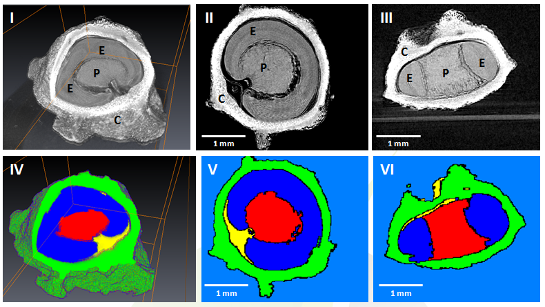
Juvenile Ovine Ex Vivo Larynges: Phonatory, Histologic, and Micro CT Based Anatomic Analyses
It is well known that the phonatory process changes during the life span. However, detailed investigations on potential factors concerned are rare. To deal with this issue, we performed extended biomechanical, macro anatomical, and histological analyses of the contributing laryngeal structures in ex vivo juvenile sheep models. Altogether twelve juvenile sheep larynges were analyzed within the phonatory experiments. Three different elongation levels and 16 different flow levels were applied to... Read more
Michael Döllinger, Olaf Wendler, Claus Gerstenberger, Tanja Grossmann, Marion Semmler, Hossein Sadeghi, and Markus Gugatschka
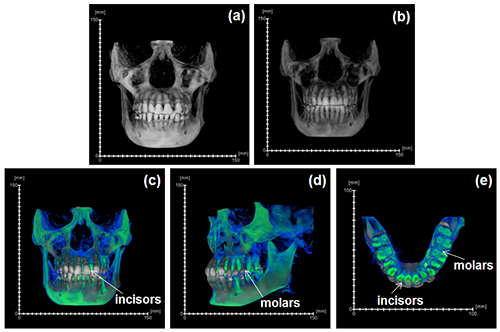
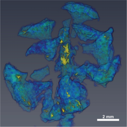
During the acts of biting and chewing, the muscles of the jaw (consisting of the masseter, temporalis, and medial pterygoid in the elevator group, and lateral pterygoid as the main depressor) generate forces that dictate jaw kinematics . The movement of jaws hinges about the temporomandibular joint and are brought together by the muscles attached to respective bones through bone-tendon interfaces known as entheses . Thus, chewing forces affect aspects of craniofacial structure as well as bo... Read more
Kyle H.-Y. Chan, fourth-year undergraduate student in Molecular and Cell Biology, and Public Health at UC Berkeley, FeiFei Yang, Ph.D., postdoctoral scholar in the Laboratory of Multiscale Biomechanics and Biomineralization, School of Dentistry, UCSF, and Sunita P. Ho, Ph.D., Division of Biomaterials and Bioengineering, Department of Preventive and Restorative Dental Sciences, School of Dentistry, University of California San Francisco

Electron Beam Melting (EBM) is an Additive Manufacturing (AM) process which can be used to fabricate complex 3D metal components. Ti-6Al-4V is one of the high value aerospace materials that has been widely used in the EBM process. However, defects such as gas pores and lack of fusion are often found in the as-built components, which can make a significant difference to the fatigue resistance of the parts. The Manufacturing Technology Centre (MTC) uses X-ray Computed Tomography (XCT) and 3D im... Read more
Dr Shuai Hou, Mélanie Bombardiere, Nathanael Turner, Jonathan Settle, Emmanuel Muzangaza, Dr David Brackett
GEVES uses Avizo software to study and control plant seeds
At GEVES, Microtomography (micro-CT), which is a technique using X-rays to investigate internal anatomy and morphology of organisms without destruction, is applied to plant seed quality control and phenotyping. Several applications have been developed using micro-CT coupled with Avizo software development, such as for example in the Measurements of internal and external sugar beet seed structures for seed phenotyping
Read more
Ghassen Trigui, Laurence Le Corre, Dominique Honoré and Karima Boudehri-Giresse - 2D/3D Imaging - R&D team, GEVES
Computed tomography is an increasingly popular technique for the non-destructive study of fossils. Whilst the science of X-ray computed tomography (CT) has greatly matured since its first fossil applications in the early 1980s, the applications and limitations of neutron tomography (NT) remain relatively unexplored in palaeontology. These highest resolution neutron tomographic scans in palaeontology to date were conducted on a specimen of Austrosequoia novae-zeelandiae (Ettingshausen) Mays an... Read more
Chris Mays, Joseph J. Bevitt, and Jeffrey D. Stilwell
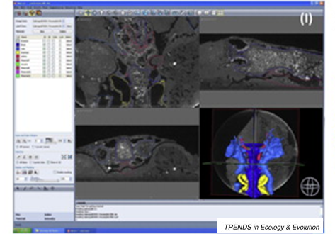
A virtual world of paleontology
Computer-aided visualization and analysis has revolutionized the study of fossils. Fossils can now be characterized in three dimensions and in unprecedented detail. The resulting digital reconstructions can be used in rigorous functional analyses. Hypotheses regarding the function of extinct organisms can therefore be tested.
Read more
Trends in Ecology & Evolution
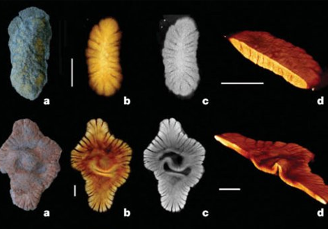
2 BILLION years old fossils appear to represent a first experiment in megascopic multicellularity
The Paleoproterozoic Era witnessed crucial steps in the evolution of Earth’s surface environments following the first appreciable rise of free atmospheric oxygen concentrations ∼2.3 to 2.1 Ga ago, and concomitant shallow ocean oxygenation. Combined microtomography, geochemistry, and sedimentary analysis suggest a biota fossilized during early diagenesis. The emergence of this biota follows a rise in atmospheric oxygen, which is consistent with the idea that surface oxygenation allowe... Read more
Abderrazak El Albani, Laboratoire HYDRASA, UMR 6269 CNRS-INSU, Université de Poitiers, France
This paper used the X-ray three-dimensional (3D) microscope and acquired, through CT scanning, the 3D data of the long-frame coal sample from the Daliuta Coal Mine.
Then, the 3D datacube reconstructed from the coal’s CT scanning data was visualized with the use of Avizo, an advanced visualization software. By means of a gray-scale segmentation technique, the model of the coal’s micro-pore structure was extracted from the object region, and the precise characterization was then cond... Read more
Gang Zhou, Qi Zhang, Ruonan Baiand, Guanhua Ni
Aetosauria is a clade of heavily armored, quadrupedal omnivorous to herbivorous archosaurs known from the Late Triassic across what was the supercontinent of Pangea. Their abundance in many deposits relative to the paucity of other Triassic herbivores indicates that they were key components of Late Triassic ecosystems. However, their evolutionary relationships remain contentious due, in large part, to their extensive dermal armor, which often obstructs observation of internal skeletal anatomy... Read more
Devin K. Hoffman, Andrew B. Heckert, Lindsay E. Zanno
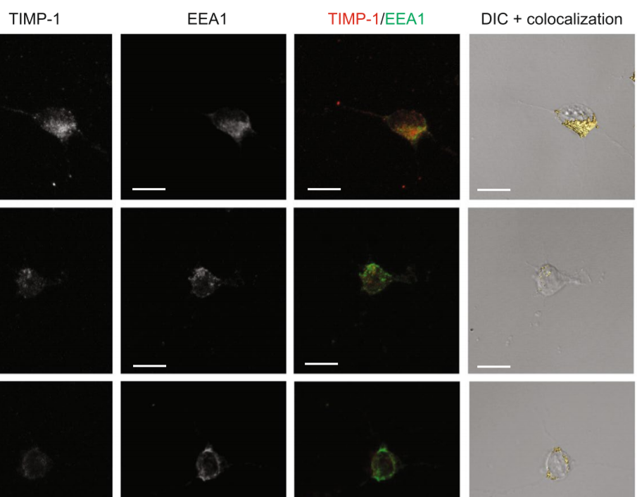
The tissue inhibitor of metalloproteinases-1 (TIMP-1) exerts inhibitory activity against matrix metalloproteinases and cytokine-like effects. We previously showed that TIMP-1 reduces neurite outgrowth in mouse cortical neurons and that this cytokine-like effect depends on TIMP-1 endocytosis mediated by the low-density lipoprotein receptor-related protein-1 (LRP-1). To gain insight into the interaction between TIMP-1 and LRP-1, we considered conformational changes that occur when a ligand bind... Read more
Laurie Verzeaux, Nicolas Belloy, Jessica Thevenard-Devy, Jérôme Devy, Géraldine Ferracci, Laurent Martiny, Stéphane Dedieu, Manuel Dauchez, Hervé Emonard, Nicolas Etique & Emmanuelle Devarenne-Charpentier