Welcome to the Amira-Avizo Software Use Case Gallery
Below you will find a collection of use cases of our 3D data visualization and analysis software. These use cases include scientific publications, articles, papers, posters, presentations or even videos that show how Amira-Avizo Software is used to address various scientific and industrial research topics.
Use the Domain selector to filter by main application area, and use the Search box to enter keywords related to specific topics you are interested in.
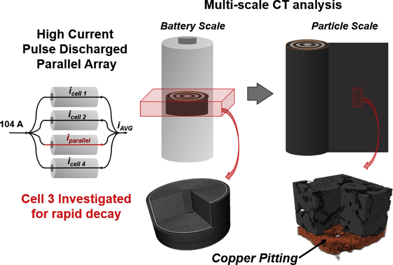
X-ray computed tomography (X-ray CT) across multiple length scales is utilized for the first time to investigate the physical abuse of high C-rate pulsed discharge on cells wired individually and in parallel.. Manufactured lithium iron phosphate cells boasting high rate capability were pulse power tested in both wiring conditions with high discharge currents of 10C for a high number of cycles (up to 1200) until end of life (<80% of initial discharge capacity retained). The parallel ass... Read more
Rachel Carter, Brett Huhman, Corey T. Love, Iryna V. Zenyuk
High performance anode with dendritic porous structure for low temperature solid oxide fuel cells
A dendritic porous supported microstructure simultaneously creates small pore size and broad gas diffusion pathways in a solid oxide fuel cell anode membrane. This microstructure also achieves pore sizes that reduce with increasing depth within the membrane without increasing the structure tortuosity. Such a microstructure supplies high triple phase boundary density, fast gas diffusion and low polarization resistance. Here we characterise the performance of a porous anode with such a dendriti... Read more
Xin Shao, William D.A. Rickard, Dehua Dong, Huu Dang , Martin Saunders, Aaron Dodd, Gordon Parkinson, Chun-Zhu Li
Growing popularity and rapid development of Solid Oxide Fuel Cells (SOFCs) stem for their potential to become a gamechanger in the field of clean power generation technologies.
In this paper, a transient microstructure-oriented numerical simulation of a planar Direct Internal Reforming Solid Oxide Fuel Cell (DIR-SOFC) is delivered. The performance criteria in a direct steam reforming for a fuel starvation scenario are analyzed in order to optimize the underlying process. The proposed t... Read more
Maciej Chalusiak, Michal Wrobel, Marcin Mozdzierz, Katarzyna Berent, Janusz S. Szmyd, Shinji Kimijima, Grzegorz Brus
Mesoscale characterization of local property distributions in heterogeneous electrodes
The performance of electrochemical devices depends on the three-dimensional (3D) distributions of microstructural features in their electrodes. Several mature methods exist to characterize 3D microstructures over the microscale (tens of microns), which are useful in understanding homogeneous electrodes. However, methods that capture mesoscale (hundreds of microns) volumes at appropriate resolution (tens of nm) are lacking, though they are needed to understand more common, less ideal electrode... Read more
Tim Hsu, William K. Epting, Rubayyat Mahbub, Noel T. Nuhfer, Sudip Bhattachary, Yinkai Lei, Herbert M. Miller, Paul R. Ohodnicki, Kirk R. Gerdes, Harry W. Abernathy, Gregory A. Hackett, Anthony D. Rollett, Marc De Graef, Shawn Litster, Paul A. Salvador
Laser processing of metal surfaces by ultrafast Read more
Edwin Peng, Alexander Roth, Craig A. Zuhlke, Soodabeh Azadehranjbar, Dennis R. Alexander, George Gogos, Jeffrey E. Shield

Alternative battery technologies are required to meet growing energy demands and address the limitations of present technologies. As such, it is necessary to look beyond lithium-ion batteries. Zinc batteries enable high power density while being sourced from ubiquitous and cost-effective materials. This paper presents, for the first time known to the authors, multi-length scale tomography studies of failure mechanisms in zinc batteries with and without commercial microporous separators. In bo... Read more
Vladimir Yufit, Farid Tariq David S. Eastwood Moshiel Biton Billy Wu Peter D. Lee Nigel P. Brandon
Microstructural analysis of TRISO particles using multi-scale X-ray computed tomography
TRISO particles, a composite nuclear fuel built up by ceramic and graphitic layers, have outstanding high temperature resistance. TRISO fuel is the key technology for High Temperature Reactors (HTRs) and the Generation IV Very High Temperature Reactor (VHTR) variant.
TRISO offers unparalleled containment of fission products and is extremely robust during accident conditions. An understanding of the thermal performance and mechanical properties of TRISO fuel requires a detailed knowledg... Read more
T. Lowe, R.S. Bradley, S. Yue, K. Barii, J. Gelb, N. Rohbeck, J. Turner, P.J. Withers
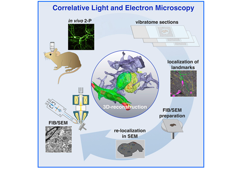
Label-free 3D-CLEM using endogenous tissue landmarks
We demonstrate feasibility of the workflow by combining in vivo 2-photon microscopy and focused ion beam scanning electron microscopy (FIB/SEM) to dissect the role of astrocytic coverage in the persistence of dendritic spines.
Emerging 3D correlative light and electron microscopy (CLEM) approaches enable studying neuronal structure-function relations at unprecedented depth and precision. However, established protocols for the correlation of light and electron micrographs rely ... Read more
Manja Luckner,Steffen Burgold, Severin Filser, Maximilian Scheungrab, Yilmaz Niyaz, Eric Hummel, Gerhard Wanner, Jochen Herms
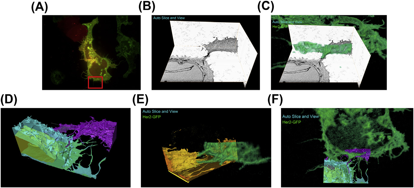
A fully integrated, three-dimensional fluorescence to electron microscopy correlative workflow
While fluorescence microscopy provides tools for highly specific labeling and sensitive detection, its resolution limit and lack of general contrast has hindered studies of cellular structure and protein localization. Recent advances in correlative light and electron microscopy (CLEM), including the fully integrated CLEM workflow instrument, the Thermo Scientific CorrSight with MAPS, have allowed for a more reliable, reproducible, and quicker approach to correlate three-dimensional time-lapse... Read more
Claudia S. Lopez, Cedric Bouchet-Marquis, Christopher P. Arthur, Jessica L. Riesterer, Gregor Heiss, Guillaume Thibault, Lee Pullan, Sunjong Kwon, Joe W. Gray
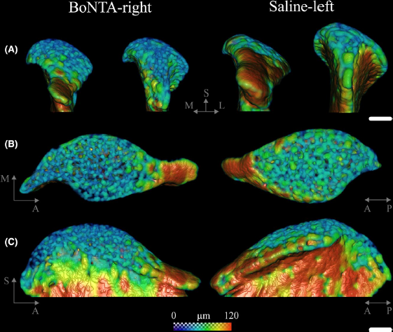
Masseter muscle function influences mandibular bone homeostasis. As previously reported, bone resorption markers increased in the mouse mandibular condyle two days after masseter paralysis induced with botulinum toxin type A (BoNTA), followed by local bone loss.
This study aimed to evaluate the bone quality of both the mandibular condyle and alveolar process in the mandible of adult mice during the early stage of a BoNTA‐induced masseter muscle atrophy, using a combined 3D histomorpho... Read more
Julián Balanta‐Melo, María Angélica Torres‐Quintana, Maximilian Bemmann, Carolina Vega, Constanza González, Kornelius Kupczik, Viviana Toro‐Ibacache, Sonja Buvinic

Synergistic role of nucleotides and lipids for the self-assembly of Shs1 septin oligomers
Amira capacities for membranes and filaments segmentation in cryo-TEM images are featured on the front cover of Biochemical Journal, July 2020.
Budding yeast septins are essential for cell division and polarity. (…) [The authors] have dissected, here, for the first time, the behavior of the Shs1 protomer bound to membranes at nanometer resolution, in complex with the other septins. Using electron microscopy, [the authors] have shown that on membranes, Shs1 protomers self-assembl... Read more
Cyntia Taveneau, Rémi Blanc, Gerard Pehau-Arnaudet, Aurélie Cicco, Aurélie Bertin
Microscopic organisms that penetrate calcareous structures by actively dissolving the carbonate matrix, namely microendoliths, have an important influence on the breakdown of marine carbonates.
Microscopic organisms that penetrate calcareous structures by actively dissolving the carbonate matrix, namely microendoliths, have an important influence on the breakdown of marine carbonates. The study of these microorganisms and the bioerosion traces they produce is crucial for understanding ... Read more
Philipp-Konrad Schätzle, Max Wisshak, Andreas Bick, André Freiwald, Alexander Kieneke
Ovipositor of the braconid wasp Habrobracon hebetor: structural and functional aspects
The Braconidae are a megadiverse and ecologically highly important group of insects. The vast majority of braconid wasps are parasitoids of other insects, usually attacking the egg or larval stages of their hosts. The ovipositor plays a crucial role in the assessment of the potential host and precise egg laying. We used lightand electron-microscopic techniques to investigate all inherent cuticular elements of the ovipositor (the female 9th abdominal tergum, two pairs of valvifers, and three p... Read more
Michael Csader, Karin Mayer, Oliver Betz, Stefan Fischer, Benjamin Eggs
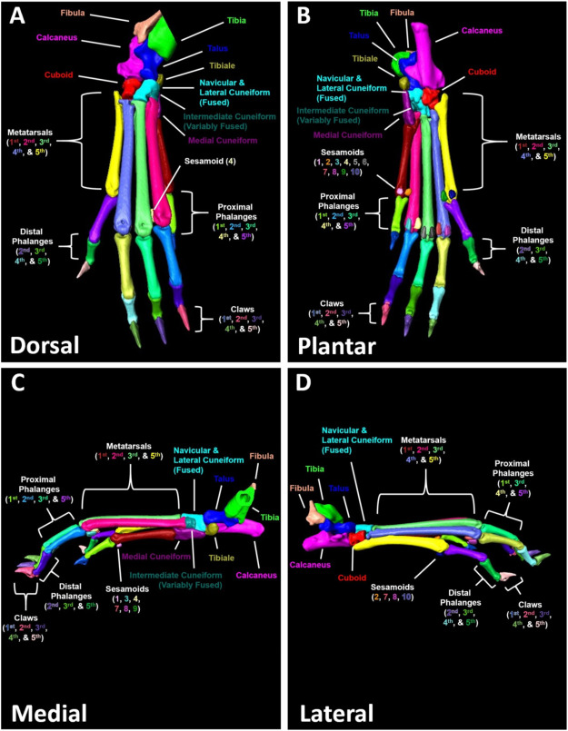
A high-throughput semi-automated bone segmentation workflow for murine hindpaw Micro-CT datasets
Micro-computed tomography (μCT) is a valuable imaging modality for longitudinal quantification of bone volumes to identify disease or treatment effects for a broad range of conditions that affect bone health. Complex structures, such as the hindpaw with up to 31 distinct bones in mice, have considerable analytic potential, but quantification is often limited to a single bone volume metric due to the intensive effort of manual segmentation. Herein, we introduce a high-throughput, user-friendl... Read more
H. Mark Kenney, Yue Peng, Kiana L.Chen, Raquel Ajalik, Lindsay Schnur, Ronald W.Wood, Edward M.Schwarz, Hani A. Awad

Protocols for Generating Surfaces and Measuring 3D Organelle Morphology Using Amira
High-resolution 3D images of organelles are of paramount importance in cellular biology. Although light microscopy and transmission electron microscopy (TEM) have provided the standard for imaging cellular structures, they cannot provide 3D images.
However, recent technological advances such as serial block-face scanning electron microscopy (SBF-SEM) and focused ion beam scanning electron microscopy (FIB-SEM) provide the tools to create 3D images for the ultrastructural analysis of org... Read more
Edgar Garza-Lopez, Zer Vue, Prasanna Katti, Kit Neikirk, Michelle Biete, Jacob Lam, Heather K. Beasley, Andrea G. Marshall, Taylor A. Rodman, Trace A. Christensen, Jeffrey L. Salisbury, Larry Vang, Margaret Mungai, Salma Ash Shareef, Sandra A. Murray, Jianqiang Shao, Jennifer Streeter, Brian Glancy, Renata O. Pereira1, E. Dale Abel, and Antentor Hinton, Jr.
Automatic whole cell organelle segmentation in volumetric electron microscopy
Cells contain hundreds of different organelle and macromolecular assemblies intricately organized relative to each other to meet any cellular demands. Obtaining a complete understanding of their organization is challenging and requires nanometer-level, three-dimensional reconstruction of whole cells. Even then, the immense size of datasets and large number of structures to be characterized requires generalizable, automatic methods.
To meet this challenge, we developed an analy... Read more
Larissa Heinrich, Davis Bennett, David Ackerman, Woohyun Park, John Bogovic, View ORCID ProfileNils Eckstein, Alyson Petruncio, Jody Clements, C. Shan Xu, Jan Funke, Wyatt Korff, Harald F. Hess, Jennifer Lippincott-Schwartz, Stephan Saalfeld, Aubrey V. Weigel, COSEM Project Team
Precise methods for quantifying drug accumulation in brain tissue are currently very limited, challenging the development of new therapeutics for brain disorders. Transcardial perfusion is instrumental for removing the intravascular fraction of an injected compound, thereby allowing for ex vivo assessment of extravasation into the brain. However, pathological remodeling of tissue microenvironment can affect the efficiency of transcardial perfusion, which has been largely overlooked.
We... Read more
Serhii Kostrikov, Kasper B. Johnsen, Thomas H. Braunstein, Johann M. Gudbergsson, Frederikke P. Fliedner, Elisabeth A. A. Obara, Petra Hamerlik, Anders E. Hansen, Andreas Kjaer, Casper Hempel & Thomas L. Andresen
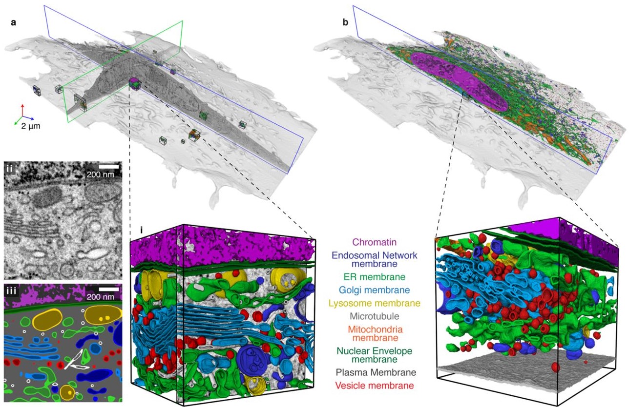
Integrative Imaging Reveals SARS-CoV-2-Induced Reshaping of Subcellular Morphologies
Cortese et al. use integrative imaging techniques to generate a publicly available repository of morphological alterations induced by SARS-CoV-2 in lung cells. Accumulation of ER-derived double-membrane vesicles, the viral replication organelle, occurs concomitantly with cytoskeleton remodeling and Golgi fragmentation. Pharmacological alteration of cytoskeleton dynamics restricts viral replication and spread.
Pathogenesis induced by SARS-CoV-2 is thought to result from both an inflamma... Read more
Mirko Cortese, Ji-Young Lee, Berati Cerikan, ..., Laurent Chatel-Chaix, Yannick Schwab, Ralf Bartenschlager
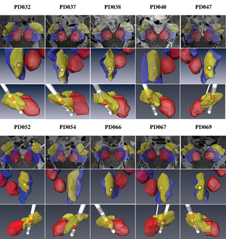
Patient-specific anatomical model for deep brain stimulation based on 7 Tesla MRI
Deep brain stimulation (DBS) requires accurate localization of the anatomical target structure, and the precise placement of the DBS electrode within it. Ultra-high field 7 Tesla (T) MR images can be utilized to create patient-specific anatomical 3D models of the subthalamic nuclei (STN) to enhance pre-surgical DBS targeting as well as post-surgical visualization of the DBS lead position and orientation. We validated the accuracy of the 7T imaging-based patient-specific model of the STN and m... Read more
Yuval Duchin, Reuben R. Shamir, Remi Patriat, Jinyoung Kim, Jerrold L. Vitek, Guillermo Sapiro, Noam Harel
Influenza A matrix protein M1 is sufficient to induce lipid membrane deformation
The matrix protein M1 of the Influenza A virus is considered to mediate viral assembly and budding at the plasma membrane (PM) of infected cells. In order for a new viral particle to form, the PM lipid bilayer has to bend into a vesicle towards the extracellular side. Studies in cellular models have proposed that different viral proteins might be responsible for inducing membrane curvature in this context (including M1), but a clear consensus has not been reached. In this study, we use a comb... Read more
Ismail Dahmani, Kai Ludwig, Salvatore Chiantia
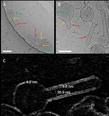
Membrane architecture of pulmonary lamellar bodies revealed by post-correlation on-lamella cryo-CLEM
Lamellar bodies (LBs) are surfactant rich organelles in alveolar type 2 cells. LBs disassemble into a lipid-protein network that reduces surface tension and facilitates gas exchange at the air-water interface in the alveolar cavity. Current knowledge of LB architecture is predominantly based on electron microscopy studies using disruptive sample preparation methods. We established a post-correlation on-lamella cryo-correlative light and electron microscopy approach for cryo-FIB milled lung ce... Read more
Steffen Klein, Benedikt H. Wimmer, Sophie L. Winter, Androniki Kolovou, Vibor Laketa, Petr Chlanda