Welcome to the Amira-Avizo Software Use Case Gallery
Below you will find a collection of use cases of our 3D data visualization and analysis software. These use cases include scientific publications, articles, papers, posters, presentations or even videos that show how Amira-Avizo Software is used to address various scientific and industrial research topics.
Use the Domain selector to filter by main application area, and use the Search box to enter keywords related to specific topics you are interested in.
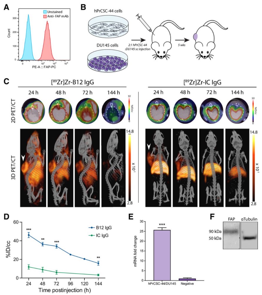
Malignant cells are surrounded by a complex and supportive tumor microenvironment that consists of immune cells, extracellular matrix, vasculature, and fibroblasts. Cancer-associated fibroblasts (CAF) are the major cell type in the reactive stroma and are known to promote tumorigenesis and metastasis. Fibroblast activation protein alpha (FAP) is a transmembrane serine protease expressed by CAFs in the microenvironment of epithelial tumors.
Meta-analysis of FAP expression and clinical ... Read more
Hallie M. Hintz, Joseph P. Gallant, Donald J. Vander Griend, Ilsa M. Coleman, Peter S. Nelson and Aaron M. LeBeau
Precise methods for quantifying drug accumulation in brain tissue are currently very limited, challenging the development of new therapeutics for brain disorders. Transcardial perfusion is instrumental for removing the intravascular fraction of an injected compound, thereby allowing for ex vivo assessment of extravasation into the brain. However, pathological remodeling of tissue microenvironment can affect the efficiency of transcardial perfusion, which has been largely overlooked.
We... Read more
Serhii Kostrikov, Kasper B. Johnsen, Thomas H. Braunstein, Johann M. Gudbergsson, Frederikke P. Fliedner, Elisabeth A. A. Obara, Petra Hamerlik, Anders E. Hansen, Andreas Kjaer, Casper Hempel & Thomas L. Andresen
Adipose models have been applied to mechanistic studies of metabolic diseases (such as diabetes) and the subsequent discovery of new therapeutics. However, typical models are either insufficiently complex (2D cell cultures) or expensive and labor intensive (mice/in vivo). To bridge the gap between these models and in order to better inform pre-clinical studies we have developed a drug-responsive 3D model of white adipose tissue (WAT).
Here, spheroids (680 ± 60 μm) comp... Read more
Alexander D Graham, Rajesh Pandey, Viktoriya S Tsancheva, Alessia Candeo, Stanley W Botchway, Alasdair J Allan, Lydia Teboul4, Kamel Madi, Tahkur S Babra, Louisa A K Zolkiewski, Xuan Xue, Liz Bentley, Joan Gannon, Sam N Olof and Roger D Cox
Optical Resolution Photoacoustic Microscopy of Ovary and Fallopian Tube
Ovarian cancer is the leading cause of death among gynecological cancers, but is poorly amenable to preoperative diagnosis. In this study, we investigate the feasibility of “optical biopsy,” using high-optical-resolution photoacoustic microscopy (OR-PAM) to quantify the microvasculature of ovarian and fallopian tube tissue. The technique is demonstrated using excised human ovary and fallopian tube specimens imaged immediately after surgery.
This report describes the first applicatio... Read more
Bin Rao, Xiandong Leng, Yifeng Zeng, Yixiao Lin, Ruimin Chen, Qifa Zhou, Andrea R. Hagemann, Lindsay M. Kuroki, Carolyn K. McCourt, David G. Mutch, Matthew A. Powell, Ian S. Hagemann & Quing Zhu
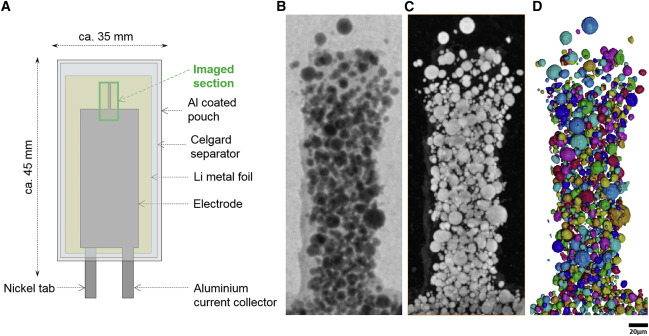
Nickel-rich transition metal oxide materials […] are of great interest for achieving immediate improvements in the energy density of Li-ion batteries and for risk reduction within the Li-ion battery supply chain.
[…] An increase in Ni content in NMC materials leads to accelerated degradation […]. This potentially complicates their adoption in applications requiring extended cycle life such as in electric vehicles.
Recent developments in X-ray characterization to... Read more
ChunTan, Andrew S.Leach, Thomas M.M. Heenan, Huw Parks, Rhodri Jervis, Johanna Nelson Weker, Daniel J.L. Brett, Paul R.Shearing
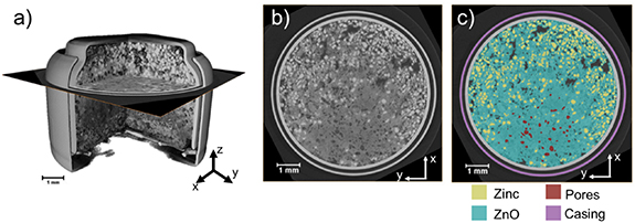
Given the urgent need to move to low-carbon technologies, batteries are being increasingly used in a range of applications. Lithium-ion batteries are the most widely used chemistry, but to meet the growing demand, there is a need to move beyond lithium towards alternative battery chemistries. Metal–air batteries are a group of such battery alternatives that hold promise, especially for stationary power and flexible electronics applications.
However, barriers to their widespread adopt... Read more
Jennifer Hack, Drasti Patel, Josh J Bailey, Francesco Iacoviello, Paul R Shearing, and Dan J L Brett
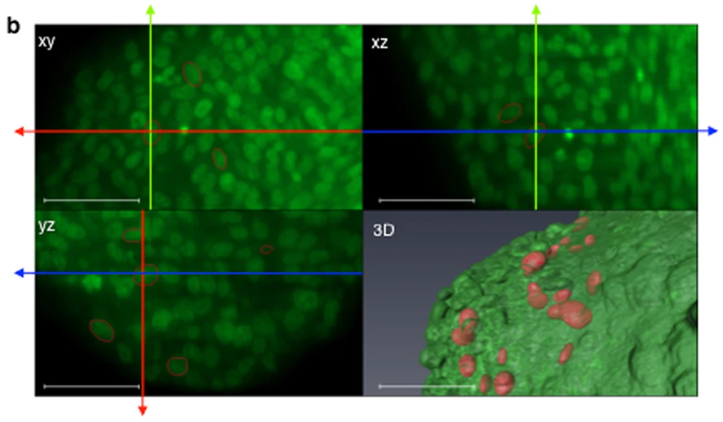
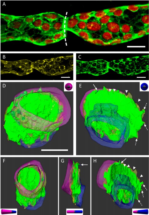
Impact of physical confinement on nuclei geometry and cell division dynamics in 3D spheroids
Multicellular tumour spheroids are used as a culture model to reproduce the 3D architecture, proliferation gradient and cell interactions of a tumour micro-domain. However, their 3D characterization at the cell scale remains challenging due to size and cell density issues. In this study, we developed a methodology based on 3D light sheet fluorescence microscopy (LSFM) image analysis and convex hull calculation that allows characterizing the 3D shape and orientation of cell nuclei relative to ... Read more
Annaïck Desmaison, Ludivine Guillaume, Sarah Triclin, Pierre Weiss, Bernard Ducommun & Valérie Lobjois
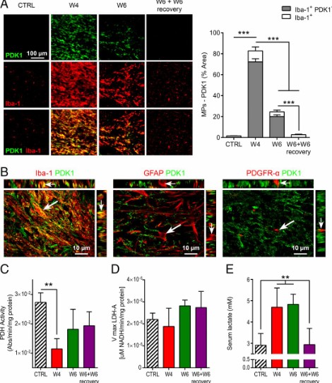
Proinflammatory mononuclear phagocytes (MPs) play a crucial role in the progression of multiple sclerosis (MS) and other neurodegenerative diseases. Despite advances in neuroimaging, there are currently limited available methods enabling noninvasive detection of MPs in vivo. Interestingly, upon activation and subsequent differentiation toward a proinflammatory phenotype MPs undergo metabolic reprogramming that results in increased glycolysis and production of lactate. Hyperpolarized (HP)
Caroline Guglielmetti, Chloé Najac, Alessandro Didonna, Annemie Van der Linden, Sabrina M. Ronen, and Myriam M. Chaumeil
Insights into data with the KAUST Visualization Core Lab
Through collaboration, the KAUST Visualization Core Lab (KVL) team augments the efforts and domain expertise of KAUST researchers by providing complimentary technical knowledge with exploratory visualization and analytic tools.
KVL’s multi-year collaboration with KAUST Distinguished Professor P. Magistretti and research scientist C. Calì’s KAUST-EPFL Alliance for Integrative Modelling of Brain Energy Metabolism project—itself a collaboration with the Swiss Blue Br... Read more
By the KAUST Visualization Core Lab team and Caitlin Clark
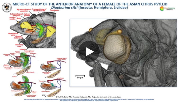
The Asian citrus psyllid (ACP), Diaphorina citri, is a harmful pest of citrus trees that transmits Candidatus Liberibacter spp. which causes Huanglongbing (HLB) (citrus greening disease); this is considered to be the most serious bacterial disease of citrus plants.
Here we detail an anatomical study of the external and internal anatomy (excluding the reproductive system) using micro-computed tomography (micro-CT). This is the first complete 3D micro-CT reconstruction o... Read more
Javier Alba-Tercedor, Wayne B. Hunter & Ignacio Alba-Alejandre
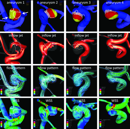
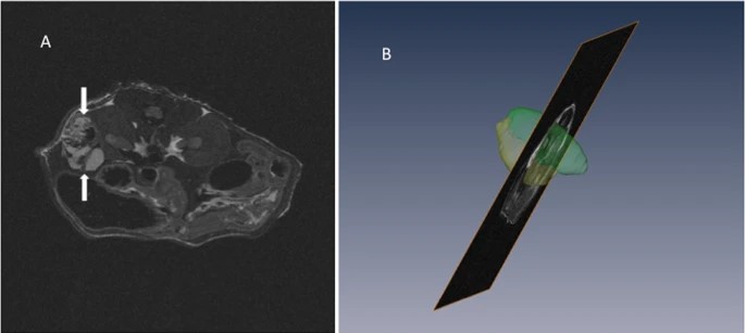
Regional Aneurysm Wall Enhancement is Affected by Local Hemodynamics: A 7T MRI Study
Aneurysm wall enhancement has been proposed as a biomarker for inflammation and instability. However, the mechanisms of aneurysm wall enhancement remain unclear. We used 7T MR imaging to determine the effect of flow in different regions of the wall.
Twenty-three intracranial aneurysms imaged with 7T MR imaging and 3D angiography were studied with computational fluid dynamics. Local flow conditions were compared between aneurysm wall enhancement and nonenhanced regions. Aneurysm wall en... Read more
S. Hadad, F. Mut, B.J. Chung, J.A. Roa, A.M. Robertson, D.M. Hasan, E.A. Samaniego and J.R. Cebral
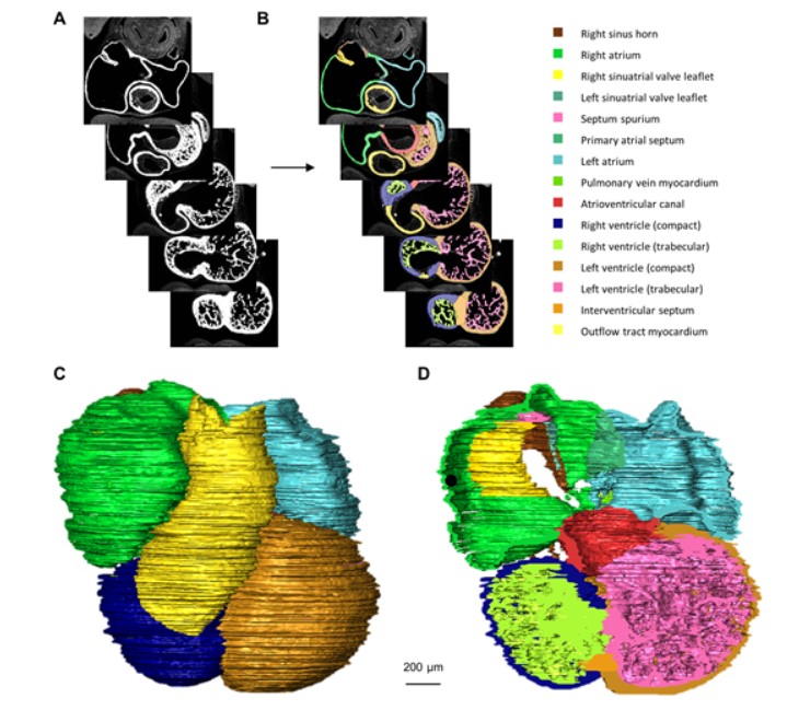
Quantified growth of the human embryonic heart
The size and growth patterns of the components of the human embryonic heart have remained largely undefined.
To provide these data, three-dimensional heart models were generated from immunohistochemically stained sections of ten human embryonic hearts ranging from Carnegie stage 10 to 23. Fifty-eight key structures were annotated and volumetrically assessed. Sizes of the septal foramina and atrioventricular canal opening were also measured. The heart grows exponentially throughout embr... Read more
Jaeike W. Faber, Jaco Hagoort, Antoon F. M. Moorman, Vincent M. Christoffels, Bjarke Jensen
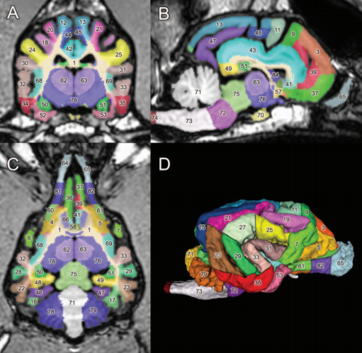
Patient-specific anatomical model for deep brain stimulation based on 7 Tesla MRI
Deep brain stimulation (DBS) requires accurate localization of the anatomical target structure, and the precise placement of the DBS electrode within it. Ultra-high field 7 Tesla (T) MR images can be utilized to create patient-specific anatomical 3D models of the subthalamic nuclei (STN) to enhance pre-surgical DBS targeting as well as post-surgical visualization of the DBS lead position and orientation. We validated the accuracy of the 7T imaging-based patient-specific model of the STN and m... Read more
Yuval Duchin, Reuben R. Shamir, Remi Patriat, Jinyoung Kim, Jerrold L. Vitek, Guillermo Sapiro, Noam Harel
A detailed canine brain label map for neuroimaging analysis
Dogs have recently become an important model species for comparative social and cognitive neuroscience. Brain template-related label maps are essential for functional magnetic resonance imaging (fMRI) data analysis, to localize neural responses. In this study, we present a detailed, individual-based, T1-weighted MRI-based brain label map used in dog neuroimaging analysis. Methods: A typical, medium-headed dog (a 7.5-year-old
male Golden Retriever) was selected from a cohort of ... Read more
Czeibert Kálmán, Andics Attila, Petneházy Örs, Kubinyi Enikő, Kálmán Czeibert
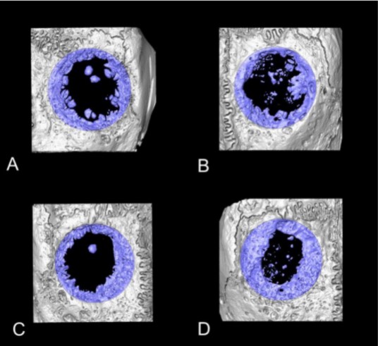
Collagen-Based Matrices for Osteoconduction: A Preclinical In Vivo Study
The aim of this study was to evaluate the influence of additional hydroxyapatite (HA) in collagen-based matrices (CM) and membrane placement on bone formation in calvarial defects.
Critical size defects in the calvaria of 16 New Zealand White Rabbits were randomly treated with CM or mineralized collagen-based matrices (mCM). Half of the sites were covered with a collagen membrane. Animals were euthanized after 12 weeks of healing. The samples were studied by micro-CT and histology. New... Read more
Hiroki Katagiri, Yacine El Tawil, Niklaus P. Lang, Jean-Claude Imber, Anton Sculean, Masako Fujioka-Kobayashi and Nikola Saulacic
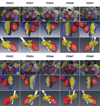
Recent treatment developments for metastatic renal cell carcinoma offer combinations of immunotherapies or immunotherapy associated with tyrosine kinase inhibitors (TKI). There is currently no argument to choose one solution or another. Easy-to-use markers to assess longitudinal responses to TKI are necessary to determine when to switch to immunotherapies. These new markers will enable an earlier adaptation of therapeutic strategy in order to prevent tumor development, unnecessary toxicity an... Read more
Alexandre Ingels, Ingrid Leguerney, Paul-Henry Cournède, Jacques Irani, Sophie Ferlicot, Catherine Sébrié, Baya Benatsou, Laurène Jourdain, Stephanie Pitre-Champagnat, Jean-Jacques Patard & Nathalie Lassau
Influenza A matrix protein M1 is sufficient to induce lipid membrane deformation
The matrix protein M1 of the Influenza A virus is considered to mediate viral assembly and budding at the plasma membrane (PM) of infected cells. In order for a new viral particle to form, the PM lipid bilayer has to bend into a vesicle towards the extracellular side. Studies in cellular models have proposed that different viral proteins might be responsible for inducing membrane curvature in this context (including M1), but a clear consensus has not been reached. In this study, we use a comb... Read more
Ismail Dahmani, Kai Ludwig, Salvatore Chiantia
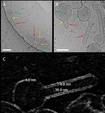
Membrane architecture of pulmonary lamellar bodies revealed by post-correlation on-lamella cryo-CLEM
Lamellar bodies (LBs) are surfactant rich organelles in alveolar type 2 cells. LBs disassemble into a lipid-protein network that reduces surface tension and facilitates gas exchange at the air-water interface in the alveolar cavity. Current knowledge of LB architecture is predominantly based on electron microscopy studies using disruptive sample preparation methods. We established a post-correlation on-lamella cryo-correlative light and electron microscopy approach for cryo-FIB milled lung ce... Read more
Steffen Klein, Benedikt H. Wimmer, Sophie L. Winter, Androniki Kolovou, Vibor Laketa, Petr Chlanda
3D Dissection of Structural Membrane-Wall Contacts in Filamentous Moss Protonemata
Cell-to-cell contact is essential for communication and development of multicellular organisms. A prerequisite is the passage through membranes. That way, molecular exchange and information flow is regulated via hormones, membrane proteins and pores.
In plants, the rigid cell walls prevent large membrane contact areas between protoplasts. Only plasmodesmata, minute channels between adjacent cells, form direct connections. Often, molecular data of the proteins involved are manifold but t... Read more
Dominik Harant and Ingeborg Lang
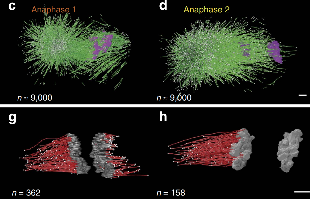
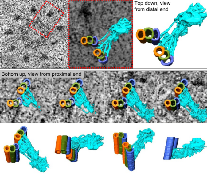
C. elegans chromosomes connect to centrosomes by anchoring into the spindle network
The mitotic spindle ensures the faithful segregation of chromosomes. Here we combine the first large-scale serial electron tomography of whole mitotic spindles in early C. elegans embryos with live-cell imaging to reconstruct all microtubules in 3D and identify their plus- and minus-ends. We classify them as kinetochore (KMTs), spindle (SMTs) or astral microtubules (AMTs) according to their positions, and quantify distinct properties of each class. While our light microscopy and muta... Read more
Stefanie Redemann, Johannes Baumgart, Norbert Lindow, Michael Shelley, Ehssan Nazockdast, Andrea Kratz, Steffen Prohaska, Jan Brugués, Sebastian Fürthauer & Thomas Müller-Reichert
Centrioles are vital cellular structures that form centrosomes and cilia. The formation and function of cilia depends on a set of centriole’s distal appendages. In this study, we use correlative super resolution and electron microscopy to precisely determine where distal appendage proteins localize in relation to the centriole microtubules and appendage electron densities. Here we characterize a novel distal appendage protein ANKRD26 and detail, in high resolution, the initial steps of dist... Read more
Mathew Bowler, Dong Kong, Shufeng Sun, Rashmi Nanjundappa, Lauren Evans, Veronica Farmer, Andrew Holland, Moe R. Mahjoub, Haixin Sui & Jadranka Loncarek