Welcome to the Amira-Avizo Software Use Case Gallery
Below you will find a collection of use cases of our 3D data visualization and analysis software. These use cases include scientific publications, articles, papers, posters, presentations or even videos that show how Amira-Avizo Software is used to address various scientific and industrial research topics.
Use the Domain selector to filter by main application area, and use the Search box to enter keywords related to specific topics you are interested in.
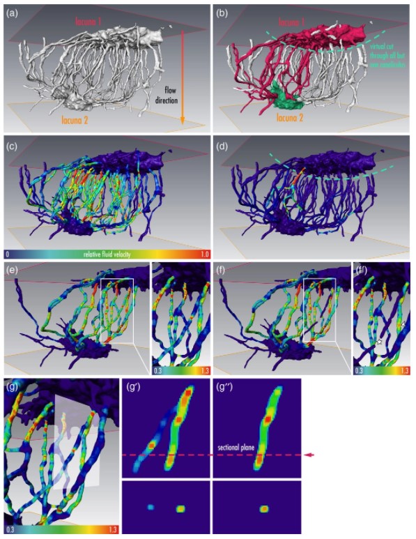
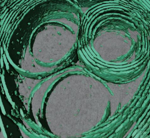
Osteocytes are the most frequent bone cells connected with each other through cell processes within tiny tubular-shaped canaliculi. The so-called osteocyte lacunar-canalicular network (LCN) plays a crucial role in bone remodeling and mineral homeostasis. Given the critical nature of these functions, it is herein hypothesized that the LCN must be structurally “overengineered” to provide network resilience.
This hypothesis is tested by characterizing canalicular networks in human bon... Read more
Emely Bortel, Liam M Grover, Neil Eisenstein, Christian Seim, Heikki Suhonen, Alexandra Pacureanu, Peter Westenberger, Kay Raum, Max Langer, Francoise Peyrin, Owen Addison, Bernhard Hesse
Precise methods for quantifying drug accumulation in brain tissue are currently very limited, challenging the development of new therapeutics for brain disorders. Transcardial perfusion is instrumental for removing the intravascular fraction of an injected compound, thereby allowing for ex vivo assessment of extravasation into the brain. However, pathological remodeling of tissue microenvironment can affect the efficiency of transcardial perfusion, which has been largely overlooked.
We... Read more
Serhii Kostrikov, Kasper B. Johnsen, Thomas H. Braunstein, Johann M. Gudbergsson, Frederikke P. Fliedner, Elisabeth A. A. Obara, Petra Hamerlik, Anders E. Hansen, Andreas Kjaer, Casper Hempel & Thomas L. Andresen
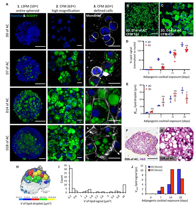
Adipose models have been applied to mechanistic studies of metabolic diseases (such as diabetes) and the subsequent discovery of new therapeutics. However, typical models are either insufficiently complex (2D cell cultures) or expensive and labor intensive (mice/in vivo). To bridge the gap between these models and in order to better inform pre-clinical studies we have developed a drug-responsive 3D model of white adipose tissue (WAT).
Here, spheroids (680 ± 60 μm) comp... Read more
Alexander D Graham, Rajesh Pandey, Viktoriya S Tsancheva, Alessia Candeo, Stanley W Botchway, Alasdair J Allan, Lydia Teboul4, Kamel Madi, Tahkur S Babra, Louisa A K Zolkiewski, Xuan Xue, Liz Bentley, Joan Gannon, Sam N Olof and Roger D Cox
Optical Resolution Photoacoustic Microscopy of Ovary and Fallopian Tube
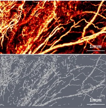
Ovarian cancer is the leading cause of death among gynecological cancers, but is poorly amenable to preoperative diagnosis. In this study, we investigate the feasibility of “optical biopsy,” using high-optical-resolution photoacoustic microscopy (OR-PAM) to quantify the microvasculature of ovarian and fallopian tube tissue. The technique is demonstrated using excised human ovary and fallopian tube specimens imaged immediately after surgery.
This report describes the first applicatio... Read more
Bin Rao, Xiandong Leng, Yifeng Zeng, Yixiao Lin, Ruimin Chen, Qifa Zhou, Andrea R. Hagemann, Lindsay M. Kuroki, Carolyn K. McCourt, David G. Mutch, Matthew A. Powell, Ian S. Hagemann & Quing Zhu
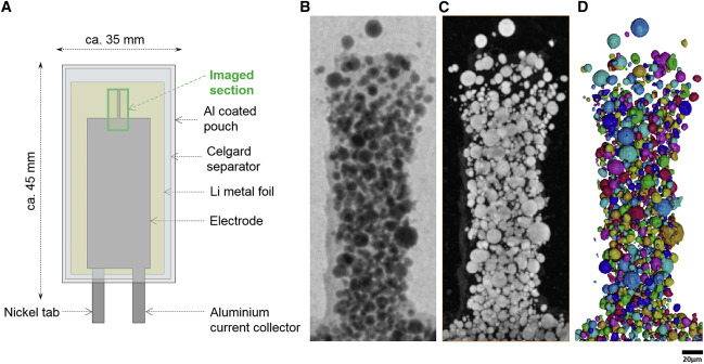
Nickel-rich transition metal oxide materials […] are of great interest for achieving immediate improvements in the energy density of Li-ion batteries and for risk reduction within the Li-ion battery supply chain.
[…] An increase in Ni content in NMC materials leads to accelerated degradation […]. This potentially complicates their adoption in applications requiring extended cycle life such as in electric vehicles.
Recent developments in X-ray characterization to... Read more
ChunTan, Andrew S.Leach, Thomas M.M. Heenan, Huw Parks, Rhodri Jervis, Johanna Nelson Weker, Daniel J.L. Brett, Paul R.Shearing
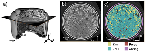
Given the urgent need to move to low-carbon technologies, batteries are being increasingly used in a range of applications. Lithium-ion batteries are the most widely used chemistry, but to meet the growing demand, there is a need to move beyond lithium towards alternative battery chemistries. Metal–air batteries are a group of such battery alternatives that hold promise, especially for stationary power and flexible electronics applications.
However, barriers to their widespread adopt... Read more
Jennifer Hack, Drasti Patel, Josh J Bailey, Francesco Iacoviello, Paul R Shearing, and Dan J L Brett
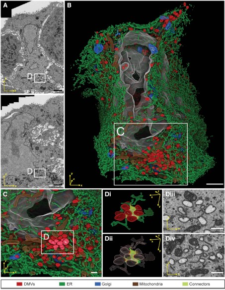
Integrative Imaging Reveals SARS-CoV-2-Induced Reshaping of Subcellular Morphologies
Cortese et al. use integrative imaging techniques to generate a publicly available repository of morphological alterations induced by SARS-CoV-2 in lung cells. Accumulation of ER-derived double-membrane vesicles, the viral replication organelle, occurs concomitantly with cytoskeleton remodeling and Golgi fragmentation. Pharmacological alteration of cytoskeleton dynamics restricts viral replication and spread.
Pathogenesis induced by SARS-CoV-2 is thought to result from both an inflamma... Read more
Mirko Cortese, Ji-Young Lee, Berati Cerikan, ..., Laurent Chatel-Chaix, Yannick Schwab, Ralf Bartenschlager
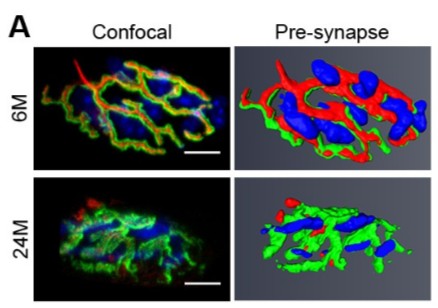
Loss of adult skeletal muscle stem cells drives age-related neuromuscular junction degeneration
Neuromuscular junction degeneration is a prominent aspect of sarcopenia, the age-associated loss of skeletal muscle integrity. Previously, we showed that muscle stem cells activate and contribute to mouse neuromuscular junction regeneration in response to denervation (Liu et al., 2015). Here, we examined gene expression profiles and neuromuscular junction integrity in aged mouse muscles, and unexpectedly found limited denervation despite a high level of degenerated neuromuscular junctions. In... Read more
Wenxuan Liu, Alanna Klose, Sophie Forman, Nicole D Paris, Lan Wei-LaPierre, Mariela Cortés-Lopéz, Aidi Tan, Morgan Flaherty, Pedro Miura, Robert T Dirksen, Joe V Chakkalakal
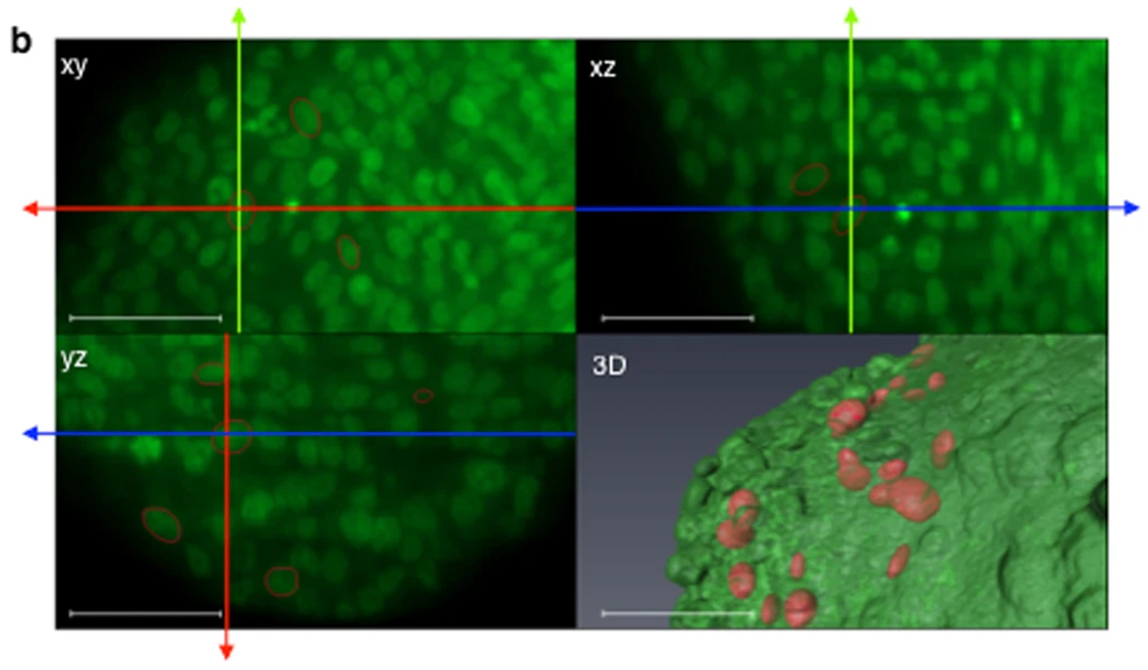
Impact of physical confinement on nuclei geometry and cell division dynamics in 3D spheroids
Multicellular tumour spheroids are used as a culture model to reproduce the 3D architecture, proliferation gradient and cell interactions of a tumour micro-domain. However, their 3D characterization at the cell scale remains challenging due to size and cell density issues. In this study, we developed a methodology based on 3D light sheet fluorescence microscopy (LSFM) image analysis and convex hull calculation that allows characterizing the 3D shape and orientation of cell nuclei relative to ... Read more
Annaïck Desmaison, Ludivine Guillaume, Sarah Triclin, Pierre Weiss, Bernard Ducommun & Valérie Lobjois
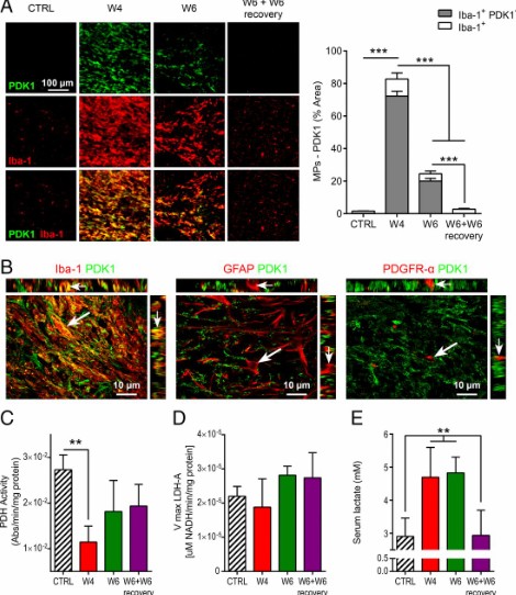
Proinflammatory mononuclear phagocytes (MPs) play a crucial role in the progression of multiple sclerosis (MS) and other neurodegenerative diseases. Despite advances in neuroimaging, there are currently limited available methods enabling noninvasive detection of MPs in vivo. Interestingly, upon activation and subsequent differentiation toward a proinflammatory phenotype MPs undergo metabolic reprogramming that results in increased glycolysis and production of lactate. Hyperpolarized (HP)
Caroline Guglielmetti, Chloé Najac, Alessandro Didonna, Annemie Van der Linden, Sabrina M. Ronen, and Myriam M. Chaumeil
Regional Aneurysm Wall Enhancement is Affected by Local Hemodynamics: A 7T MRI Study
Aneurysm wall enhancement has been proposed as a biomarker for inflammation and instability. However, the mechanisms of aneurysm wall enhancement remain unclear. We used 7T MR imaging to determine the effect of flow in different regions of the wall.
Twenty-three intracranial aneurysms imaged with 7T MR imaging and 3D angiography were studied with computational fluid dynamics. Local flow conditions were compared between aneurysm wall enhancement and nonenhanced regions. Aneurysm wall en... Read more
S. Hadad, F. Mut, B.J. Chung, J.A. Roa, A.M. Robertson, D.M. Hasan, E.A. Samaniego and J.R. Cebral
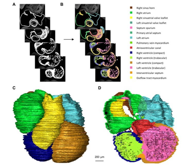
Quantified growth of the human embryonic heart
The size and growth patterns of the components of the human embryonic heart have remained largely undefined.
To provide these data, three-dimensional heart models were generated from immunohistochemically stained sections of ten human embryonic hearts ranging from Carnegie stage 10 to 23. Fifty-eight key structures were annotated and volumetrically assessed. Sizes of the septal foramina and atrioventricular canal opening were also measured. The heart grows exponentially throughout embr... Read more
Jaeike W. Faber, Jaco Hagoort, Antoon F. M. Moorman, Vincent M. Christoffels, Bjarke Jensen
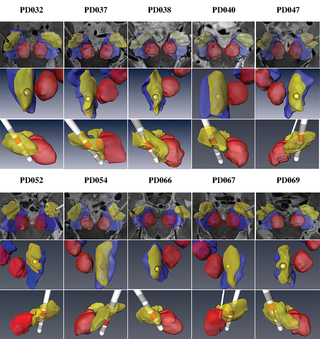
Patient-specific anatomical model for deep brain stimulation based on 7 Tesla MRI
Deep brain stimulation (DBS) requires accurate localization of the anatomical target structure, and the precise placement of the DBS electrode within it. Ultra-high field 7 Tesla (T) MR images can be utilized to create patient-specific anatomical 3D models of the subthalamic nuclei (STN) to enhance pre-surgical DBS targeting as well as post-surgical visualization of the DBS lead position and orientation. We validated the accuracy of the 7T imaging-based patient-specific model of the STN and m... Read more
Yuval Duchin, Reuben R. Shamir, Remi Patriat, Jinyoung Kim, Jerrold L. Vitek, Guillermo Sapiro, Noam Harel
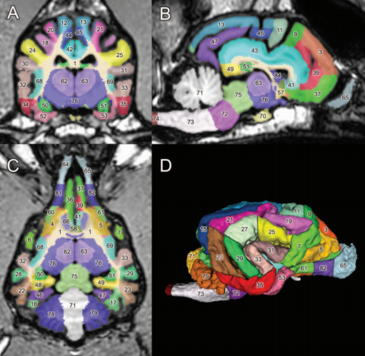
A detailed canine brain label map for neuroimaging analysis
Dogs have recently become an important model species for comparative social and cognitive neuroscience. Brain template-related label maps are essential for functional magnetic resonance imaging (fMRI) data analysis, to localize neural responses. In this study, we present a detailed, individual-based, T1-weighted MRI-based brain label map used in dog neuroimaging analysis. Methods: A typical, medium-headed dog (a 7.5-year-old
male Golden Retriever) was selected from a cohort of ... Read more
Czeibert Kálmán, Andics Attila, Petneházy Örs, Kubinyi Enikő, Kálmán Czeibert
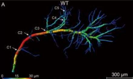
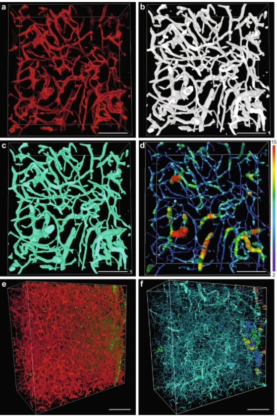
Accumulating evidence indicates the critical importance of cerebrovascular dysfunction in the pathogenesis of Alzheimer’s disease (AD). However, systematic comparative studies on the precise brain vasculature of wild-type and AD model mice are still rare. Using an image optimization method for analyzing Micro-Optical Sectioning Tomography (MOST) data, we generated cross-scale whole-brain 3D atlases that cover the entire vascular system from large vessels down to smallest capillaries at ... Read more
Xiaochuan Zhang, Xianzhen Yin, Jingjing Zhang, Anan Li, Hui Gong, Qingming Luo, Haiyan Zhang, Zhaobing Gao, Hualiang Jiang
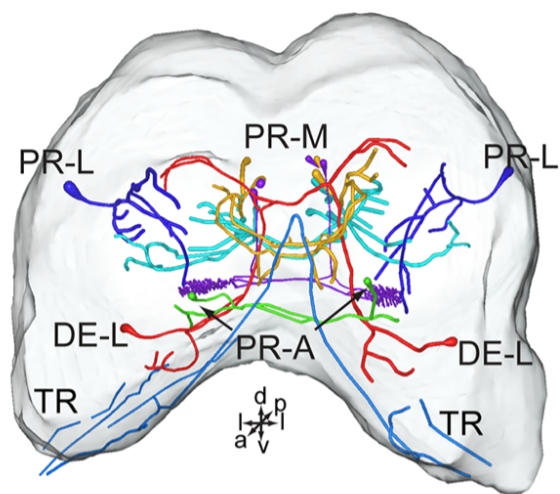
Serotonin (5-hydroxytryptamine, 5-HT) is an important biogenic amine that acts as a neural circuit modulator. It is widespread in the central nervous system of insects. However, little is known about the distribution of serotonin in the nervous system of the cotton bollworm Helicoverpa armigera. In the present study, we performed immunohistochemical experiments with anti-serotonin serum to examine the distribution of serotonin in the central nervous system of H. armigera larv... Read more
Qing-Bo Tang, Wei-Wei Song, Ya-Jun Chang, Gui-Ying Xie, Wen-Bo Chen* and Xin-Cheng Zhao
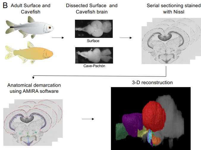
A shift in environmental conditions impacts the evolution of complex developmental and behavioral traits. The Mexican cavefish, Astyanax mexicanus, is a powerful model for examining the evolution of development, physiology, and behavior because multiple cavefish populations can be compared to an extant and ancestral-like surface population of the same species. Many behaviors have diverged in cave populations of A. mexicanus, and previous studies have shown that cavefish ha... Read more
Cody Loomis, View ORCID ProfileRobert Peuß, James Jaggard, Yongfu Wang, Sean McKinney, Stephen Raftopoulos, Austin Raftopoulos, Daniel Whu, Matthew Green, Suzanne E. McGaugh, Nicolas Rohner, Alex C. Keene, Erik R. Duboue
Collagen-Based Matrices for Osteoconduction: A Preclinical In Vivo Study
The aim of this study was to evaluate the influence of additional hydroxyapatite (HA) in collagen-based matrices (CM) and membrane placement on bone formation in calvarial defects.
Critical size defects in the calvaria of 16 New Zealand White Rabbits were randomly treated with CM or mineralized collagen-based matrices (mCM). Half of the sites were covered with a collagen membrane. Animals were euthanized after 12 weeks of healing. The samples were studied by micro-CT and histology. New... Read more
Hiroki Katagiri, Yacine El Tawil, Niklaus P. Lang, Jean-Claude Imber, Anton Sculean, Masako Fujioka-Kobayashi and Nikola Saulacic
Influenza A matrix protein M1 is sufficient to induce lipid membrane deformation
The matrix protein M1 of the Influenza A virus is considered to mediate viral assembly and budding at the plasma membrane (PM) of infected cells. In order for a new viral particle to form, the PM lipid bilayer has to bend into a vesicle towards the extracellular side. Studies in cellular models have proposed that different viral proteins might be responsible for inducing membrane curvature in this context (including M1), but a clear consensus has not been reached. In this study, we use a comb... Read more
Ismail Dahmani, Kai Ludwig, Salvatore Chiantia
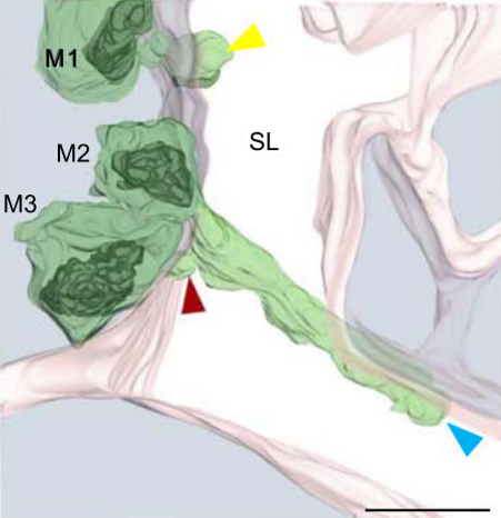
Multiple membrane extrusion sites drive megakaryocyte migration into bone marrow blood vessels
Platelets, cells central to hemostasis and thrombosis, are formed from parent cell megakaryocytes. Although the process is highly efficient in vivo, our ability to generate them in vitro is still remarkably inefficient. We proposed that greater understanding of the process in vivo is needed and used an imaging approach, intravital correlative light electron microscopy, to visualize platelet generation in bone marrow in the living mouse. In contrast to current understanding, we found that most... Read more
Edward Brown, Leo M Carlin, Claus Nerlov, Cristina Lo Celso, Alastair W Poole
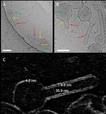
Membrane architecture of pulmonary lamellar bodies revealed by post-correlation on-lamella cryo-CLEM
Lamellar bodies (LBs) are surfactant rich organelles in alveolar type 2 cells. LBs disassemble into a lipid-protein network that reduces surface tension and facilitates gas exchange at the air-water interface in the alveolar cavity. Current knowledge of LB architecture is predominantly based on electron microscopy studies using disruptive sample preparation methods. We established a post-correlation on-lamella cryo-correlative light and electron microscopy approach for cryo-FIB milled lung ce... Read more
Steffen Klein, Benedikt H. Wimmer, Sophie L. Winter, Androniki Kolovou, Vibor Laketa, Petr Chlanda