Welcome to the Amira-Avizo Software Use Case Gallery
Below you will find a collection of use cases of our 3D data visualization and analysis software. These use cases include scientific publications, articles, papers, posters, presentations or even videos that show how Amira-Avizo Software is used to address various scientific and industrial research topics.
Use the Domain selector to filter by main application area, and use the Search box to enter keywords related to specific topics you are interested in.
Objectives
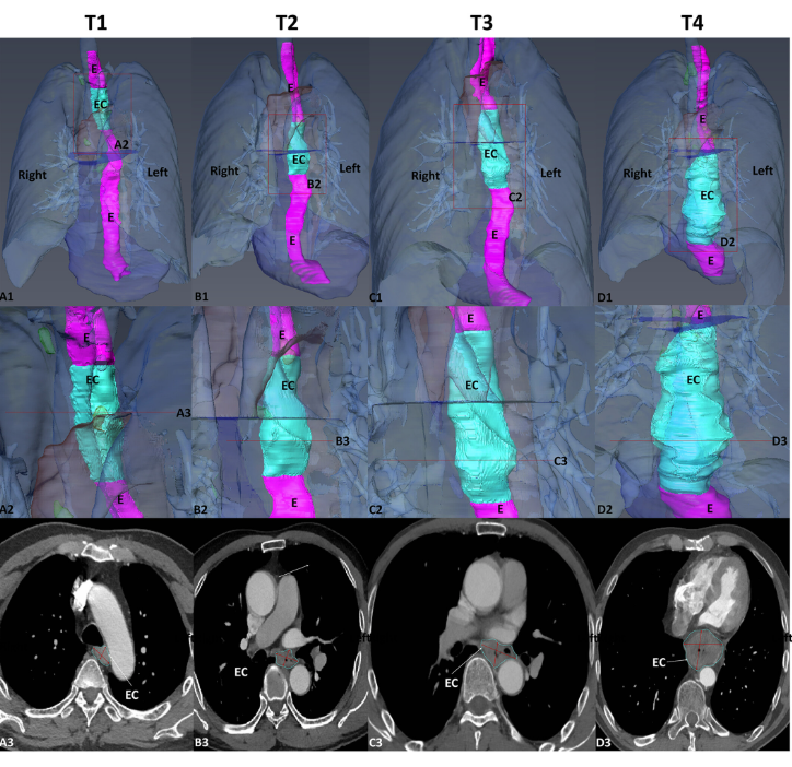
To statistically study the 3D shape of oesophageal cancer (EC) and its spatial relationships based on computed tomography angiography (CTA) 3D reconstruction, to determine its relationship with T-stages, and to create an optimal T-stage diagnosis protocol based on CTA calculation.
Runyuan Wang, Xiaoqin Zhang, Wei Wu, Jinfeng Ma, Jincheng Chen, Zhu Zhang, Liqun Liu, Shanshan Xu, Ximei Cao, Yi Wu, Huilin Cui
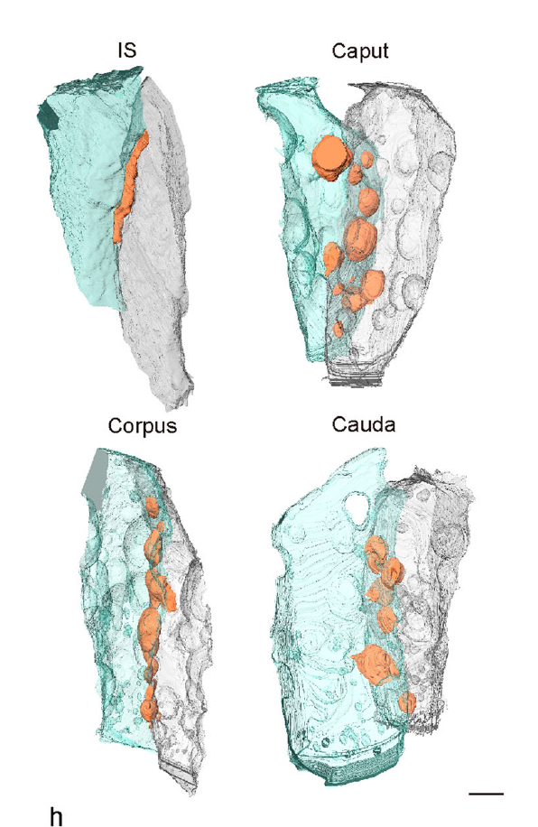
Mammalian epididymal epithelial cells are crucial for sperm maturation. Historically,
vacuole-like ultrastructures in epididymal epithelial cells were
observed via transmission electron microscopy but were undefined. Here, we
utilize volume electron microscopy (vEM) to generate 3D reconstructions of
epididymal epithelial cells and identify these vacuoles as intercellular organelle
reservoirs (IORs) in the lateral intercellular space (LIS), which contains
pr... Read more
Xia Li, Feng Qiao, Jiansheng Guo, Ting Jiang, Huifang Lou, Huixia Li, Gangcai Xie, Hangjun Wu, Weizhen Wang, Ruoyu Pei, Sha Liu, Mei Ye, Jin Li, Shiqin Huang, Mengya Zhang, Chaoye Ma, Yiwen Huang, Shushu Xu, Xiaofeng Li, Xiao Sun, Jun Yu, Kin Lam Fok, Shumin Duan & Hao Chen
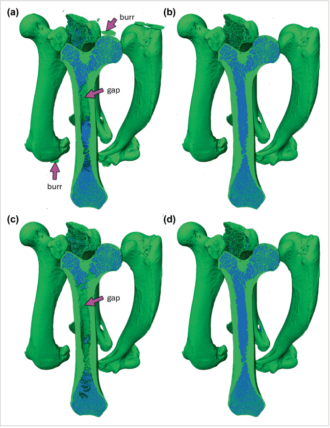
Computed tomography (CT) enables rapid imaging of large-scale studies of bone, but those datasets typically require manual segmentation, which is time-consuming and prone to error. Convolutional neural networks (CNNs) offer an automated solution, achieving superior performance on image data. In this methodology-focused paper, we used CNNs to train segmentation models from scratch on 2D and 3D patches from micro-CT scans of otter long bones. These new models, collectively called BONe (Bone One... Read more
Andrew H. Lee, Julian M. Moore, Brandon Vera Covarrubias, Leigha M. Lynch
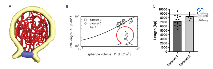
Architecture of the chikungunya virus replication organelle
Alphaviruses are mosquito-borne viruses that cause serious disease in humans and other mammals. Along with its mosquito vector, the Alphavirus chikungunya virus (CHIKV) has spread explosively in the last 20 years, and there is no approved treatment for chikungunya fever. On the plasma membrane of the infected cell, CHIKV generates dedicated organelles for viral RNA replication, so-called spherules. Whereas structures exist for several viral proteins that make up the spherule, ... Read more
Timothée Laurent, Pravin Kumar, Susanne Liese, Farnaz Zare, Mattias Jonasson, Andreas Carlson, Lars-Anders Carlson.
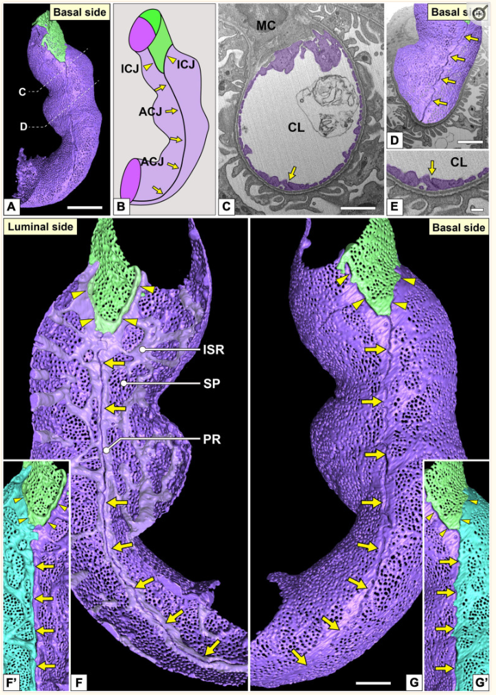
Three-Dimensional Architecture of Glomerular Endothelial Cells Revealed by FIB-SEM Tomography
Focused-ion beam-scanning electron microscopic (FIB-SEM) tomography enables easier acquisition of a series of ultrastructural, sectional images directly from resin-embedded biological samples. In this study, to clarify the three-dimensional (3D) architecture of glomerular endothelial cells (GEnCs) in adult rats, we manually extracted GEnCs from serial FIB-SEM images and reconstructed them on an Amira reconstruction software. The luminal and basal surface structures were clearly visualized in ... Read more
Yuto Kawasaki, Yasue Hosoyamada, Takayuki Miyaki, Junji Yamaguchi, Soichiro Kakuta, Tatsuo Sakai, and Koichiro Ichimura
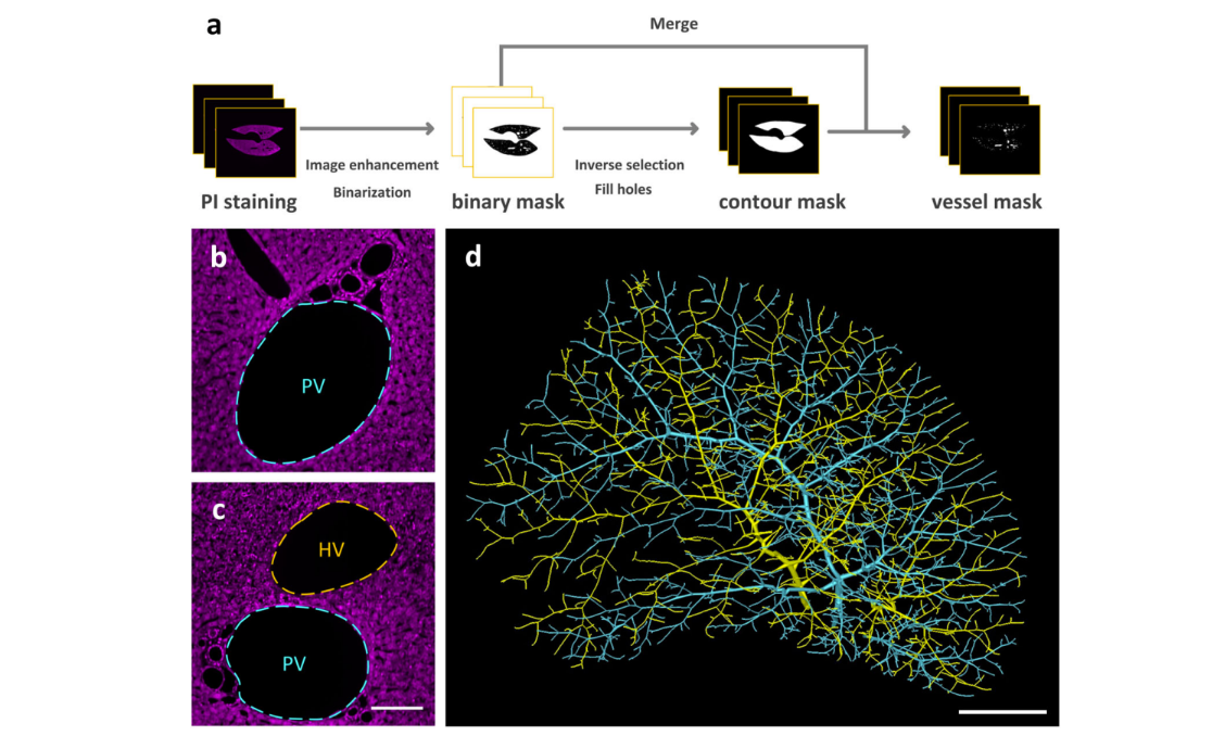
Multiscale reconstruction of various vessels in the intact murine liver lobe
The liver contains a variety of vessels and participates in miscellaneous physiological functions. While past studies generally focused on certain hepatic vessels, we simultaneously obtained all the vessels and cytoarchitectural information of the intact mouse liver lobe at single-cell resolution. […] providing a technology roadmap for studying the fine hepatic vascular structures and their spatial relationship, which will help research into liver diseases and evaluation of medical effi... Read more
Qi Zhang, Anan Li, Siqi Chen, Jing Yuan, Tao Jiang, Xiangning Li, Qingming Luo,Zhao Feng & Hui Gon
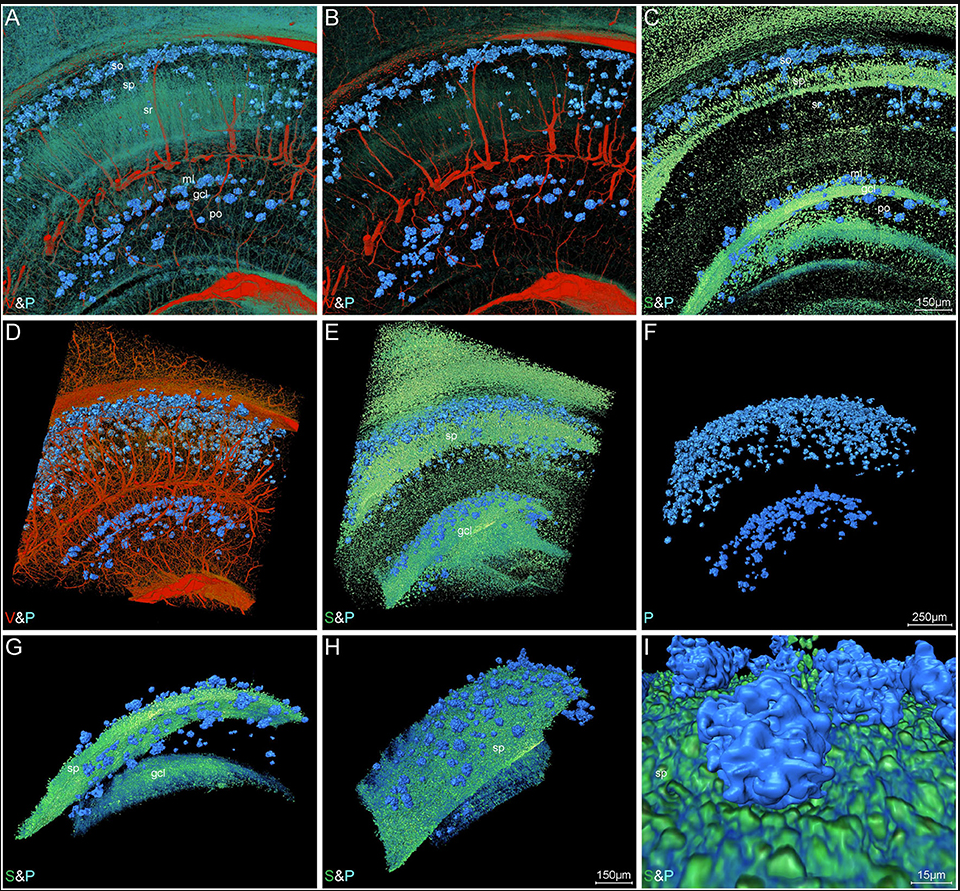
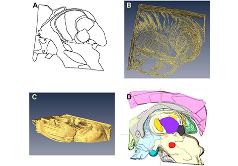
High-Resolution Digital Panorama of Multiple Structures in Whole Brain of Alzheimer's Disease Mice
Our study placed emphasis on solving problems in processing high-throughput bright field images and made attempt in developing a method for the extraction and reconstruction of multiple structures. This will facilitate a better understanding of the cerebral anatomical features under the pathological state of AD and shows extensive application prospect in drug efficacy assessment from brain-wide level.
Simultaneously visualizing Amyloid-β (Aβ) plaque with its surrounding brain structu... Read more
Xianzhen Yin, Xiaochuan Zhang, Jingjing Zhang, Weicheng Yang, Xian Sun, Haiyan Zhang, Zhaobing Gao, Hualiang Jiang
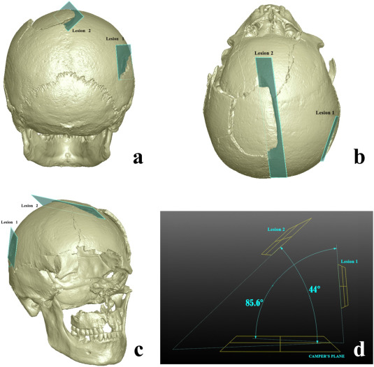
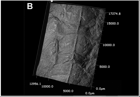
Human skeletal remains from archaeological contexts occasionally present signs of traumatic injuries from weapons, revealing, for example, the degree of interpersonal violence, the type of weapon and the sequence of events of a specific historical context.
Traumatic lesions are generally analyzed using macroscopic and microscopic methods, which are not necessarily integrated in the same study. In this study, we employed a multi-analytical approach to determine i... Read more
Antonino Vazzana, Lucia Martina Scalise, Mirko Traversari, Carla Figus, Salvatore Andrea Apicella, Laura Buti, Gregorio Oxilia, Rita Sorrentino, Silvia Pellegrini, Chiara Matteucci, Lucio Calcagnile, Raffaele Savigni, Robin N.M.Feeney, Giorgio Gruppioni, Stefano Benazziah
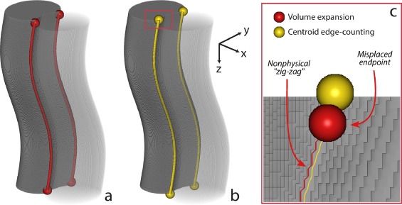
Improving microstructural quantification in FIB/SEM nanotomography
Advanced nanotomographic analysis is still far from routine, and a number of challenges remain in data acquisition and post-processing. In this work, we present a number of techniques to improve the quality of the acquired data, together with easy-to-implement methods to obtain “advanced” microstructural quantifications. The techniques are applied to a solid oxide fuel cell cathode of interest to the electrochemistry community, but the methodologies are easily adaptable to a wide range of... Read more
Joshua A.Taillon, Christopher Pellegrinelli, Yi-Lin Huang, Eric D.Wachsman, Lourdes G. Salamanca-Riba
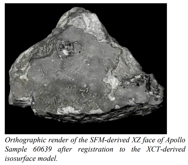
Our team is developing a modern, cross-disciplinary approach to documentation and preservation of astromaterials, specifically lunar and meteorite samples stored at the Johnson Space Center (JSC) Lunar Sample Laboratory Facility.
Apollo Lunar Sample 60639, collected as part of rake sample 60610 during the 3rd Extra-Vehicular Activity of the Apollo 16 mission in 1972, served as the first NASA preserved lunar sample to be examined by our team in the development of a novel approach to int... Read more
K.R. Beaulieu , E.H. Blumenfeld , D.A. Liddle , E.R. Oshel , C.A. Evans , R.A. Zeigler , K. Righter , R.D. Hanna , R.A. Ketcham
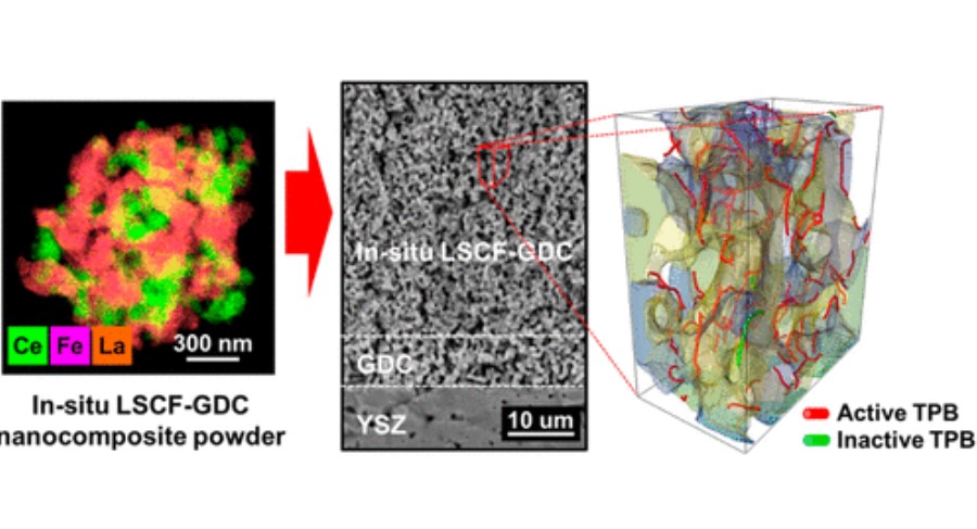
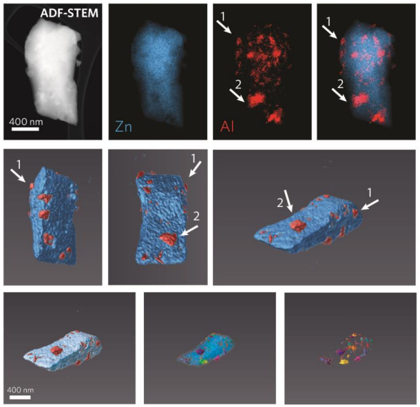
Composite cathodes comprising nanoscale
powders are expected to impart with high specific surface
area and triple phase boundary (TPB) density, which will lead
to better performance.
However, uniformly mixing nanosized heterophase powders remains a challenge due to their high surface energy and thus ease with which they agglomerate into their individual phases during the mixing and sintering
processes. In this study, we successfully synthesized La0.6Sr0.4Co0.2Fe... Read more
Dong Woo Joh, Areum Cha, Jeong Hwa Park, Kyeong Joon Kim, Kyung Taek Bae, Doyeub Kim, Young Ki Choi, Hyegsoon An, Ji Su Shin, Kyung Joong Yoon, and Kang Taek Lee
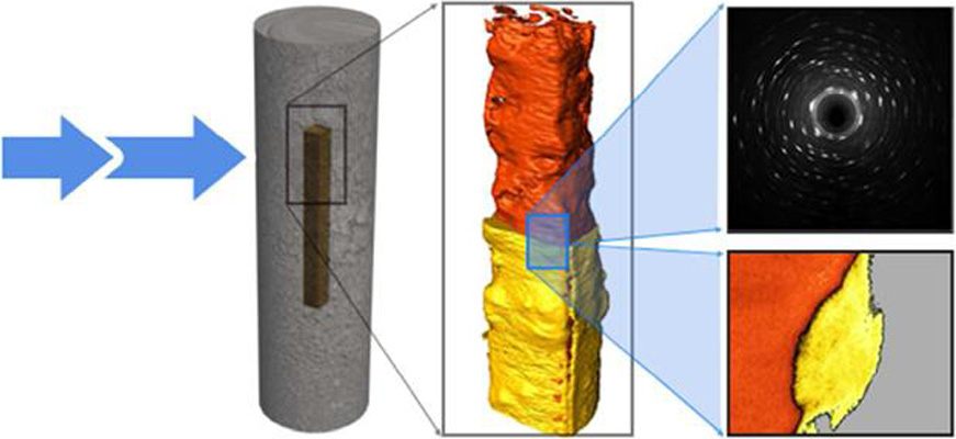
Nuclear waste viewed in a new light; a synchrotron study of uranium encapsulated in grout
How do you characterise the contents of a sealed nuclear waste package without breaking it open?
This question is important when the contained corrosion products are potentially reactive with air and radioactive. Synchrotron X-rays have been used to perform micro-scale in-situ observation and characterisation of uranium encapsulated in grout; a simulation for a typical intermediate level waste storage packet. X-ray tomography and X-ray powder diffraction generated both qualitative and ... Read more
C.A. Stitt, M. Hart, N.J. Harker, K.R. Hallam, J. MacFarlane, A. Banos, C. Paraskevoulakos, E. Butcher, C. Padovani, T.B. Scott
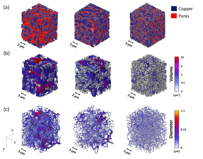
Porous materials have attracted great attention for various applications, e.g., catalysis, novel materials, energy related topics, acoustics, microelectronics, actuator, bioengineering and biomimetic. Recently, porous materials have gained interest as interconnect materials for power semiconductor device.
The trend in the semiconductor industry goes towards eco-friendliness and higher energy efficiency. Semiconductor compound materials, such as silicon carbide (SiC) or gallium nitride... Read more
A.Wijaya, B.Eichinger, F.F.Chamasemani, B.Sartory, R.Hammer, V.Maier-Kiener, D.Kiener, M.Mischitz, R.Brunner
Metal-Organic Framework Crystal-Glass Composites
The majority of research into metal-organic frameworks (MOFs) focuses on their crystalline nature. However, in recent research the vitrification of a number of MOFs has been revealed. We propose that the solid-liquid phase transitions involved in MOF-glass formation can provide unique opportunities for the creation of a new class of functional, stable and porous composite materials. Described herein is the design, synthesis, and characterisation of novel metal-organic framework (MOF) crystal-... Read more
Jingwei Hou, Christopher W. Ashling, Sean M. Collins, Andraž Krajnc, Chao Zhou, Louis Longley, Duncan N. Johnstone, Philip A. Chater, Shichun Li, François-Xavier Coudert, David A. Keen, Paul A. Midgley, Gregor Mali, Vicki Chen, Thomas Bennett
Classification and delineation of the motor-related nuclei in the human thalamus have been the focus of numerous discussions for a long time. Difficulties in finding consensus have for the most part been caused by paucity of direct experimental data on connections of individual nuclear entities. Kultas-Ilinsky et al. (2011, J Comp Neurol, 519:2811-2837) showed that distribution of the isoform 65 of glutamic acid decarboxylase (GAD65), the enzyme that synthesizes inhibitory neurotransmitt... Read more
Igor Ilinsky, Andreas Horn, Perrine Paul-Gilloteaux, Pierre Gressens, Catherine Verney, Kristy Kultas-Ilinsky
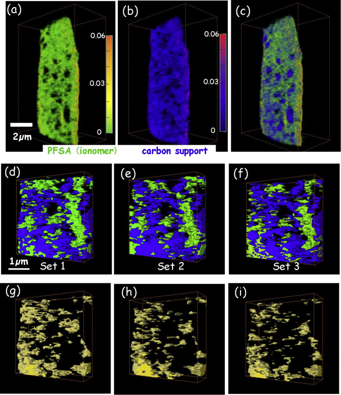
4D imaging – the three-dimensional distributions of chemical species determined using multi-energy X-ray tomography – of cathode catalyst layers of polymer electrolyte membrane fuel cells (PEM-FC) has been measured by scanning transmission x-ray microscopy (STXM) spectro-tomography at the C 1s and F 1s edges. In order to monitor the effects of radiation damage on the composition and 3D structure of the perfluorosulfonic acid (PFSA) ionomer, the same volume was measu... Read more
Juan Wu, Lis G.A.Melo, Xiaohui Zhu, Marcia M.West, Viatcheslav Berejnov, Darija Susac, Juergen Stumper, Adam P.Hitchcock
Paleozoic Nymphal Wing Pads Support Dual Model of Insect Wing Origins
The appearance of wings in insects, early in their evolution [1], has been one of the more critical innovations contributing to their extraordinary diversity. Despite the conspicuousness and importance of wings, the origin of these structures has been difficult to resolve and represented one of the “abominable mysteries” in evolutionary biology [2]. More than a century of debate has boiled the matter down to two competing alternatives—one of wings representing an extension of the thorac... Read more
Department of Zoology, Faculty of Science, Charles University, Praha, Czech Republic and al.
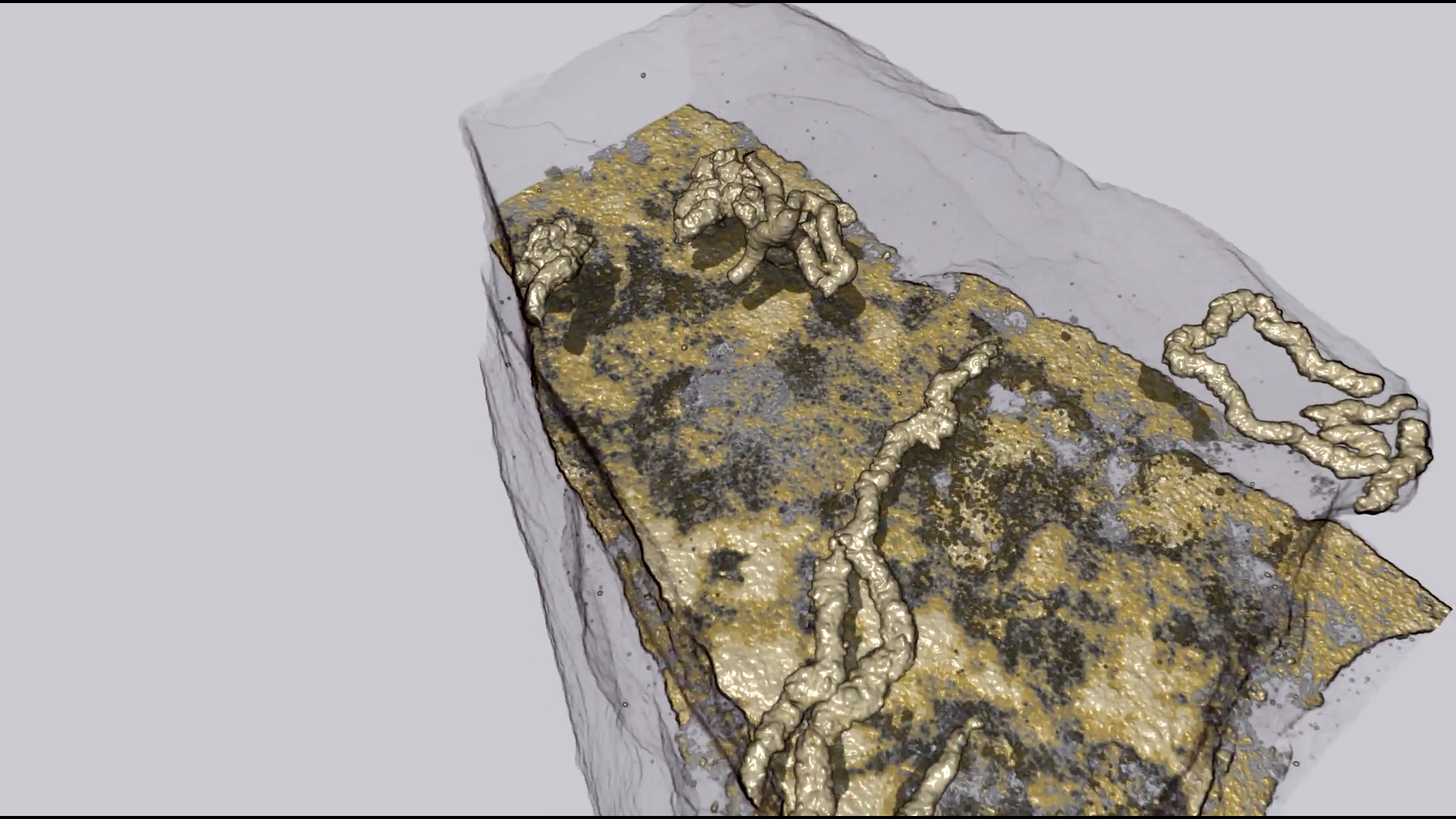
Organism motility in an oxygenated shallow-marine environment 2.1 billion years ago
Evidence for macroscopic life in the Paleoproterozoic Era comes from 1.8 billion-year-old (Ga) compression fossils [Han TM, Runnegar B (1992) Science 257:232–235; Knoll et al. (2006) Philos Trans R Soc Lond B 361:1023–1038], Stirling biota [Bengtson S et al. (2007) Paleobiology 33:351–381], and large colonial organisms exhibiting signs of coordinated growth from the 2.1-Ga Francevillian series, Gabon. Here we report on pyritized string-shaped structures from... Read more
Abderrazak El Albani, M. Gabriela Mangano, Luis A. Buatois, Stefan Bengtson, Armelle Riboulleau, Andrey Bekker, Kurt Konhauser, Timothy Lyons, Claire Rollion-Bard, Olabode Bankole, Stellina Gwenaelle Lekele Baghekema, Alain Meunier, Alain Trentesaux, Arnaud Mazurier, Jeremie Aubineau, Claude Laforest, Claude Fontaine, Philippe Recourt, Ernest Chi Fru, Roberto Macchiarelli, Jean Yves Reynaud, François Gauthier-Lafaye, and Donald E. Canfield
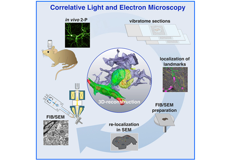
Label-free 3D-CLEM using endogenous tissue landmarks
We demonstrate feasibility of the workflow by combining in vivo 2-photon microscopy and focused ion beam scanning electron microscopy (FIB/SEM) to dissect the role of astrocytic coverage in the persistence of dendritic spines.
Emerging 3D correlative light and electron microscopy (CLEM) approaches enable studying neuronal structure-function relations at unprecedented depth and precision. However, established protocols for the correlation of light and electron micrographs rely ... Read more
Manja Luckner,Steffen Burgold, Severin Filser, Maximilian Scheungrab, Yilmaz Niyaz, Eric Hummel, Gerhard Wanner, Jochen Herms
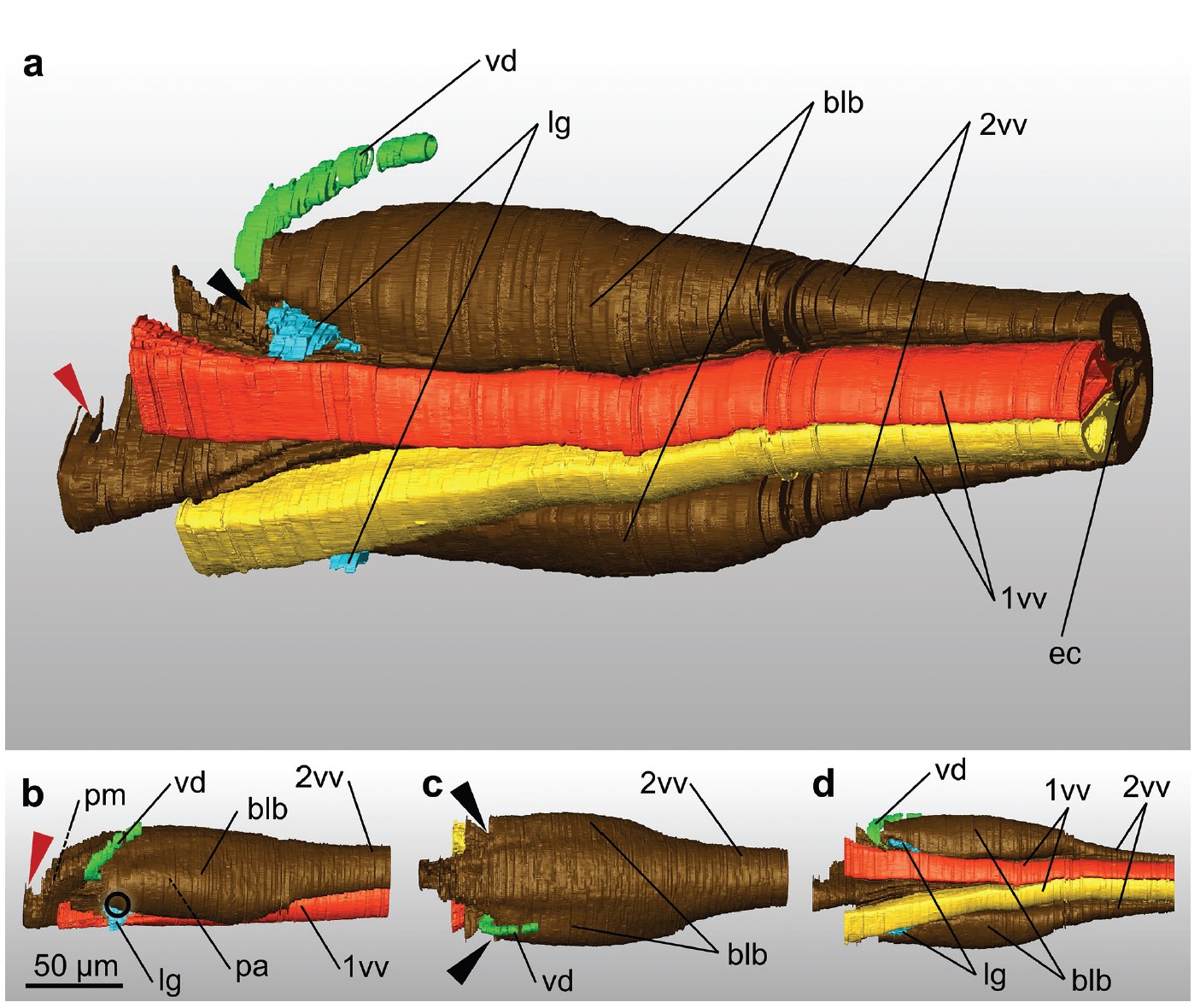
Ovipositor of the braconid wasp Habrobracon hebetor: structural and functional aspects
The Braconidae are a megadiverse and ecologically highly important group of insects. The vast majority of braconid wasps are parasitoids of other insects, usually attacking the egg or larval stages of their hosts. The ovipositor plays a crucial role in the assessment of the potential host and precise egg laying. We used lightand electron-microscopic techniques to investigate all inherent cuticular elements of the ovipositor (the female 9th abdominal tergum, two pairs of valvifers, and three p... Read more
Michael Csader, Karin Mayer, Oliver Betz, Stefan Fischer, Benjamin Eggs

Protocols for Generating Surfaces and Measuring 3D Organelle Morphology Using Amira
High-resolution 3D images of organelles are of paramount importance in cellular biology. Although light microscopy and transmission electron microscopy (TEM) have provided the standard for imaging cellular structures, they cannot provide 3D images.
However, recent technological advances such as serial block-face scanning electron microscopy (SBF-SEM) and focused ion beam scanning electron microscopy (FIB-SEM) provide the tools to create 3D images for the ultrastructural analysis of org... Read more
Edgar Garza-Lopez, Zer Vue, Prasanna Katti, Kit Neikirk, Michelle Biete, Jacob Lam, Heather K. Beasley, Andrea G. Marshall, Taylor A. Rodman, Trace A. Christensen, Jeffrey L. Salisbury, Larry Vang, Margaret Mungai, Salma Ash Shareef, Sandra A. Murray, Jianqiang Shao, Jennifer Streeter, Brian Glancy, Renata O. Pereira1, E. Dale Abel, and Antentor Hinton, Jr.