Welcome to the Amira-Avizo Software Use Case Gallery
Below you will find a collection of use cases of our 3D data visualization and analysis software. These use cases include scientific publications, articles, papers, posters, presentations or even videos that show how Amira-Avizo Software is used to address various scientific and industrial research topics.
Use the Domain selector to filter by main application area, and use the Search box to enter keywords related to specific topics you are interested in.
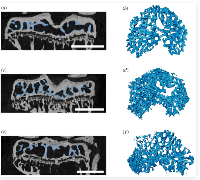
Many physiological, biomechanical, evolutionary and clinical studies that explore skeletal structure and function require successful separation of trabecular from cortical compartments of a bone that has been imaged by X-ray micro-computed tomography (micro-CT) prior to analysis. Separation often involves manual subdivision of these two similarly radio-opaque compartments, which can be time-consuming and subjective. We have developed an objective, semi-automated protocol which reduces user bi... Read more
Eva C. Herbst, Alessandro A. Felder, Lucinda A. E. Evans, Sara Ajami, Behzad Javaheri and Andrew A. Pitsillides
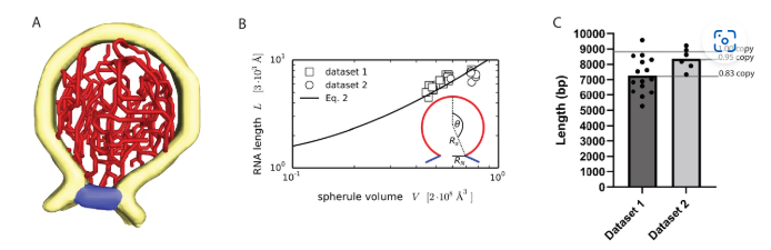
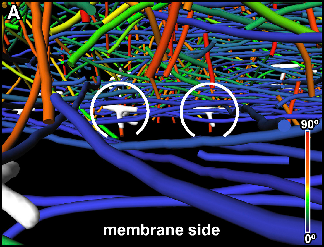
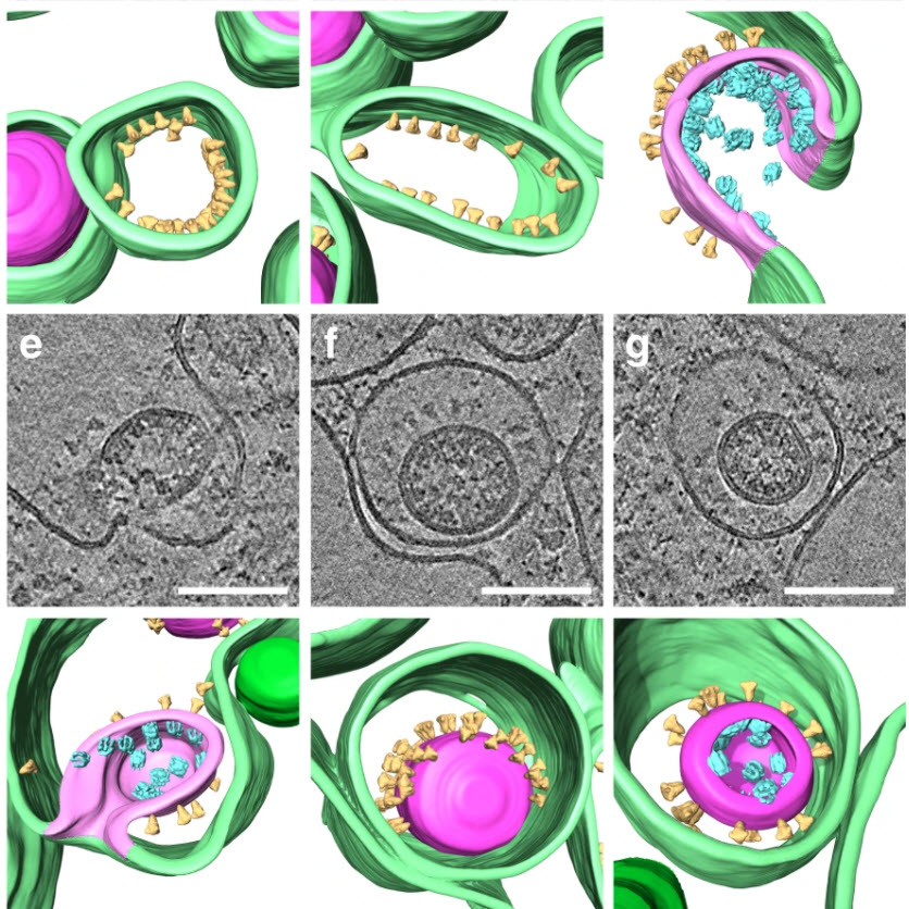
Architecture of the chikungunya virus replication organelle
Alphaviruses are mosquito-borne viruses that cause serious disease in humans and other mammals. Along with its mosquito vector, the Alphavirus chikungunya virus (CHIKV) has spread explosively in the last 20 years, and there is no approved treatment for chikungunya fever. On the plasma membrane of the infected cell, CHIKV generates dedicated organelles for viral RNA replication, so-called spherules. Whereas structures exist for several viral proteins that make up the spherule, ... Read more
Timothée Laurent, Pravin Kumar, Susanne Liese, Farnaz Zare, Mattias Jonasson, Andreas Carlson, Lars-Anders Carlson.
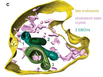
The Ebola virus VP40 matrix layer undergoes endosomal disassembly essential for membrane fusion
Ebola viruses (EBOVs) assemble into filamentous virions, whose shape and stability are determined by the matrix viral protein 40 (VP40). Virus entry into host cells occurs via membrane fusion in late endosomes; however, the mechanism of how the remarkably long virions undergo uncoating, including virion disassembly and nucleocapsid release into the cytosol, remains unknown. Here, we investigate the structural architecture of EBOVs entering host cells and discover that the VP40 matrix disassem... Read more
Sophie L Winter, Gonen Golani, Fabio Lolicato, Melina Vallbracht, Keerthihan Thiyagarajah, Samy Sid Ahmed, Christian Lüchtenborg, Oliver T Fackler, Britta Brügger, Thomas Hoenen, Walter Nickel Ulrich S Schwarz, Petr Chlanda
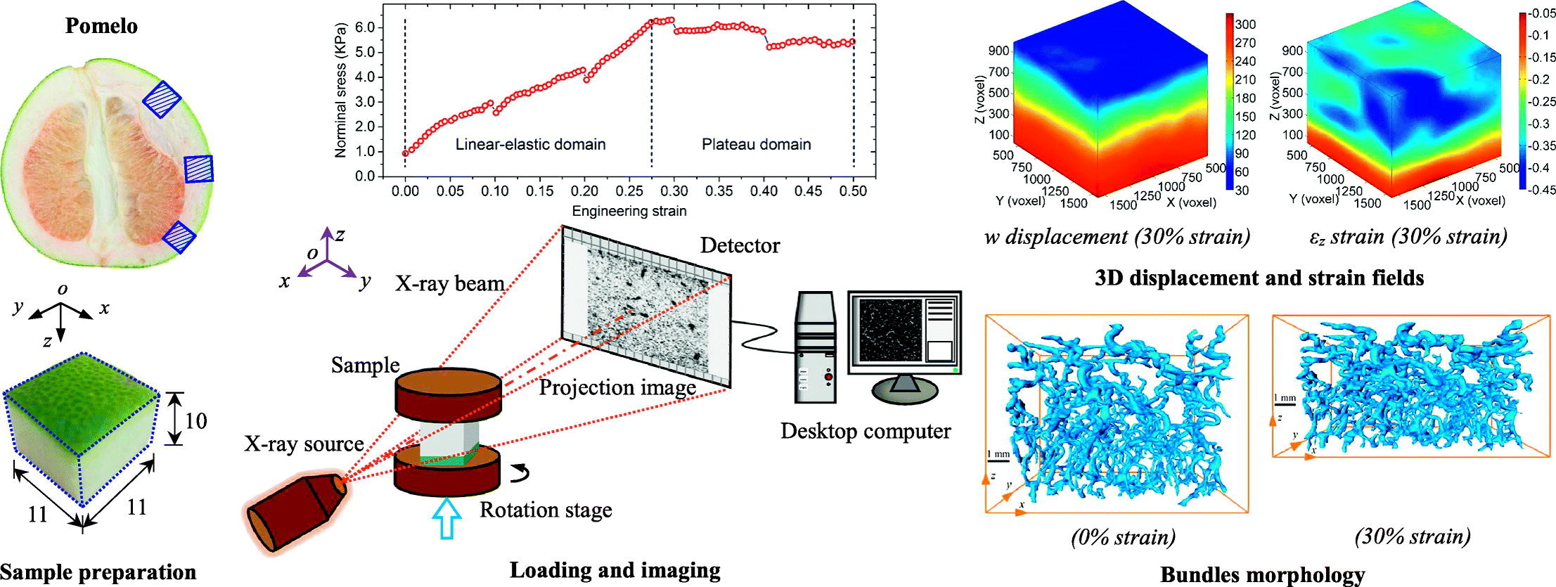
Among natural cellular materials, pomelo peels, having a foam-like hierarchical microstructure, represent an ideal model for developing materials with high energy absorption efficiency. In this work, by combining X-ray tomographic imaging technique and digital volume correlation (DVC), in-situ stepwise uniaxial compression tests were performed to quantify the internal morphological evolution and kinematic responses of pomelo peel samples during compression.
Read more
B.Wang, B.Pan, G.Lubineau
Improving microstructural quantification in FIB/SEM nanotomography
Advanced nanotomographic analysis is still far from routine, and a number of challenges remain in data acquisition and post-processing. In this work, we present a number of techniques to improve the quality of the acquired data, together with easy-to-implement methods to obtain “advanced” microstructural quantifications. The techniques are applied to a solid oxide fuel cell cathode of interest to the electrochemistry community, but the methodologies are easily adaptable to a wide range of... Read more
Joshua A.Taillon, Christopher Pellegrinelli, Yi-Lin Huang, Eric D.Wachsman, Lourdes G. Salamanca-Riba
The three-dimensional (3D) characterization of nuclear fuel with X-ray microscopy has historically proven difficult, due to uranium’s high attenuation of easily accessible X-rays, both in a laboratory setting and at a synchrotron user facility. However, this imaging modality provides nondestructive information that can be used to investigate morphological changes arising from external stimuli (e.g., neutron irradiation, high-temperature testing).
Using an appropriate X-ray energy spe... Read more
Nikolaus L. Cordes, William C. Chuirazzi & Joshua J. Kane - John D. Stempien
Microstructural analysis of TRISO particles using multi-scale X-ray computed tomography
TRISO particles, a composite nuclear fuel built up by ceramic and graphitic layers, have outstanding high temperature resistance. TRISO fuel is the key technology for High Temperature Reactors (HTRs) and the Generation IV Very High Temperature Reactor (VHTR) variant.
TRISO offers unparalleled containment of fission products and is extremely robust during accident conditions. An understanding of the thermal performance and mechanical properties of TRISO fuel requires a detailed knowledg... Read more
T. Lowe, R.S. Bradley, S. Yue, K. Barii, J. Gelb, N. Rohbeck, J. Turner, P.J. Withers
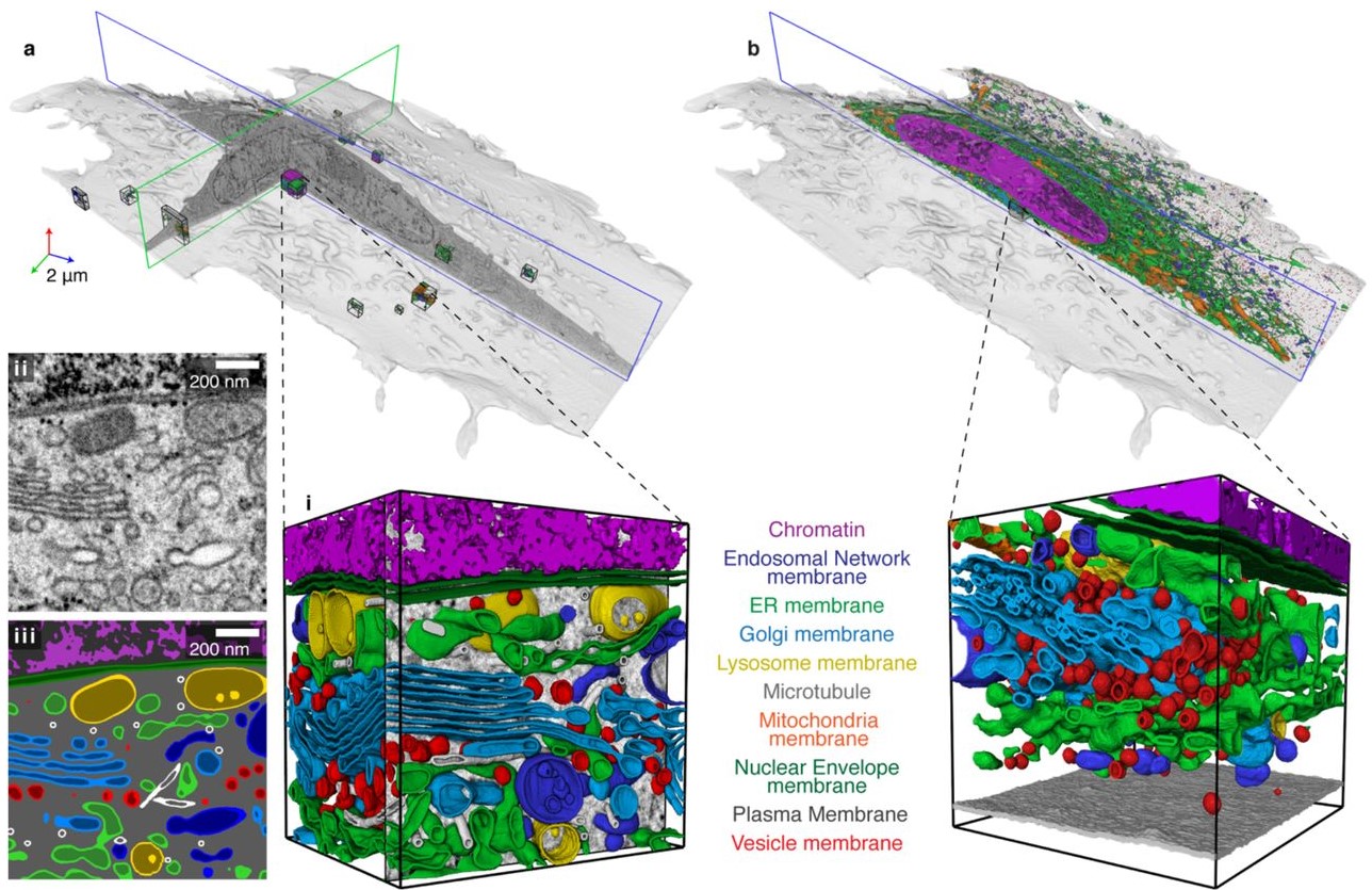
Automatic whole cell organelle segmentation in volumetric electron microscopy
Cells contain hundreds of different organelle and macromolecular assemblies intricately organized relative to each other to meet any cellular demands. Obtaining a complete understanding of their organization is challenging and requires nanometer-level, three-dimensional reconstruction of whole cells. Even then, the immense size of datasets and large number of structures to be characterized requires generalizable, automatic methods.
To meet this challenge, we developed an analy... Read more
Larissa Heinrich, Davis Bennett, David Ackerman, Woohyun Park, John Bogovic, View ORCID ProfileNils Eckstein, Alyson Petruncio, Jody Clements, C. Shan Xu, Jan Funke, Wyatt Korff, Harald F. Hess, Jennifer Lippincott-Schwartz, Stephan Saalfeld, Aubrey V. Weigel, COSEM Project Team
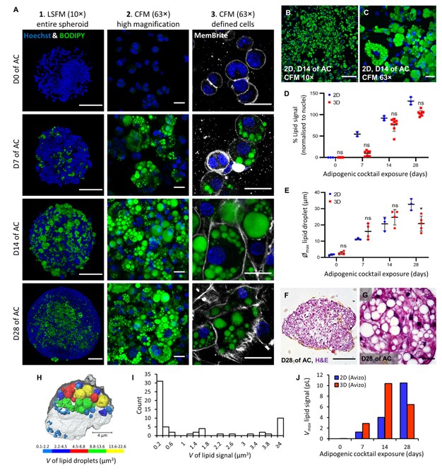
Adipose models have been applied to mechanistic studies of metabolic diseases (such as diabetes) and the subsequent discovery of new therapeutics. However, typical models are either insufficiently complex (2D cell cultures) or expensive and labor intensive (mice/in vivo). To bridge the gap between these models and in order to better inform pre-clinical studies we have developed a drug-responsive 3D model of white adipose tissue (WAT).
Here, spheroids (680 ± 60 μm) comp... Read more
Alexander D Graham, Rajesh Pandey, Viktoriya S Tsancheva, Alessia Candeo, Stanley W Botchway, Alasdair J Allan, Lydia Teboul4, Kamel Madi, Tahkur S Babra, Louisa A K Zolkiewski, Xuan Xue, Liz Bentley, Joan Gannon, Sam N Olof and Roger D Cox
Optical Resolution Photoacoustic Microscopy of Ovary and Fallopian Tube
Ovarian cancer is the leading cause of death among gynecological cancers, but is poorly amenable to preoperative diagnosis. In this study, we investigate the feasibility of “optical biopsy,” using high-optical-resolution photoacoustic microscopy (OR-PAM) to quantify the microvasculature of ovarian and fallopian tube tissue. The technique is demonstrated using excised human ovary and fallopian tube specimens imaged immediately after surgery.
This report describes the first applicatio... Read more
Bin Rao, Xiandong Leng, Yifeng Zeng, Yixiao Lin, Ruimin Chen, Qifa Zhou, Andrea R. Hagemann, Lindsay M. Kuroki, Carolyn K. McCourt, David G. Mutch, Matthew A. Powell, Ian S. Hagemann & Quing Zhu
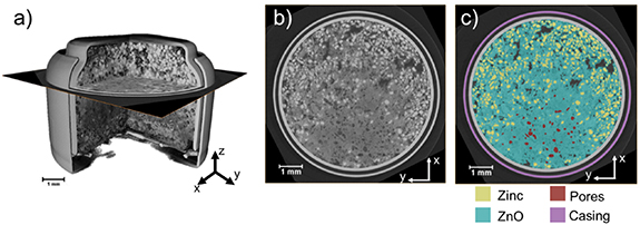
Given the urgent need to move to low-carbon technologies, batteries are being increasingly used in a range of applications. Lithium-ion batteries are the most widely used chemistry, but to meet the growing demand, there is a need to move beyond lithium towards alternative battery chemistries. Metal–air batteries are a group of such battery alternatives that hold promise, especially for stationary power and flexible electronics applications.
However, barriers to their widespread adopt... Read more
Jennifer Hack, Drasti Patel, Josh J Bailey, Francesco Iacoviello, Paul R Shearing, and Dan J L Brett
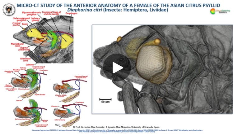
The Asian citrus psyllid (ACP), Diaphorina citri, is a harmful pest of citrus trees that transmits Candidatus Liberibacter spp. which causes Huanglongbing (HLB) (citrus greening disease); this is considered to be the most serious bacterial disease of citrus plants.
Here we detail an anatomical study of the external and internal anatomy (excluding the reproductive system) using micro-computed tomography (micro-CT). This is the first complete 3D micro-CT reconstruction o... Read more
Javier Alba-Tercedor, Wayne B. Hunter & Ignacio Alba-Alejandre
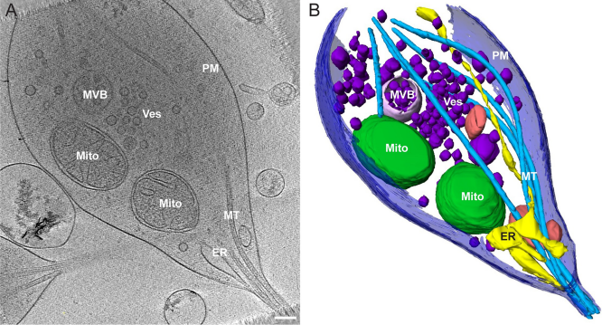
The Architecture of Traveling Actin Waves Revealed by Cryo-Electron Tomography
Actin waves are dynamic supramolecular structures involved in cell migration, cytokinesis, adhesion, and neurogenesis. Although wave-like propagation of actin networks is a widespread phenomenon, the actin architecture underlying wave propagation remained unknown. In situ cryo-electron tomography of Dictyostelium cells unveils the wave architecture and provides evidence for wave progression by de novo actin nucleation. Subtomogram averaging reveals the structu... Read more
Marion Jasnin, Florian Beck, Mary Ecke, Yoshiyuki Fukuda, Antonio Martinez-Sanchez, Wolfgang Baumeister, Günther Gerisch
Morphology of mitochondria in spatially restricted axons revealed by cryo-electron tomography
Neurons project axons to local and distal sites and can display heterogeneous morphologies with limited physical dimensions that may influence the structure of large organelles such as mitochondria. Using cryo-electron tomography (cryo-ET), we characterized native environments within axons and presynaptic varicosities to examine whether spatial restrictions within these compartments influence the morphology of mitochondria. Segmented tomographic reconstructions revealed distinctive morphologi... Read more
Tara D. Fischer, Pramod K. Dash, Jun Liu, M. Neal Waxham
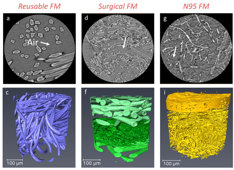
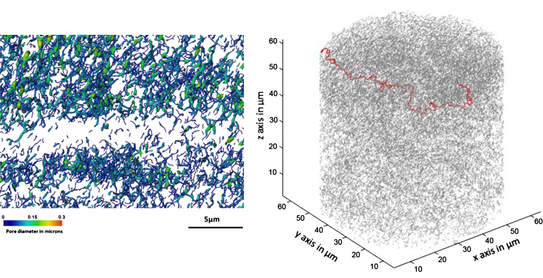
Microstructure analysis and image-based modelling of face masks for COVID-19 virus protection
Until March 2021, around 120 million coronavirus disease (COVID-19) infected cases and over 2.6 million deaths have been reported worldwide. […] Recent investigations have implied that face masks help to reduce the disease transmission and therefore slow down the growth of the epidemic curve. However, there are still ongoing debates on the efficacy of wearing masks […] since there is a general lack of information relating to the material structure of commonly used face masks.Read more
Wenjia Du, Francesco Iacoviello, Tacson Fernandez, Rui Loureiro, Daniel J. L. Brett & Paul R. Shearing
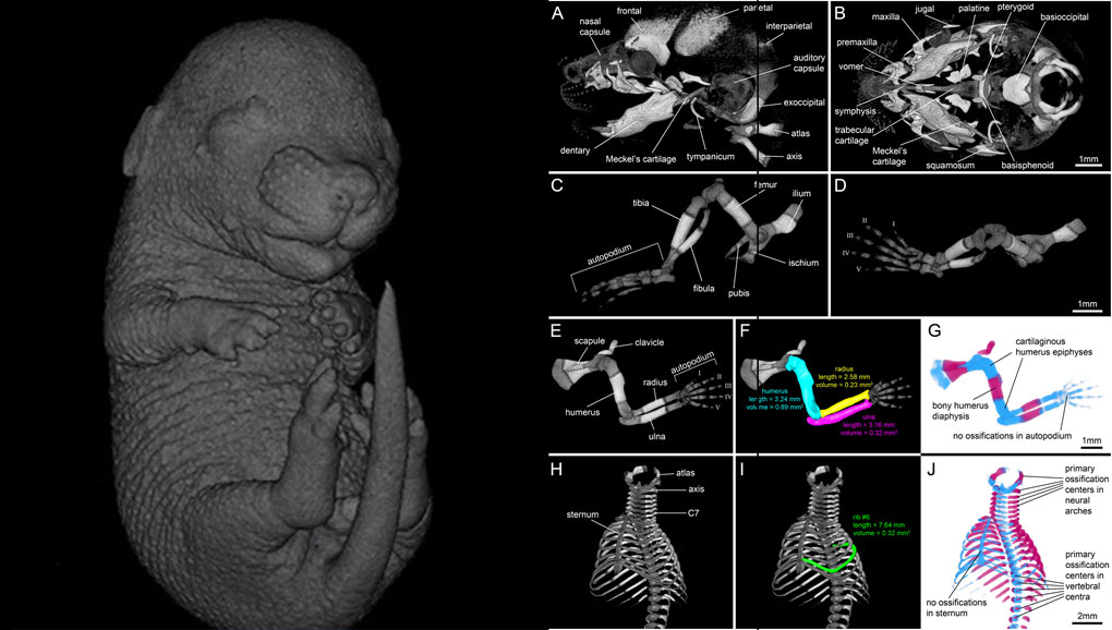
For decades, clearing and staining with Alcian Blue and Alizarin Red has been the gold standard to image vertebrate skeletal development. Here, we present an alternate approach to visualise bone and cartilage based on X-ray microCT imaging, which allows the collection of genuine 3D data of the entire developing skeleton at micron resolution.
Our novel protocol is based on ethanol fixation and staining with Ruthenium Red, and efficiently contrasts cartilage matrix, as demonstrated in wh... Read more
Simone Gabner, Peter Böck, Dieter Fink, Martin Glösmann, Stephan Handschuh
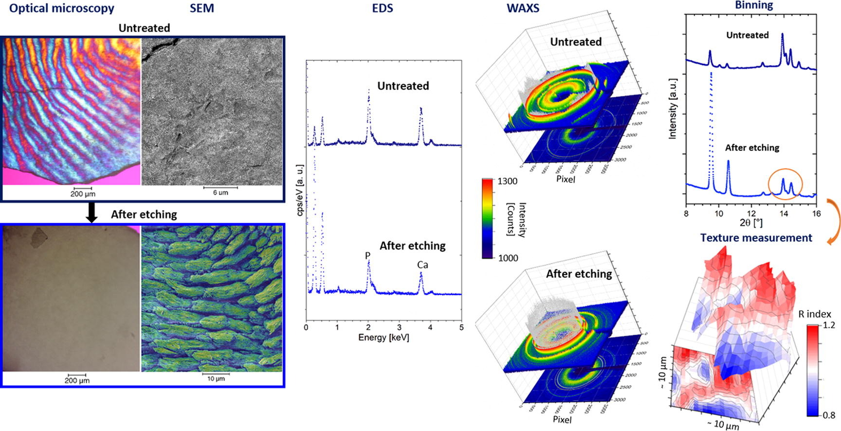
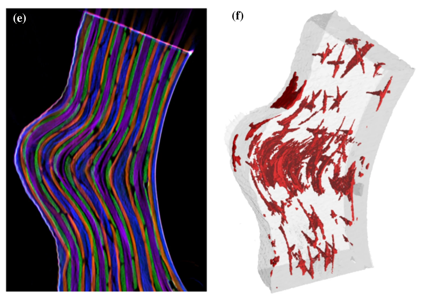
Enamel caries is a highly prevalent worldwide disease that involves the demineralisation of the outer tooth structure. In this study, we report the analysis of artificially demineralised human enamel sections (‘slices’) etched using lactic acid (2% v/v) in comparison with healthy enamel using correlative techniques of optical and electron microscopy, as well as scanning diffraction. Demineralisation of the enamel was characterised at the micron to sub-micron scale. The structure of the he... Read more
Cyril Besnard, Robert A. Harper, Thomas E. J. Moxham, Jonathan D. James, Malte Storm, Enrico Salvati, Gabriel Landini, Richard M. Shelton, Alexander M.Korsunsky
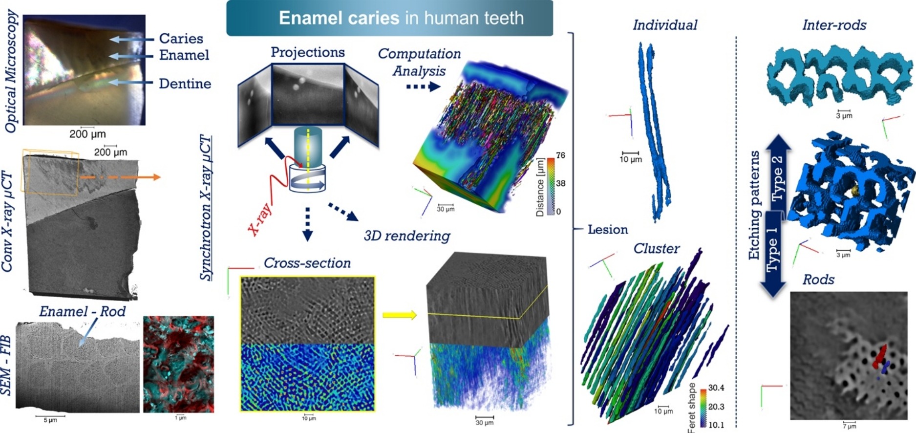
Unprecedented combination of resolution, field of view and contrast for the analysis human enamel carious lesions was achieved. Synchrotron X-ray micro-computed tomography revealed sub-micron details of enamel rod and inter-rod regions inaccessible by laboratory tomography. Successful segmentation and labelling allowed the extraction of enamel etching patterns and statistics. Correlation was obtained between synchrotron X-ray micro-tomography and FIB-SEM cross-sec... Read more
Cyril Besnard, Robert A. Harper, Thomas E. J. Moxham, Jonathan D. James, Malte Storm, Enrico Salvati, Gabriel Landini, Richard M. Shelton, Alexander M.Korsunsky
The high strength at moderate weight in combination with superior corrosion and fatigue properties makes carbon-fiber-reinforced polymer (CFRP) an attractive material for lightweight applications in aerospace. Nonetheless, besides several benefits, CFRP components also bear significant risks like a low resistance to impact damage. […] In this work, we present a multimodal approach to three-dimensionally quantify and visualize fiber orientation and resin-rich areas in carbon-fiber-reinf... Read more
Jonathan Glinz, Jan Šleichrt, Daniel Kytýř, Santhosh Ayalur-Karunakaran, Simon Zabler, Johann Kastner & Sascha Senck
Thermal Runaway of a Li-Ion Battery Studied by Combined ARC and Multi-Length Scale X-ray CT
Lithium ion battery failure occurs across multiple length scales. In this work, the properties of thermal failure and its effects on electrode materials were investigated in a commercial battery using a combination of accelerating rate calorimetry (ARC) and multi-length scale X-ray computed tomography (CT). ARC measured the heat dissipated from the cell during thermal runaway and enabled the identification of key thermal failure characteristics such as onset temperature and the rate of heat g... Read more
Drasti Patel, James B. Robinson, Sarah Ball, Daniel J. L. Brett and Paul R. Shearing
SARS-CoV-2 structure and replication characterized by in situ cryo-electron tomography
Severe acute respiratory syndrome coronavirus 2 (SARS-CoV-2), the causative agent of the COVID19 pandemic, is a highly pathogenic β-coronavirus. As other coronaviruses, SARS-CoV-2 is enveloped, replicates in the cytoplasm and assembles at intracellular membranes. Here, we structurally characterize the viral replication compartment and report critical insights into the budding mechanism of the virus, and the structure of extracellular virions close to their native state by in situ cryo-electr... Read more
Steffen Klein, Mirko Cortese, Sophie L. Winter, Moritz Wachsmuth-Melm, Christopher J. Neufeldt, Berati Cerikan, Megan L. Stanifer, Steeve Boulant, Ralf Bartenschlager, Petr Chlanda