Welcome to the Amira-Avizo Software Use Case Gallery
Below you will find a collection of use cases of our 3D data visualization and analysis software. These use cases include scientific publications, articles, papers, posters, presentations or even videos that show how Amira-Avizo Software is used to address various scientific and industrial research topics.
Use the Domain selector to filter by main application area, and use the Search box to enter keywords related to specific topics you are interested in.
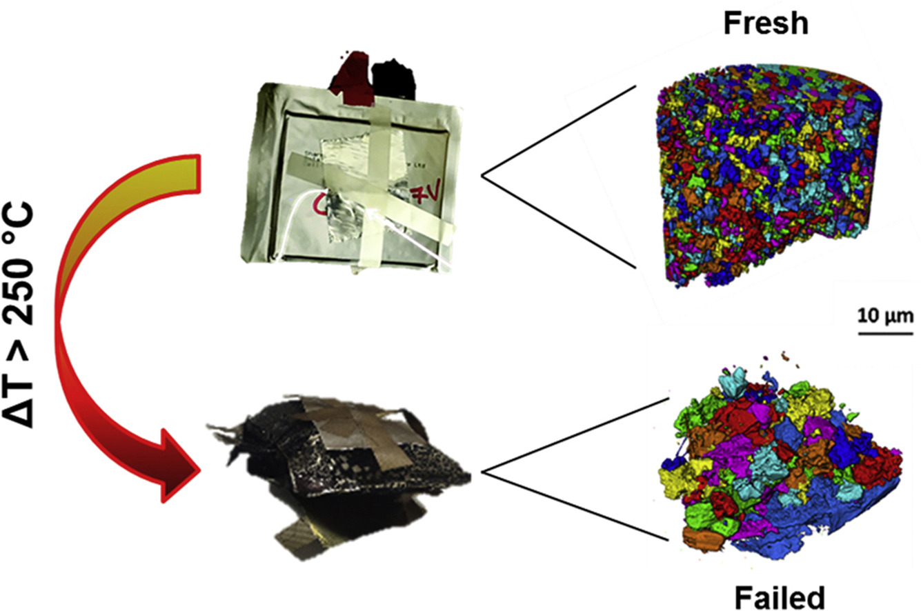
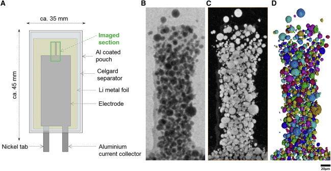
Multiscale tomographic analysis of the thermal failure of Na-Ion batteries
In recent years, the ability to examine the processes that cause the catastrophic failure of batteries as a result of thermal runaway has improved substantially. In this work, the effect of thermal runaway on the microstructure of the electrodes of a Na-ion battery is examined using X-ray computed tomography for the first time. The thermal failure induced via accelerating rate calorimetry enabled the examination of failed electrodes, which were subsequently compared with fresh s... Read more
Robinson, J. B., Heenan, T. M. M., Jervis, J. R., Tan, C., Kendrick, E., Brett, D. J. L., & Shearing, P. R.
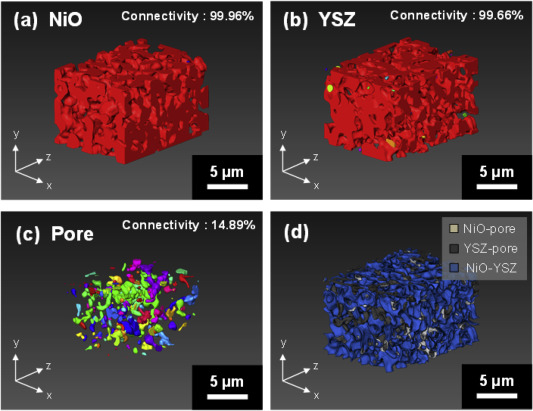
Nickel-yttria-stabilized zirconia (Ni-YSZ) cermet is widely used as an anode material in solid oxide fuel cells (SOFCs); however, Ni re-oxidation causes critical problems due to volume expansion, which causes high thermal stress. We fabricated a Ni-YSZ anode functional layer (AFL), which is an essential component in high-performance SOFCs, and re-oxidized it to investigate the related three-dimensional (3D) microstructural and thermo-mechanical effects. A 3D model of the re-oxidized AFL ... Read more
Jun Woo Kim, Kiho Bae, Hyun Joong Kim, Ji-won Son, Namkeun Kim, Stefan Stenfelt, Fritz B. Prinz, Joon Hyung Shim
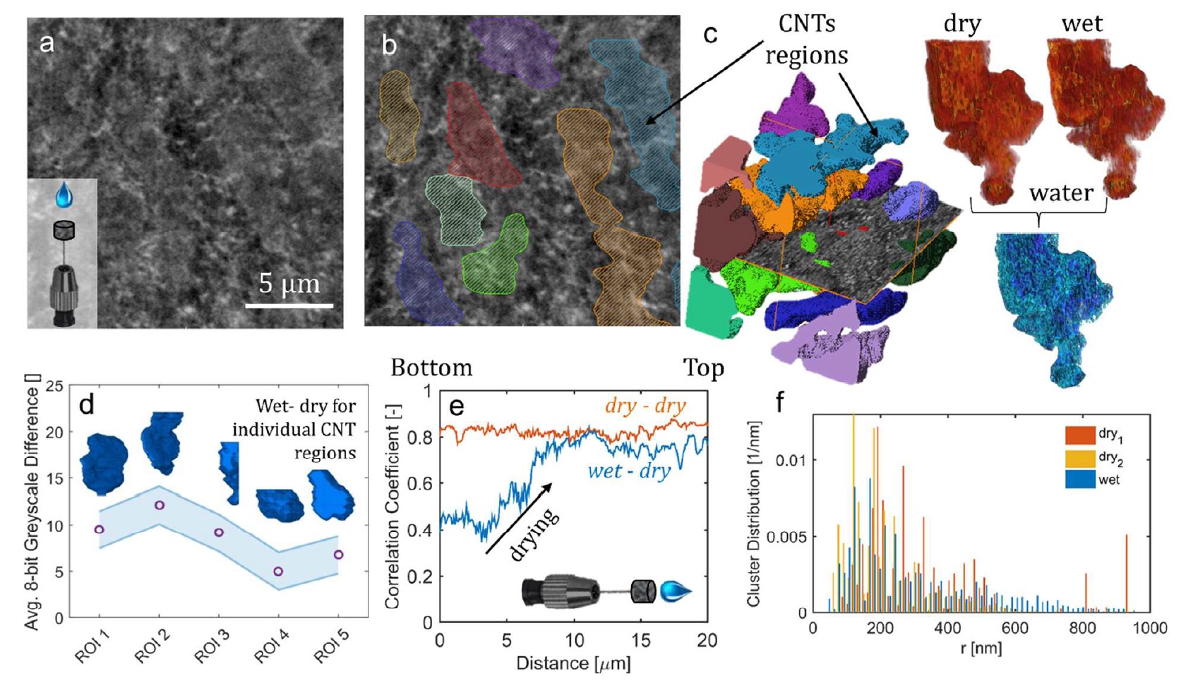
This work describes the performance improvement of a polymer electrolyte fuel cell with a novel class of microporous layers (MPLs) that incorporates hydrophilic additives: one with 30 μm aluminosilicate fibers and another with multiwalled carbon nanotubes with a domain size of 5 μm. Higher current densities at low potentials were observed for cells with the additive-containing MPLs compared to a baseline cell with a conventional MPL, which correlate with improvements in water management. Th... Read more
Dusan Spernjak, Rangachary Mukundan, Rodney L. Borup, Liam Connolly, Benjamin Zackin, Vincent De Andrade, Michael Wojcik, Dilworth Y. Parkinson, David Jacobson, Daniel Seth Hussey, Karren L More, Thomas Chan, Adam Z Weber, and Iryna V. Zenyuk
Cryo-STEM mapping of solid–liquid interfaces and dendrites in lithium-metal batteries
Solid–liquid interfaces are important in a range of chemical, physical and biological processes but are often not fully understood owing to the lack of high-resolution characterization methods that are compatible with both solid and liquid components. For example, the related processes of dendritic deposition of lithium metal and the formation of solid–electrolyte interphase layers are known to be key determinants of battery safety and performance in high-energy-density lithium-metal bat... Read more
Michael J. Zachman, Zhengyuan Tu, Snehashis Choudhury, Lynden A. Archer & Lena F. Kourkoutis
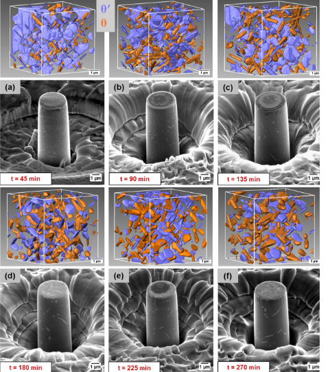
A unique approach to correlating an evolving 3D microstructure in an Al-Cu alloyand its micro-scale mechanical properties has been introduced. Using these nanoscale three-dimensional microstructures derived from Transmission X-rayMicroscopy (TXM), individual contributions from different strengthening mechanisms were quantified. The spatial distribution and morphology of the individual θ′ and θ phases were seen to play an important role in influencing dislocation storage. Uniaxi... Read more
C. Shashank Kaira, Christopher Kantzos, Jason J. Williams, Vincent De Andrade, Francesco De Carlo, Nikhilesh Chawlaa
Mesoscale characterization of local property distributions in heterogeneous electrodes
The performance of electrochemical devices depends on the three-dimensional (3D) distributions of microstructural features in their electrodes. Several mature methods exist to characterize 3D microstructures over the microscale (tens of microns), which are useful in understanding homogeneous electrodes. However, methods that capture mesoscale (hundreds of microns) volumes at appropriate resolution (tens of nm) are lacking, though they are needed to understand more common, less ideal electrode... Read more
Tim Hsu, William K. Epting, Rubayyat Mahbub, Noel T. Nuhfer, Sudip Bhattachary, Yinkai Lei, Herbert M. Miller, Paul R. Ohodnicki, Kirk R. Gerdes, Harry W. Abernathy, Gregory A. Hackett, Anthony D. Rollett, Marc De Graef, Shawn Litster, Paul A. Salvador
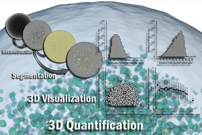
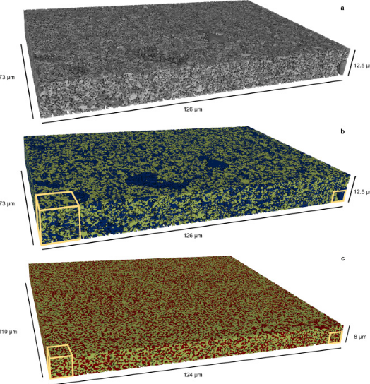
The three-dimensional (3D) characterization of nuclear fuel with X-ray microscopy has historically proven difficult, due to uranium’s high attenuation of easily accessible X-rays, both in a laboratory setting and at a synchrotron user facility. However, this imaging modality provides nondestructive information that can be used to investigate morphological changes arising from external stimuli (e.g., neutron irradiation, high-temperature testing).
Using an appropriate X-ray energy spe... Read more
Nikolaus L. Cordes, William C. Chuirazzi & Joshua J. Kane - John D. Stempien
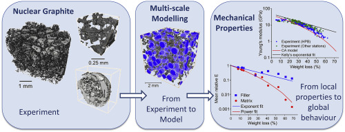
A multi-scale approach for fracture simulation, based on the Cellular Automata technique, has been developed and then applied to a nuclear graphite that is used in structural components of the UK Advanced Gas-cooled Reactors (AGR).
High resolution X-ray computed tomographs of Gilsocarbon grade
graphite, with up to 68% weight loss by radiolytic oxidation, provide quantitative descriptions of the porosity within its constitutive filler particles and their surrounding matrix. The st... Read more
Yelena Vertyagina, Thomas James Marrow
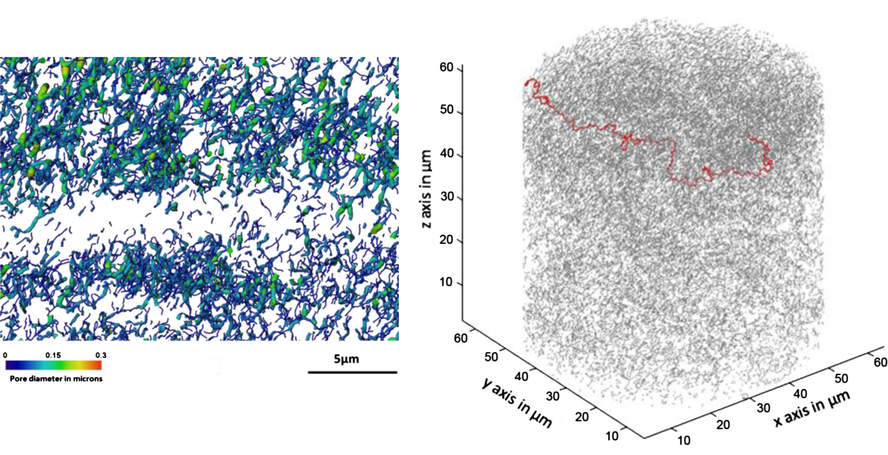
Microstructural analysis of TRISO particles using multi-scale X-ray computed tomography
TRISO particles, a composite nuclear fuel built up by ceramic and graphitic layers, have outstanding high temperature resistance. TRISO fuel is the key technology for High Temperature Reactors (HTRs) and the Generation IV Very High Temperature Reactor (VHTR) variant.
TRISO offers unparalleled containment of fission products and is extremely robust during accident conditions. An understanding of the thermal performance and mechanical properties of TRISO fuel requires a detailed knowledg... Read more
T. Lowe, R.S. Bradley, S. Yue, K. Barii, J. Gelb, N. Rohbeck, J. Turner, P.J. Withers
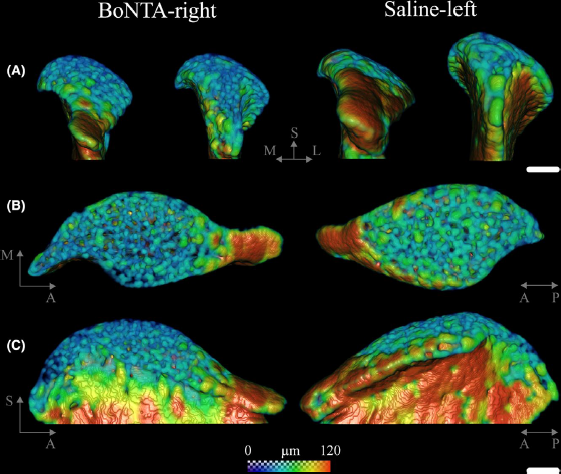
Masseter muscle function influences mandibular bone homeostasis. As previously reported, bone resorption markers increased in the mouse mandibular condyle two days after masseter paralysis induced with botulinum toxin type A (BoNTA), followed by local bone loss.
This study aimed to evaluate the bone quality of both the mandibular condyle and alveolar process in the mandible of adult mice during the early stage of a BoNTA‐induced masseter muscle atrophy, using a combined 3D histomorpho... Read more
Julián Balanta‐Melo, María Angélica Torres‐Quintana, Maximilian Bemmann, Carolina Vega, Constanza González, Kornelius Kupczik, Viviana Toro‐Ibacache, Sonja Buvinic
Synergistic role of nucleotides and lipids for the self-assembly of Shs1 septin oligomers
Amira capacities for membranes and filaments segmentation in cryo-TEM images are featured on the front cover of Biochemical Journal, July 2020.
Budding yeast septins are essential for cell division and polarity. (…) [The authors] have dissected, here, for the first time, the behavior of the Shs1 protomer bound to membranes at nanometer resolution, in complex with the other septins. Using electron microscopy, [the authors] have shown that on membranes, Shs1 protomers self-assembl... Read more
Cyntia Taveneau, Rémi Blanc, Gerard Pehau-Arnaudet, Aurélie Cicco, Aurélie Bertin
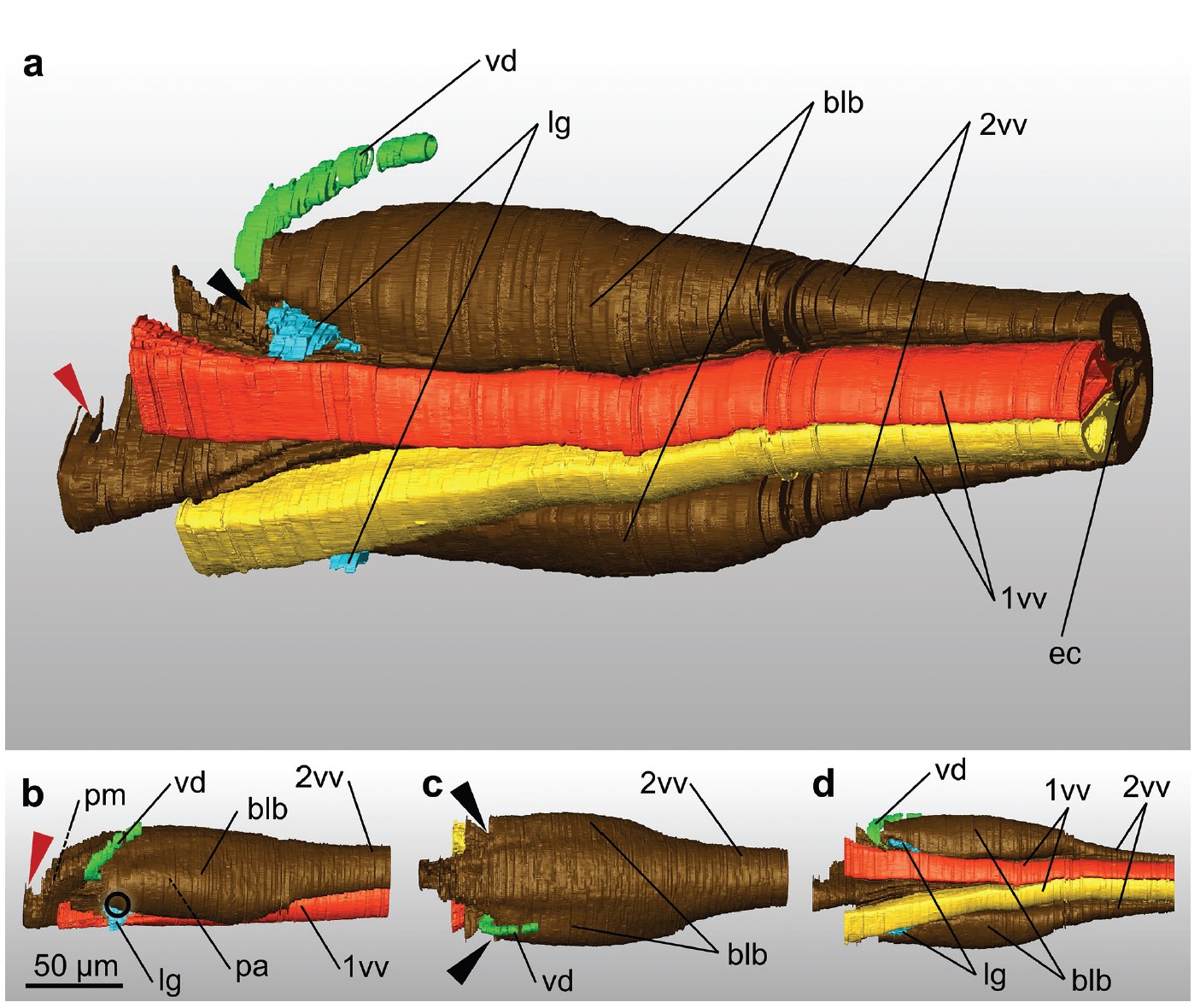
Ovipositor of the braconid wasp Habrobracon hebetor: structural and functional aspects
The Braconidae are a megadiverse and ecologically highly important group of insects. The vast majority of braconid wasps are parasitoids of other insects, usually attacking the egg or larval stages of their hosts. The ovipositor plays a crucial role in the assessment of the potential host and precise egg laying. We used lightand electron-microscopic techniques to investigate all inherent cuticular elements of the ovipositor (the female 9th abdominal tergum, two pairs of valvifers, and three p... Read more
Michael Csader, Karin Mayer, Oliver Betz, Stefan Fischer, Benjamin Eggs

Protocols for Generating Surfaces and Measuring 3D Organelle Morphology Using Amira
High-resolution 3D images of organelles are of paramount importance in cellular biology. Although light microscopy and transmission electron microscopy (TEM) have provided the standard for imaging cellular structures, they cannot provide 3D images.
However, recent technological advances such as serial block-face scanning electron microscopy (SBF-SEM) and focused ion beam scanning electron microscopy (FIB-SEM) provide the tools to create 3D images for the ultrastructural analysis of org... Read more
Edgar Garza-Lopez, Zer Vue, Prasanna Katti, Kit Neikirk, Michelle Biete, Jacob Lam, Heather K. Beasley, Andrea G. Marshall, Taylor A. Rodman, Trace A. Christensen, Jeffrey L. Salisbury, Larry Vang, Margaret Mungai, Salma Ash Shareef, Sandra A. Murray, Jianqiang Shao, Jennifer Streeter, Brian Glancy, Renata O. Pereira1, E. Dale Abel, and Antentor Hinton, Jr.
The molecular basis for sarcomere organization in vertebrate skeletal muscle
Sarcomeres are force-generating and load-bearing devices of muscles. A precise molecular picture of how sarcomeres are built underpins understanding their role in health and disease. Here, we determine the molecular architecture of native vertebrate skeletal sarcomeres by electron cryo-tomography.
Our reconstruction reveals molecular details of the three-dimensional organization and interaction of actin and myosin in the A-band, I-band, and Z-disc and demonstrates that α-actinin cros... Read more
Zhexin Wang, Michael Grange, Thorsten Wagner, Ay Lin Kho, Mathias Gautel, Stefan Raunser
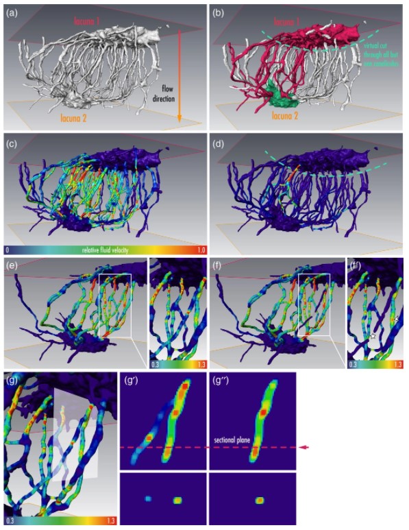
Osteocytes are the most frequent bone cells connected with each other through cell processes within tiny tubular-shaped canaliculi. The so-called osteocyte lacunar-canalicular network (LCN) plays a crucial role in bone remodeling and mineral homeostasis. Given the critical nature of these functions, it is herein hypothesized that the LCN must be structurally “overengineered” to provide network resilience.
This hypothesis is tested by characterizing canalicular networks in human bon... Read more
Emely Bortel, Liam M Grover, Neil Eisenstein, Christian Seim, Heikki Suhonen, Alexandra Pacureanu, Peter Westenberger, Kay Raum, Max Langer, Francoise Peyrin, Owen Addison, Bernhard Hesse
Nickel-rich transition metal oxide materials […] are of great interest for achieving immediate improvements in the energy density of Li-ion batteries and for risk reduction within the Li-ion battery supply chain.
[…] An increase in Ni content in NMC materials leads to accelerated degradation […]. This potentially complicates their adoption in applications requiring extended cycle life such as in electric vehicles.
Recent developments in X-ray characterization to... Read more
ChunTan, Andrew S.Leach, Thomas M.M. Heenan, Huw Parks, Rhodri Jervis, Johanna Nelson Weker, Daniel J.L. Brett, Paul R.Shearing
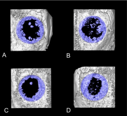
Collagen-Based Matrices for Osteoconduction: A Preclinical In Vivo Study
The aim of this study was to evaluate the influence of additional hydroxyapatite (HA) in collagen-based matrices (CM) and membrane placement on bone formation in calvarial defects.
Critical size defects in the calvaria of 16 New Zealand White Rabbits were randomly treated with CM or mineralized collagen-based matrices (mCM). Half of the sites were covered with a collagen membrane. Animals were euthanized after 12 weeks of healing. The samples were studied by micro-CT and histology. New... Read more
Hiroki Katagiri, Yacine El Tawil, Niklaus P. Lang, Jean-Claude Imber, Anton Sculean, Masako Fujioka-Kobayashi and Nikola Saulacic
Membrane architecture of pulmonary lamellar bodies revealed by post-correlation on-lamella cryo-CLEM
Lamellar bodies (LBs) are surfactant rich organelles in alveolar type 2 cells. LBs disassemble into a lipid-protein network that reduces surface tension and facilitates gas exchange at the air-water interface in the alveolar cavity. Current knowledge of LB architecture is predominantly based on electron microscopy studies using disruptive sample preparation methods. We established a post-correlation on-lamella cryo-correlative light and electron microscopy approach for cryo-FIB milled lung ce... Read more
Steffen Klein, Benedikt H. Wimmer, Sophie L. Winter, Androniki Kolovou, Vibor Laketa, Petr Chlanda
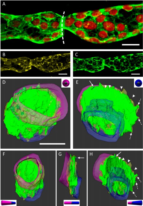
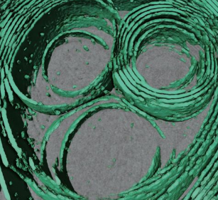
3D Dissection of Structural Membrane-Wall Contacts in Filamentous Moss Protonemata
Cell-to-cell contact is essential for communication and development of multicellular organisms. A prerequisite is the passage through membranes. That way, molecular exchange and information flow is regulated via hormones, membrane proteins and pores.
In plants, the rigid cell walls prevent large membrane contact areas between protoplasts. Only plasmodesmata, minute channels between adjacent cells, form direct connections. Often, molecular data of the proteins involved are manifold but t... Read more
Dominik Harant and Ingeborg Lang
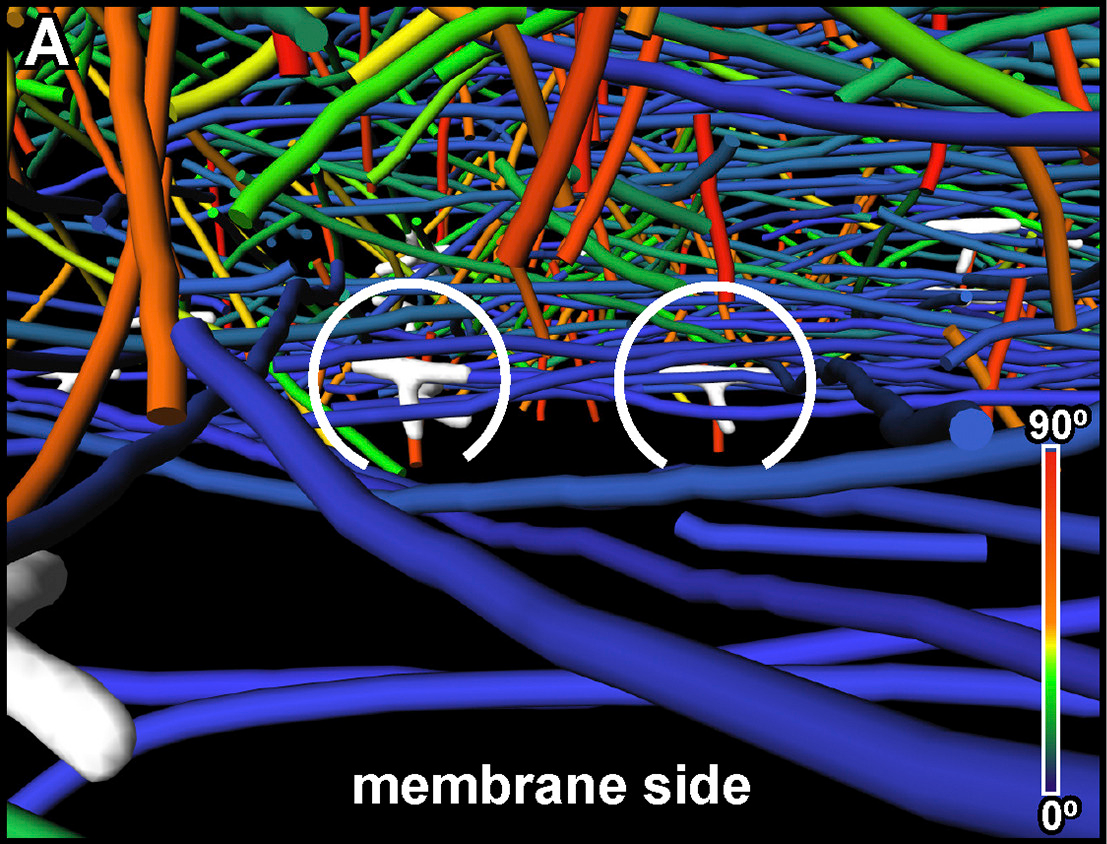
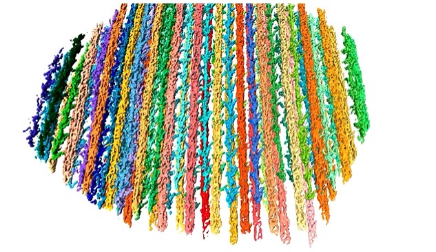
The Architecture of Traveling Actin Waves Revealed by Cryo-Electron Tomography
Actin waves are dynamic supramolecular structures involved in cell migration, cytokinesis, adhesion, and neurogenesis. Although wave-like propagation of actin networks is a widespread phenomenon, the actin architecture underlying wave propagation remained unknown. In situ cryo-electron tomography of Dictyostelium cells unveils the wave architecture and provides evidence for wave progression by de novo actin nucleation. Subtomogram averaging reveals the structu... Read more
Marion Jasnin, Florian Beck, Mary Ecke, Yoshiyuki Fukuda, Antonio Martinez-Sanchez, Wolfgang Baumeister, Günther Gerisch
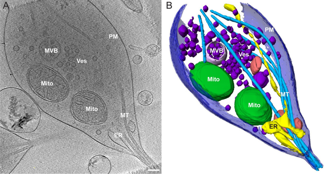
Morphology of mitochondria in spatially restricted axons revealed by cryo-electron tomography
Neurons project axons to local and distal sites and can display heterogeneous morphologies with limited physical dimensions that may influence the structure of large organelles such as mitochondria. Using cryo-electron tomography (cryo-ET), we characterized native environments within axons and presynaptic varicosities to examine whether spatial restrictions within these compartments influence the morphology of mitochondria. Segmented tomographic reconstructions revealed distinctive morphologi... Read more
Tara D. Fischer, Pramod K. Dash, Jun Liu, M. Neal Waxham