Welcome to the Amira-Avizo Software Use Case Gallery
Below you will find a collection of use cases of our 3D data visualization and analysis software. These use cases include scientific publications, articles, papers, posters, presentations or even videos that show how Amira-Avizo Software is used to address various scientific and industrial research topics.
Use the Domain selector to filter by main application area, and use the Search box to enter keywords related to specific topics you are interested in.
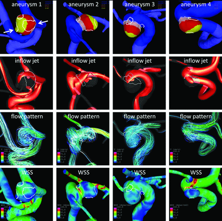
Regional Aneurysm Wall Enhancement is Affected by Local Hemodynamics: A 7T MRI Study
Aneurysm wall enhancement has been proposed as a biomarker for inflammation and instability. However, the mechanisms of aneurysm wall enhancement remain unclear. We used 7T MR imaging to determine the effect of flow in different regions of the wall.
Twenty-three intracranial aneurysms imaged with 7T MR imaging and 3D angiography were studied with computational fluid dynamics. Local flow conditions were compared between aneurysm wall enhancement and nonenhanced regions. Aneurysm wall en... Read more
S. Hadad, F. Mut, B.J. Chung, J.A. Roa, A.M. Robertson, D.M. Hasan, E.A. Samaniego and J.R. Cebral
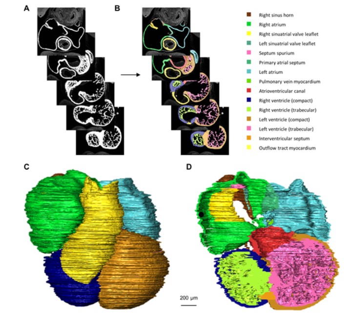
Quantified growth of the human embryonic heart
The size and growth patterns of the components of the human embryonic heart have remained largely undefined.
To provide these data, three-dimensional heart models were generated from immunohistochemically stained sections of ten human embryonic hearts ranging from Carnegie stage 10 to 23. Fifty-eight key structures were annotated and volumetrically assessed. Sizes of the septal foramina and atrioventricular canal opening were also measured. The heart grows exponentially throughout embr... Read more
Jaeike W. Faber, Jaco Hagoort, Antoon F. M. Moorman, Vincent M. Christoffels, Bjarke Jensen
Patient-specific anatomical model for deep brain stimulation based on 7 Tesla MRI
Deep brain stimulation (DBS) requires accurate localization of the anatomical target structure, and the precise placement of the DBS electrode within it. Ultra-high field 7 Tesla (T) MR images can be utilized to create patient-specific anatomical 3D models of the subthalamic nuclei (STN) to enhance pre-surgical DBS targeting as well as post-surgical visualization of the DBS lead position and orientation. We validated the accuracy of the 7T imaging-based patient-specific model of the STN and m... Read more
Yuval Duchin, Reuben R. Shamir, Remi Patriat, Jinyoung Kim, Jerrold L. Vitek, Guillermo Sapiro, Noam Harel
A detailed canine brain label map for neuroimaging analysis
Dogs have recently become an important model species for comparative social and cognitive neuroscience. Brain template-related label maps are essential for functional magnetic resonance imaging (fMRI) data analysis, to localize neural responses. In this study, we present a detailed, individual-based, T1-weighted MRI-based brain label map used in dog neuroimaging analysis. Methods: A typical, medium-headed dog (a 7.5-year-old
male Golden Retriever) was selected from a cohort of ... Read more
Czeibert Kálmán, Andics Attila, Petneházy Örs, Kubinyi Enikő, Kálmán Czeibert
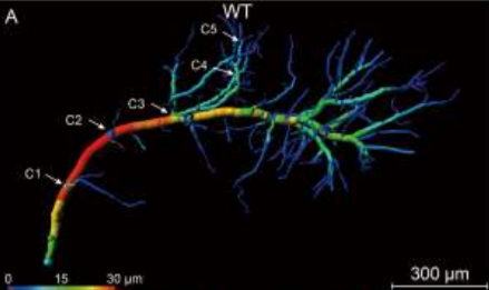
Accumulating evidence indicates the critical importance of cerebrovascular dysfunction in the pathogenesis of Alzheimer’s disease (AD). However, systematic comparative studies on the precise brain vasculature of wild-type and AD model mice are still rare. Using an image optimization method for analyzing Micro-Optical Sectioning Tomography (MOST) data, we generated cross-scale whole-brain 3D atlases that cover the entire vascular system from large vessels down to smallest capillaries at ... Read more
Xiaochuan Zhang, Xianzhen Yin, Jingjing Zhang, Anan Li, Hui Gong, Qingming Luo, Haiyan Zhang, Zhaobing Gao, Hualiang Jiang
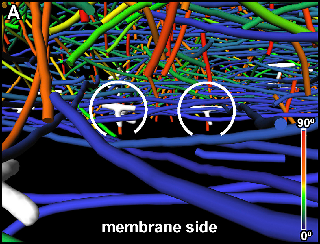
Collagen-Based Matrices for Osteoconduction: A Preclinical In Vivo Study
The aim of this study was to evaluate the influence of additional hydroxyapatite (HA) in collagen-based matrices (CM) and membrane placement on bone formation in calvarial defects.
Critical size defects in the calvaria of 16 New Zealand White Rabbits were randomly treated with CM or mineralized collagen-based matrices (mCM). Half of the sites were covered with a collagen membrane. Animals were euthanized after 12 weeks of healing. The samples were studied by micro-CT and histology. New... Read more
Hiroki Katagiri, Yacine El Tawil, Niklaus P. Lang, Jean-Claude Imber, Anton Sculean, Masako Fujioka-Kobayashi and Nikola Saulacic
Recent treatment developments for metastatic renal cell carcinoma offer combinations of immunotherapies or immunotherapy associated with tyrosine kinase inhibitors (TKI). There is currently no argument to choose one solution or another. Easy-to-use markers to assess longitudinal responses to TKI are necessary to determine when to switch to immunotherapies. These new markers will enable an earlier adaptation of therapeutic strategy in order to prevent tumor development, unnecessary toxicity an... Read more
Alexandre Ingels, Ingrid Leguerney, Paul-Henry Cournède, Jacques Irani, Sophie Ferlicot, Catherine Sébrié, Baya Benatsou, Laurène Jourdain, Stephanie Pitre-Champagnat, Jean-Jacques Patard & Nathalie Lassau
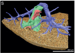
Multiple membrane extrusion sites drive megakaryocyte migration into bone marrow blood vessels
Platelets, cells central to hemostasis and thrombosis, are formed from parent cell megakaryocytes. Although the process is highly efficient in vivo, our ability to generate them in vitro is still remarkably inefficient. We proposed that greater understanding of the process in vivo is needed and used an imaging approach, intravital correlative light electron microscopy, to visualize platelet generation in bone marrow in the living mouse. In contrast to current understanding, we found that most... Read more
Edward Brown, Leo M Carlin, Claus Nerlov, Cristina Lo Celso, Alastair W Poole
The Architecture of Traveling Actin Waves Revealed by Cryo-Electron Tomography
Actin waves are dynamic supramolecular structures involved in cell migration, cytokinesis, adhesion, and neurogenesis. Although wave-like propagation of actin networks is a widespread phenomenon, the actin architecture underlying wave propagation remained unknown. In situ cryo-electron tomography of Dictyostelium cells unveils the wave architecture and provides evidence for wave progression by de novo actin nucleation. Subtomogram averaging reveals the structu... Read more
Marion Jasnin, Florian Beck, Mary Ecke, Yoshiyuki Fukuda, Antonio Martinez-Sanchez, Wolfgang Baumeister, Günther Gerisch
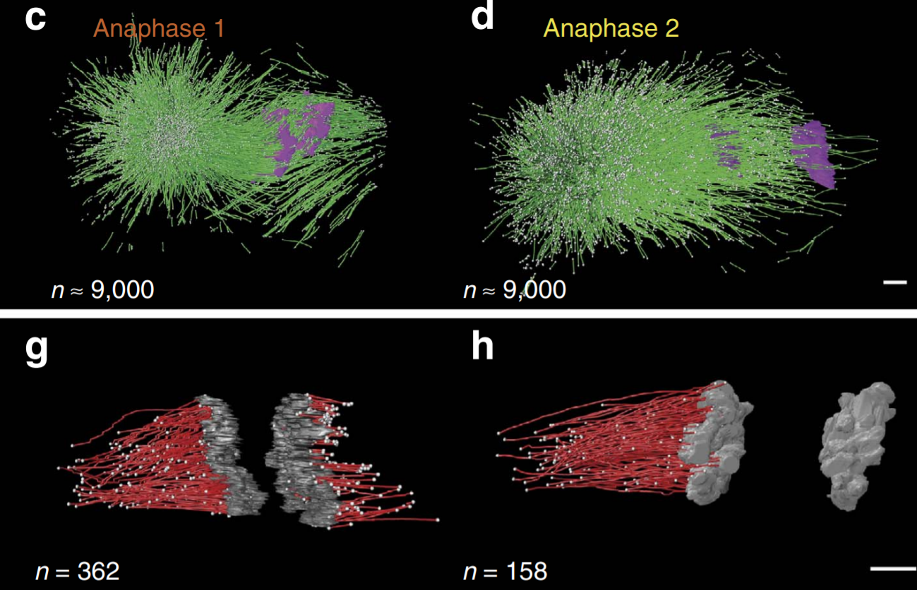
Asymmetric Centriole Numbers at Spindle Poles Cause Chromosome Missegregation in Cancer
Chromosomal instability is a hallmark of cancer and correlates with the presence of extra centrosomes, which originate from centriole overduplication.
Overduplicated centrioles lead to the formation of centriole rosettes, which mature into supernumerary centrosomes in the subsequent cell cycle. While extra centrosomes promote chromosome missegregation by clustering into pseudo-bipolar spindles, the contribution of centriole rosettes to chromosome missegregation is unknown. We us... Read more
Marco R.Cosenza, Anna Cazzola, Annik Rossberg, Nicole L. Schieber, Gleb Konotop, Elena Bausch, Alla Slynko, Tim Holland-Letz, Marc S.Raab, Taronish Dubash, Hanno Glimm, Sven Poppelreuther, Christel Herold-Mende, Yannick Schwab, Alwin Krämer
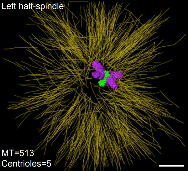
C. elegans chromosomes connect to centrosomes by anchoring into the spindle network
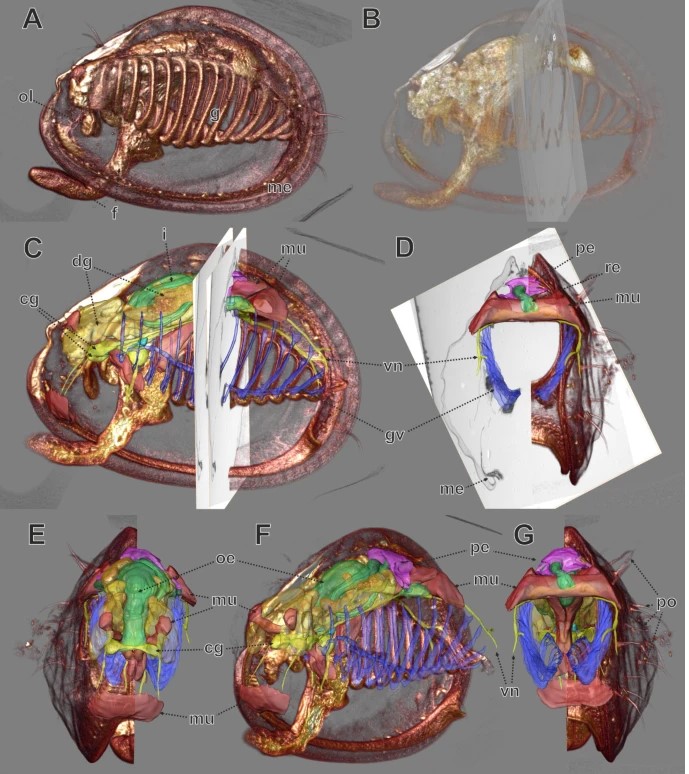
The mitotic spindle ensures the faithful segregation of chromosomes. Here we combine the first large-scale serial electron tomography of whole mitotic spindles in early C. elegans embryos with live-cell imaging to reconstruct all microtubules in 3D and identify their plus- and minus-ends. We classify them as kinetochore (KMTs), spindle (SMTs) or astral microtubules (AMTs) according to their positions, and quantify distinct properties of each class. While our light microscopy and muta... Read more
Stefanie Redemann, Johannes Baumgart, Norbert Lindow, Michael Shelley, Ehssan Nazockdast, Andrea Kratz, Steffen Prohaska, Jan Brugués, Sebastian Fürthauer & Thomas Müller-Reichert

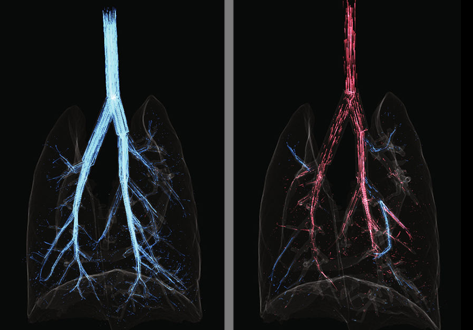
Each pulmonary segment is an anatomical and functional unit. However, it is fundamentally difficult to precisely distinguish every pulmonary segment using the conventional pulmonary intersegmental planes from computed tomography images. Building arteriopulmonary segments is likely to be an effective way to identify pulmonary segments.
The three-dimensional reconstructed images showed the branches of the pulmonary artery ramified up to their eighth order covering the entire lung as well... Read more
Huijie Gao, Chao Liu
Cardiogenic Airflow in the Lung Revealed Using Synchrotron-Based Dynamic Lung Imaging
The beating heart is known to produce pressure and airflow oscillations in the lungs of mammals. This phenomenon is often disregarded as detailed measurement of its effects in the lung have hitherto not been possible. Previous studies have attempted to measure the effect of these oscillations on gas mixing. However, the results have proven inconclusive, due to the lack of a direct measurement tool capable of flow measurement throughout the entire bronchial tree. Here we present the first deta... Read more
Stephen Dubsky, Jordan Thurgood, Andreas Fouras, Bruce R. Thompson & Gregory J. Sheard
Multiscale Co‐reconstruction of Lung Architectures and Inhalable Materials Spatial Distribution
Pulmonary diseases, such as chronic obstructive pulmonary diseases, lower respiratory infections, and lung cancer are listed in the top ten causes of deaths globally with more than 10 million mortality per year. Apart from oral administration, the Dry Powder Inhalation (DPI) for pulmonary administration provides an important alternative route for targeted treatment of these pulmonary diseases. […] Furthermore, there is a growing demand on und... Read more
Xian Sun, Xiaochuan Zhang, Xiaohong Ren, Hongyu Sun, Li Wu, Caifen Wang, Xiaohui Ye, Peter York, Zhaobing Gao, Hualiang Jiang, Jiwen Zhang, Xianzhen Yin
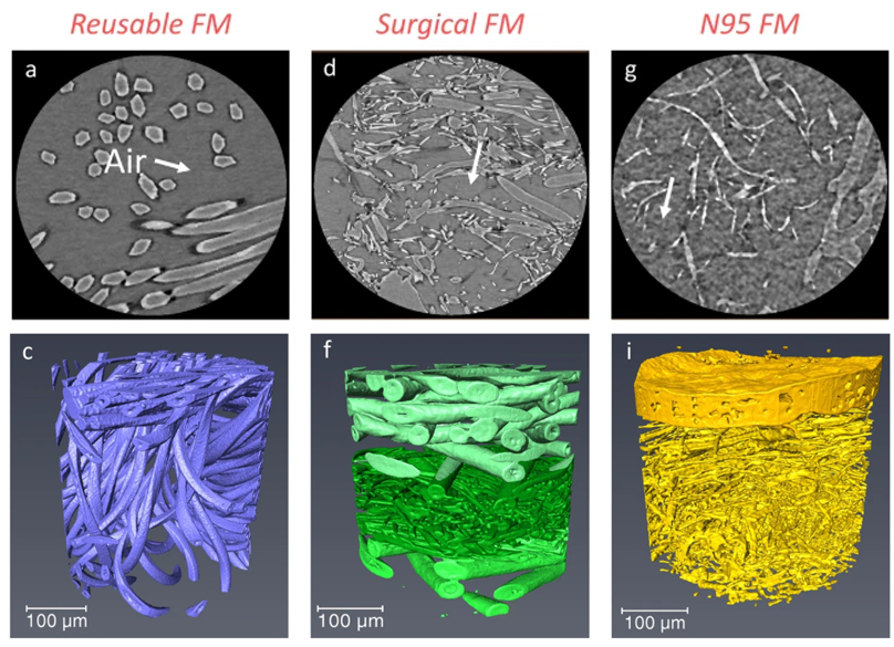
Microstructure analysis and image-based modelling of face masks for COVID-19 virus protection
Until March 2021, around 120 million coronavirus disease (COVID-19) infected cases and over 2.6 million deaths have been reported worldwide. […] Recent investigations have implied that face masks help to reduce the disease transmission and therefore slow down the growth of the epidemic curve. However, there are still ongoing debates on the efficacy of wearing masks […] since there is a general lack of information relating to the material structure of commonly used face masks.Read more
Wenjia Du, Francesco Iacoviello, Tacson Fernandez, Rui Loureiro, Daniel J. L. Brett & Paul R. Shearing
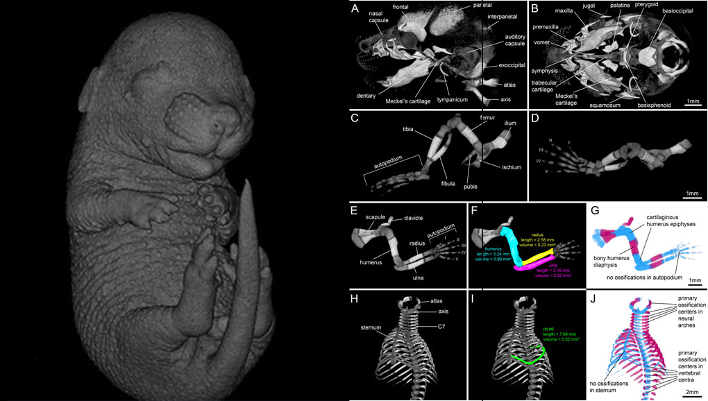
For decades, clearing and staining with Alcian Blue and Alizarin Red has been the gold standard to image vertebrate skeletal development. Here, we present an alternate approach to visualise bone and cartilage based on X-ray microCT imaging, which allows the collection of genuine 3D data of the entire developing skeleton at micron resolution.
Our novel protocol is based on ethanol fixation and staining with Ruthenium Red, and efficiently contrasts cartilage matrix, as demonstrated in wh... Read more
Simone Gabner, Peter Böck, Dieter Fink, Martin Glösmann, Stephan Handschuh
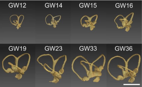
Progressive transformation of the otic placode into the functional inner ear during gestational development in humans leads to the acquisition of hearing perception via the cochlea and balance and spatial orientation via the vestibular organ.
Using a correlative approach involving micro-computerized tomography (micro-CT), transmission electron microscopy and histological techniques we were able to examine both the morphological and cellular changes associated with human inner ear devel... Read more
Lejo Johnson Chacko, David Wertjanz, Consolato Sergi, Jozsef Dudas, Natalie Fischer, Theresa Eberharter, Romed Hoermann, Rudolf Glueckert, Helga Fritsch, Helge Rask-Andersen, Anneliese Schrott-Fischer & Stephan Handschuh
In biomedical research, a huge variety of different techniques is currently available for the structural examination of small specimens, including conventional light microscopy (LM), transmission electron microscopy (TEM), confocal laser scanning microscopy (CLSM), microscopic X-ray computed tomography (microCT), and many others. Since every imaging method is physically limited by certain parameters, a correlative use of complementary methods often yields a significant broader range of inform... Read more
Stephan Handschuh, Natalie Baeumler, Thomas Schwaha & Bernhard Ruthensteiner
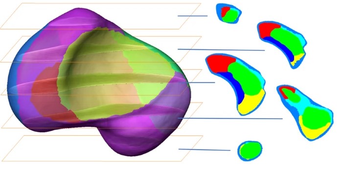
3D computational anatomy of the scaphoid and its waist for use in fracture treatment
A detailed understanding of scaphoid anatomy helps anatomic fracture reduction and optimal screw position. Therefore, we analyzed the size and shape variations of the cartilage and osseous surface, the distribution of volumetric bone mineral density (vBMD), and if the vBMD values differ between a peripheral and a central screw pathway?
Forty-three fresh frozen hand specimens (17 females, 26 males) were analysed with high-resolution peripheral quantitative computed tomography (HR-pQCT) ... Read more
Marc-Daniel Ahrend, Teun Teunis, Hansrudi Noser, Florian Schmidutz, Geoff Richards, Boyko Gueorguiev & Lukas Kamer
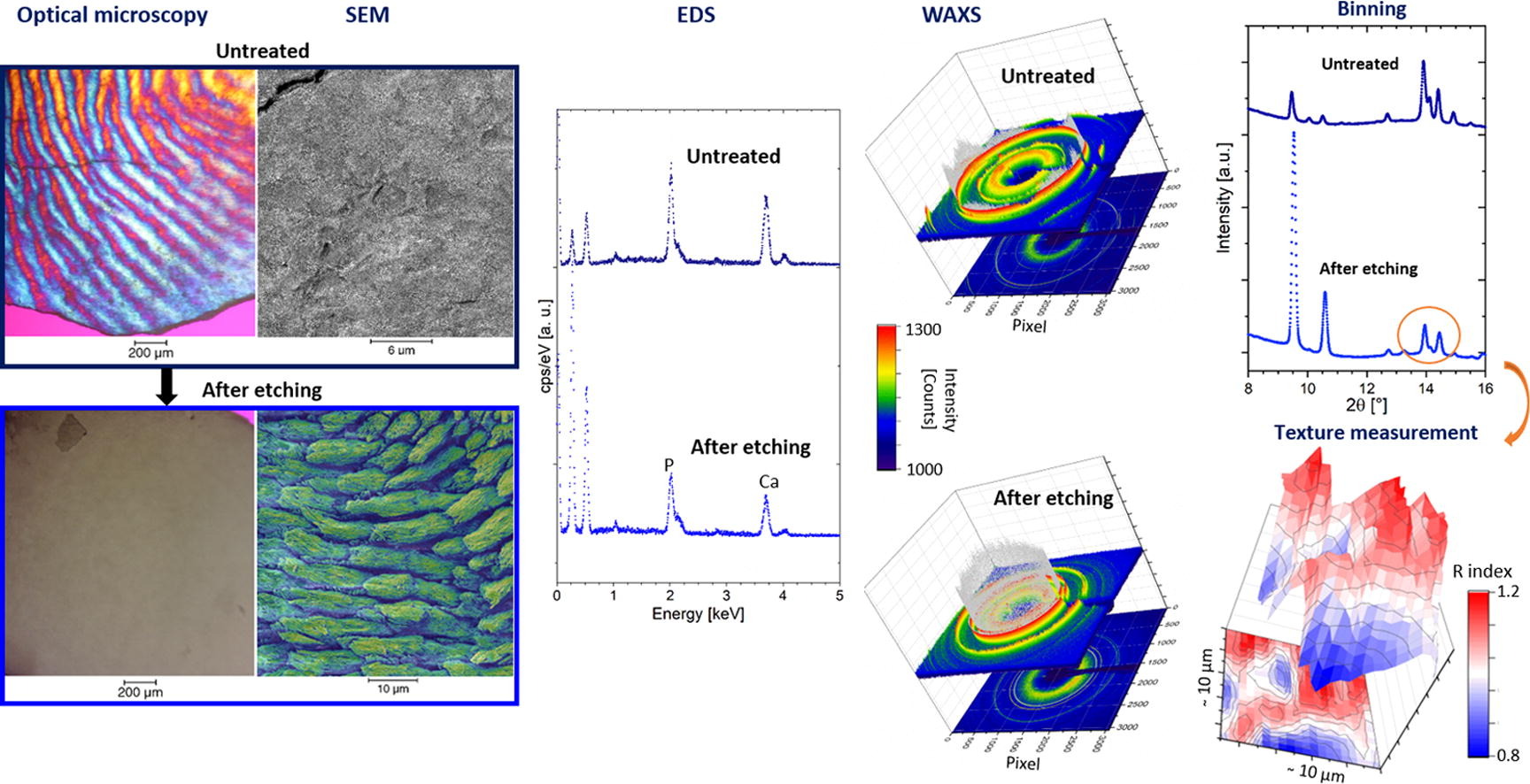
Enamel caries is a highly prevalent worldwide disease that involves the demineralisation of the outer tooth structure. In this study, we report the analysis of artificially demineralised human enamel sections (‘slices’) etched using lactic acid (2% v/v) in comparison with healthy enamel using correlative techniques of optical and electron microscopy, as well as scanning diffraction. Demineralisation of the enamel was characterised at the micron to sub-micron scale. The structure of the he... Read more
Cyril Besnard, Robert A. Harper, Thomas E. J. Moxham, Jonathan D. James, Malte Storm, Enrico Salvati, Gabriel Landini, Richard M. Shelton, Alexander M.Korsunsky
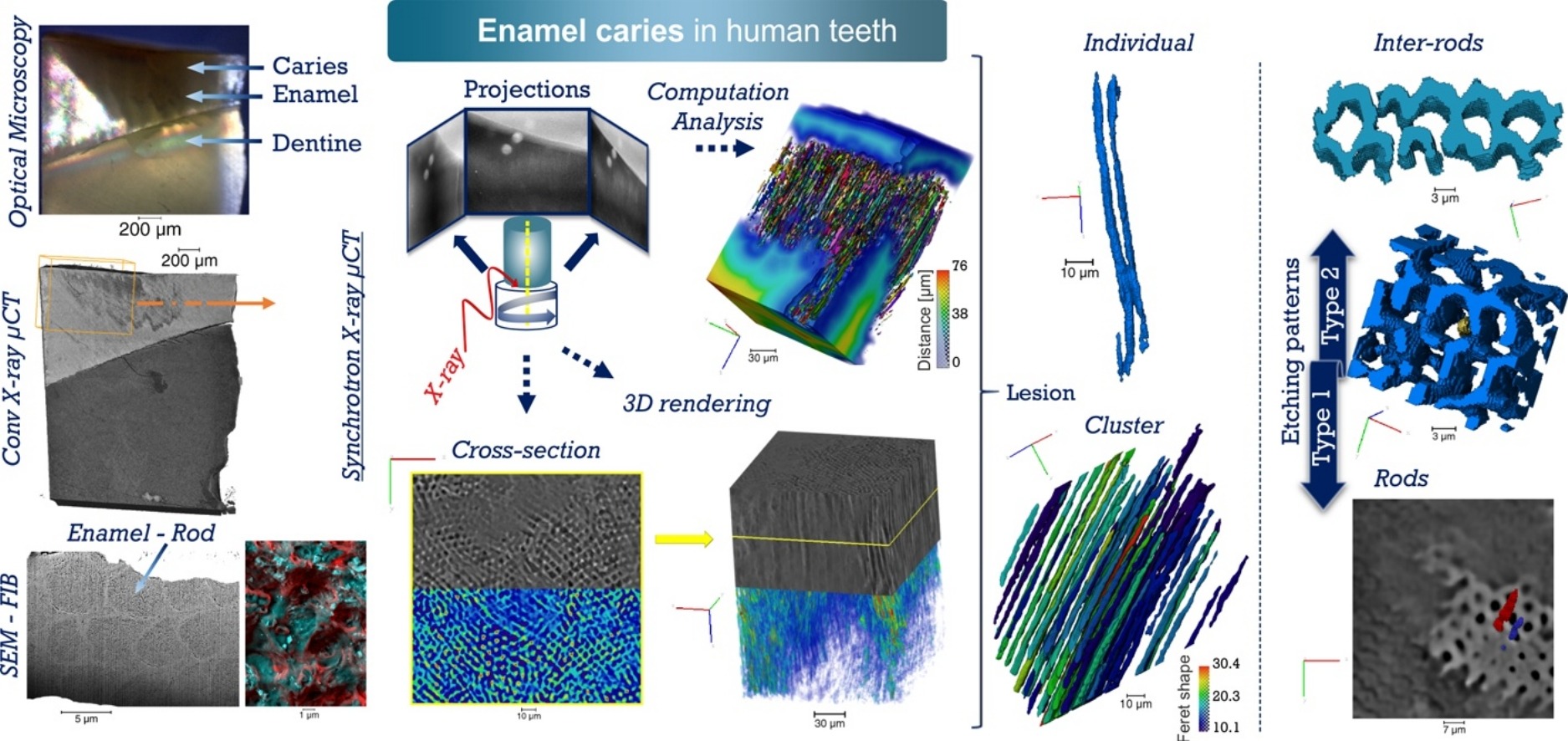
Unprecedented combination of resolution, field of view and contrast for the analysis human enamel carious lesions was achieved. Synchrotron X-ray micro-computed tomography revealed sub-micron details of enamel rod and inter-rod regions inaccessible by laboratory tomography. Successful segmentation and labelling allowed the extraction of enamel etching patterns and statistics. Correlation was obtained between synchrotron X-ray micro-tomography and FIB-SEM cross-sec... Read more
Cyril Besnard, Robert A. Harper, Thomas E. J. Moxham, Jonathan D. James, Malte Storm, Enrico Salvati, Gabriel Landini, Richard M. Shelton, Alexander M.Korsunsky